SlideShare a Scribd company logo
INTRODUCTION
Minimal access surgery is a routine surgical practice due
to its minimal invasive and associated advantages. It has a
lot of advantages but not devoid of complication, one of the
major concerned complication is the trocar site herniation
(TSH). TSH is a serious complication often requiring
emergency repair. If unattended, TSH can lead to small
bowel strangulation and incarceration.
The literature says that preventative measures should
be taken to avoid the occurrence of herniation at the portal
site. Fascial closure has been recommended as a means of
TSH prevention. One study reported a statistically higher
frequency of hernia at 12 mm port site where the fascia was
left open (8%) compared with those that were closed (0.22%)
following laparoscopy. There is a consensus that all the
port site ≥10 mm should be closed due to an increased risk
of herniation. For smaller ports, fascial closure may not be
necessary, except when manipulated extensively.
Trocar site herniation is also associated with other
technical factors other than the port site. Port location is
another factor. There are many reports suggesting that
umbilical sites are at greater risk of herniation when
compared to lateral port sites. This is due to weakness
of the fascia and absence of supporting muscle in the
area. Stretching or even extending the incision of a
port site during specimen extraction has a great risk
hernia development. Factors such as high body mass
index (BMI) are patient-related risk factors that are
associated with TSH. Here it is related to increase intra-
abdominal pressure and increase abdominal wall thickness.
Studies show that wound infection is a predisposing factor
to hernia development. Therefore, closure of fascia is
necessary for umbilical ports, ports sites that are stretched
or enlarged for specimen retrieval, and trocar sites in obese
patients.
Minimally invasive laparoscopic surgery has
revolutionized the way surgery is performed for an
increasing number of patients. Incisional hernia can occur
after any abdominal surgery and laparoscopic surgery is
not immune to this complication. The hernia that follows
laparoscopy usually occurs through the larger ports (size
>10 mm), especially the umbilicus. Fig. 1: Typical port closure needle.
Laparoscopic Port
ClosureTechnique
Predisposing factors include:
	
■ Previous laparoscopies
	
■ Extensive manipulation during surgery
	
■ Increased intra-abdominal pressure
	
■ Obesity
	
■ Use of sharp cutting-tip trocars
	
■ Rapid abdominal deflation at the end of surgery
	
■ Poor port removal techniques and defective closure of
the abdominal fascia
	
■ Wound extension
	
■ Male sex
	
■ Infection of the wound
	
■ Pre-existing umbilical defects
	
■ Postoperative chest infections
	
■ Pre-existing diseases such as diabetes mellitus
	
■ Connective tissue disorders
	
■ Job profile of the patient (weight lifting).
Among all these factors, the single most important factor
remains the improper closure of the fascial defects at the
port sites and not using proper port closure instruments
(Figs. 1 to 3). The diagnosis is often delayed because most
cases present late, and treatment might be instituted along
other lines. Computed tomography scans are helpful in its
diagnosis and will facilitate prompt treatment to avoid the
grave consequence of bowel gangrene.
Prof. Dr. R. K. Mishra
166 SECTION 1: Essentials of Laparoscopy
Fig. 2: Laparoscopic port closure Cobbler’s needle. Fig. 3: Incisional hernia development due to improper closure of
port should be repaired later by mesh.
Fig. 4: The tip of telescope should be introduced in and cannula is
pulled over telescope to prevent suction of omentum or bowel.
While surgical techniques and instrumentation have
made significant advances, it is usual that the surgical
incision is closed using invasive suturing techniques or
by the use of tapes or by the use of topical cyanoacrylate
skin adhesives (TCAs) for closure of surgical wounds. The
incidence of incisional hernia occurring at the port sites after
laparoscopic surgery lies between 0.02 and 3.6% and usually
remainsunreported,untilthedevelopmentofcomplications.
Any port closure technique should have following
characteristics:
	
■ Effective (strong and secure) surgical wound closure
	
■ Faster wound closure
	
■ Better scar cosmesis
	
■ Occlusive microbial wound dressing
	
■ Less tissue trauma, reduced inflammatory reaction
	
■ No requirement for suture/staple removal
	
■ Easy to use/simple learning curve
	
■ Reduced risk of needlestick injury—safety and costs
	
■ Cost effective.
WITHDRAWAL OF INSTRUMENTS AND PORTS
Once the surgery is finished, all the instruments should
be removed carefully under vision. All the accessory ports
should be removed and the gas is removed by releasing the
valve of 10 mm cannulas. The primary port should be taken
out in the end (Fig. 4).
If last port is suddenly withdrawn, sudden suction effect
of cannula can pull the omentum or bowel inside the port
wound, the chance of port-site hernia and adhesion is much
higher in this case. It is a good practice to insert some blunt
instrument or telescope inside the abdomen while removing
the last cannula out over that instrument, to prevent
inadvertent entrapment of omentum or bowel.
The access technique will result in breach in continuity
of abdominal wall which need to be repaired at the end of
surgery. All the 10 mm or >10 mm port should be repaired
properly to prevent any future possibility of hernia. The
rectus sheath should be sutured with Vicryl. Only one stitch
is required in middle which will convert 10 mm wound into
5 mm. The 5 mm port wounds are not necessary to repair.
Laparoscopic Port Closure Instruments
Various types of port closure instruments are available. The
suture passer is a convenient instrument for port closure.
It is used to pass the thread on the side of cannula and then it
is tied externally (Figs. 5A to D).
Port Closure Needle
This is a simple instrument just like cobblers and it can be
effectively used for closing the port. The tip of the instrument
isbluntandtheneedlefacestowardthefascia,sothechances
of injury to the bowel are less with the use of this instrument
(Fig. 6).
167
CHAPTER 12: Laparoscopic Port Closure Technique
C
A B
D
Figs. 5A to D: Port closure with the help of suture passer.
Fig. 6: Port closure needle. Fig. 7: Aneurysm needle.
Aneurysm needle can also be used for closing fascia.
The advantage of this needle is that eye is at the tip and due
to rigid structure there is no risk of bending or rotation of
needle (Fig. 7).
After closing the rectus sheath, the skin can be closed
by intradermal, skin stapler or any of the surgical skin glues
TCAs available (Fig. 8).
New Laparoscopic Port Closure Instruments
Weck®
EFx Shield Fascial Closure
System (Figs. 9A and B)
The Weck®
EFx Shield Fascial Closure System from Teleflex
is the only shielded port closure device, providing enhanced
sharps protection for uniform and consistent performance.
168 SECTION 1: Essentials of Laparoscopy
Fig. 8: Closure of skin wound by skin stapler.
A B
Figs. 9A and B: Weck®
EFx Shield Fascial Closure System.
Figs. 10A and B: NeatClose automated port closure device.
B
A
The EFx Shield®
System is designed with speed and safety in
mind. An array of enhanced features includes:
	
■ Unique shielded wing design for enhanced sharps
protection
	
■ Intuitive wing deployment
	
■ Innovative suture retrieval system for unassisted fascial
closure.
NeatClose Automated Port Closure
Device (Figs. 10A and B)
NeatStitch of Israel has come up with an automated port
closure device known as NeatClose, where it also picked
up both the Food and Drug Administration (FDA) and
European approvals in the process. It is marketed to be an
alternative to manual port closure, making it a speedy and
efficient manner to help laparoscopic surgeon save time
and money by lowering intraoperative costs. This system
lets surgeons produce a watertight seal quickly, and it goes
without saying that this would go a long way in aiding the
recovery of a patient, never mind that one does not have
Wolverine’s healing factor. When inside the operating
cavity, the surgeon can squeeze the handle leavers in order
to release a couple of blunt needle guides, where said guides
are specially positioned in a perpendicular manner to the
169
CHAPTER 12: Laparoscopic Port Closure Technique
port plane. With the activation button pressed, it will release
the needles from the guide, via the tissue and back to the
NeatClose cartridge. Once the system is pulled outside the
port, you can be sure that a safe and efficient airtight seal is
created, hence aiding the recovery of a patient for surgery
quickly. Now we are still waiting for a painless method
without the need for anesthesia.
Carter-Thomason CloseSure System—Port-site
Closure (Figs. 11A and B)
Closing any trocar site is a simple, fast and safe procedure
with the Carter-Thomason CloseSure System. The cone-
shaped Pilot®
Guide correctly angles the suture passer to
achieve full-thickness closure. It closes the port including
fascia and peritoneum (preventing Richter’s hernias)—
while maintaining pneumoperitoneum. The guide’s unique
design ensures precise placement of the suture passer for
consistent, reproducible results on any body type.
VersaOne™ Fascial Closure System (Figs. 12 and 13)
Port-site hernias are serious complications across
procedures and are a burden on patients, clinicians, and
health systems. Appropriate port-site closure is considered
to be one of the most critical factors for the prevention
of port-site herniation. The VersaOne™ Fascial Closure
System is a novel all-in-one device that serves as a trocar
and fascial closure device to deliver consistent port-site
closure and suture placement. The unique system
features a special cannula that allows for defect closure
without the need of additional devices.
As a result, the VersaOne™ Fascial Closure System:
	
■ Provides procedure efficiency
	
■ Eliminates the need to remove the trocar before closing
	
■ Makes reinsufflation unnecessary—pneumoperitoneum
can be maintained throughout the procedure
	
■ Enables tissue layers to remain aligned.
B
A
Figs. 11A and B: Carter-Thomason CloseSure System—port-site closure.
Fig. 12: VersaOne™ Fascial Closure System with its trocar. Fig. 13: VersaOne™ Fascial Closure System demonstrating
insertion of suture.
170 SECTION 1: Essentials of Laparoscopy
Fig. 14: Remove the stylet from the cannula. Fig. 15: Pass a suture material through the cannula from the tip.
Fig. 16: Take suture out from the other end. Fig. 17: Tie the loop and hide the knot in the cannula.
There are a number of methods of post site closure
but there is no gold standard. Use of traditional suturing
techniques are difficult due to blind closure of the fascial
defect. Varying degrees of success are achieved by modified
hand suturing techniques. Finding the rectus sheath and
suturing through the layers of a thicker abdominal wall
through a relatively small hole is challenging particularly
in the obese. In such cases, we need special instrument for
efficientclosureoftheportsite.Veressneedleisaninstrument
that is commonly used for creating pneumoperitoneum but
it has been used to close the port site efficiently under vision.
VERESS NEEDLE TECHNIQUE OF
PORT CLOSURE
In 1983, Janos Veress of Hungary developed a specially
designed spring-loaded needle. Interestingly, Veress did not
promote the use of his needle for laparoscopy purposes. He
used Veress needle for the induction of pneumothorax. But
now Veress needle is the most important instrument today
to create pneumoperitoneum. Veress needle consists of an
outercannulawithabeveledneedlepointforcuttingthrough
tissues. Inside the cannula of Veress needle is an inner stylet,
stylet is loaded with a spring forward in response to the
sudden decrease in pressure encountered upon crossing the
abdominal wall and entering the peritoneal cavity.
TECHNIQUE OF PORT CLOSURE BY VERESS
NEEDLE (FIGS. 14 TO 25)
Occlude the port site with a finger so that the pneumo-
peritoneum is maintained and pass the Veress beside the
fingerthroughallthelayersexcepttheskinandsubcutaneous
tissue under vision. Maintenance of pneumoperitoneum is
important as it is very difficult to close the port if vision is
compromised.
Minimal access surgeries are the present and future of
surgical procedures and no surgery is complete without port
site closure. There are a lot of methods to close the port-site
but no gold standard. This procedure with the Veress needle
is safe, efficacious, and cost-effective.
One of the preventable complications is port-site
incisional hernia (PIH), which could develop at any port
site, most frequently at the midline, possibly because of
the absence of supporting muscle. The incidence of PIH
is variable from center to center, depending on several
factors including surgical technique and of course surgical
experience.
171
CHAPTER 12: Laparoscopic Port Closure Technique
Fig. 18: Insert the suture material (that should close the port site) into
the cannula tip about 2 cm deep and bend it so that it stays in place.
Fig. 19: Now Veress needle is ready for port closure.
Fig. 20: Veress needle inserted with loop and tying
suture on one side.
Fig. 21: Retract the Veress and the suture is automatically
retained inside.
Fig. 22: Insert the Veress from the other side of the defect. Fig. 23: Entangle the suture in the loop of the Veress.
The trocar diameter, trocar design, pre-existing fascial
defects, tissue retrieval from the port site, and some
operation and patient-related factors, direction of the port
insertion, use of drain are the risk factors for development
of PIH. In obese and bariatric patients because of larger
preperitonealspaceandelevatedintra-abdominalpressure,
the risk of formation of trocar site hernia is greater. Size of
the port is another major risk factor and it is advisable to
close the hole >5 mm at the fascia level.
The meticulous closure of the laparoscopic ports is
important to prevent and reduce the chances of formation
of PIH. Port-site closure by Veress needle is an efficient
172 SECTION 1: Essentials of Laparoscopy
Fig. 24: Tighten the loop and retract the Veress along with the suture
and tie the knot outside.
Fig. 25: Thus, the port site closed under vision and is safe procedure.
and safe technique done under vision and there is no need
to buy additional equipment to close the port site thus
cost effective.
The hernia may become evident at any time following
laparoscopic surgery and the patient may either have an
uncomplicated hernia, or may be afflicted with a variety of
complications such as evisceration of the bowel or omentum
and it may become a cause of significant morbidity.
Meticulous closure of the fascia, avoidance of unnecessary
wound extension, the use of nonabsorbable sutures when
faced with defects >2 cm in size, completely defining the
extent of any pre-existing hernia and repairing this at
the time of port-site closure, are recommended to minimize
the incidence of port-site hernia after laparoscopic surgery.
BIBLIOGRAPHY
1. Ahmad SA, Schuricht AL, Azurin DJ, Arroyo LR, Paskin DL,
Bar AH, et al. Complications of laparoscopic cholecystectomy:
the experience of a university-affiliated teaching hospital.
J Laparoendosc Adv Surg Tech A. 1997;7:29-35.
2. Al-Haijar N, Duca S, Molnar G, Vasilescu A, Nicolescu N.
Incidents and postoperative complications of laparoscopic
cholecystectomies for acute cholecystitis. Rom J Gastroenterol.
2002;11:115-9.
3. Azurin DJ, Go LS, Arroyo LR, Kirkland ML. Trocar site herniation
followinglaparoscopiccholecystectomyandthesignificanceofan
incidental preexisting umbilical hernia. Am Surg. 1995;61:718-20.
4. Baird DR, Wilson JP, Mason EM, Duncan TD, Evans JS, Luke JP,
et al. An early review of 800 laparoscopic cholecystectomies at
a university-affiliated community teaching hospital. Am Surg.
1992;58:206-10.
5. Bender E, Sell H. Small bowel obstruction after laparoscopic
cholecystectomy as a result of a Maydl’s herniation of the small
bowel through a trocar site. Surgery. 1996;119:480.
6. Berthou JC, Charbonneau P. Elective laparoscopic management
of sigmoid diverticulitis: results in a series of 110 patients. Surg
Endosc. 1999;13:457-60.
7. Bhoyrul S, Payne J, Steffes B, Swanstrom L, Way LW. A randomized
prospective study of radially expanding trocars in laparoscopic
surgery. J Gastrointest Surg. 2000;4:392-7.
8. Boughey JC, Nottingham JM, Walls AC. Richter’s hernia in the
laparoscopic era: four case reports and review of the literature.
Surg Laparosc Endosc Percutan Tech. 2003;13:55-8.
9. Bowrey DJ, Blom D, Crookes PF, Bremner CG, Johansson JL, Lord
RV, et al. Risk factors and the prevalence of trocar site herniation
after laparoscopic fundoplication. Surg Endosc. 2001;15:663-6.
10. Cadeddu MO, Schlachta CM, Mamazza J, Seshadri PA, Poulin
EC. Soft-tissue images: trocar-site hernia after laparoscopic
procedures. Can J Surg. 2002;45:9-10.
11. CalleryMP,StrasbergSM,SoperNJ.Complicationsoflaparoscopic
general surgery. Gastrointest Endosc Clin N Am. 1996;6:423-44.
12. Chevallier JM, Zinzindohoue F, Elian N, Cherrak A, Blanche JP,
Berta JL, et al. Adjustable gastric banding in a public university
hospital: prospective analysis of 400 patients. Obes Surg.
2002;12:93-9.
13. Coda A, Bossotti M, Ferri F, Mattio R, Ramellini G, Poma A,
et al. Incisional hernia and fascial defect following laparo-
scopic surgery. Surg Laparosc Endosc Percutan Tech. 2000;10:
34-8.
14. CottamDR,GoreckiPJ,CurveloM,WeltmanD,AngusLD,Shaftan
G. Preperitoneal herniation into a laparoscopic port site without a
fascial defect. Obes Surg. 2002;12:121-3.
15. Crist DW, Gadacz TR. Complications of laparoscopic surgery.
Surg Clin North Am. 1993;73:265-89.
16. De Giuli M, Festa V, Denoye GC, Morino M. Large postoperative
umbilical hernia following laparoscopic cholecystectomy: a case
report. Surg Endosc. 1994;8:904-5.
17. Di Lorenzo N, Coscarella G, Lirosi F, Gaspari A. Port-site closure:
a new problem, an old device. JSLS. 2002;6:181-3.
18. Dresel A, Kuhn JA, Westmoreland MV, Talaasen LJ, McCarty TM.
Establishing a laparoscopic gastric bypass program. Am J Surg.
2002;184:617-20.
19. Duron JJ, Hay JM, Msika S, Gaschard D, Domergue J,
Gainant A, et al. Prevalence and mechanisms of small
intestinal obstruction following laparoscopic abdominal
surgery: a retrospective multicenter study. Arch Surg. 2000;
135:208-12.
20. Fear RE. Laparoscopy: a valuable aid in gynecologic diagnosis.
Obstet Gynecol. 1968;31:297-309.
21. Fitzgibbons RJ Jr, Annibali R, Litke BS. Gallbladder and
gallstone removal, open versus closed laparoscopy, and
pneumoperitoneum. Am J Surg. 1993;165:497-504.
22. Freedman AN, Sigman HH. Incarcerated paraumbilical incisional
hernia and abscess: complications of a spilled gallstone.
J Laparoendosc Surg. 1995;5:189-91.
173
CHAPTER 12: Laparoscopic Port Closure Technique
23. Hass BE, Schrager RE. Small bowel obstruction due to
Richter’s hernia after laparoscopic procedures. J Laparoendosc
Surg. 1993;3:421-3.
24. Horgan PG, O’Connell PR. Subumbilical hernia following
laparoscopic cholecystectomy. Br J Surg. 1993;80:1595.
25. Kadar N, Reich H, Liu CY, Manko GF, Gimpelson R. Incisional
hernias after major laparoscopic gynecologic procedures. Am J
Obstet Gynecol. 1993;168:1493-5.
26. KomutaK,HaraguchiM,InoueK,FuruiJ,KanematsuT.Herniation
of the small bowel through the port site following removal of
drains during laparoscopic surgery. Dig Surg. 2000;17:544-6.
27. Kopelman D, Schein M, Assalia A, Hashmonai M. Small bowel
obstruction following laparoscopic cholecystectomy: diagnosis
of incisional hernia by computed tomography. Surg Laparosc
Endosc. 1994;4:325-6.
28. Kulacoglu IH. Regarding: small bowel obstruction and incisional
hernia after laparoscopic surgery: should 5-mm trocar sites be
sutured? J Laparoendosc Adv Surg Tech A. 2000;10:227-8.
29. Lafullarde T, Van Hee R, Gys T. A safe and simple method for
routine open access in laparoscopic procedures. Surg Endosc.
1999;13:769-72.
30. Larson GM, Vitale GC, Casey J, Voyles CR. Multipractice analysis
of laparoscopic cholecystectomy in 1,983 patients. Am J Surg.
1992;163:221-6.
31. Leibl BJ, Schmedt CG, Schwarz J, Kraft K, Bittner R. Laparoscopic
surgery complications associated with trocar tip design: review
of literature and own results. J Laparoendosc Adv Surg Tech A.
1999;9:135-40.
32. Li P, Chung RS. Closure of trocar wounds using a suture carrier.
Surg Laparosc Endosc. 1996;6:469-71.
33. Liu CD, McFadden DW. Laparoscopic port sites do not require
fascial closure when nonbladed trocars are used. Am Surg.
2000;66:853-4.
34. Lumley J, Stitz R, Stevenson A, Fielding G, Luck A. Laparoscopic
colorectal surgery for cancer: intermediate to long-term
outcomes. Dis Colon Rectum. 2002;45:867-72.
35. Maio A, Ruchman RB. CT diagnosis of post laparoscopic hernia.
J Comput Assist Tomogr. 1991;15:1054-5.
36. Matter I, Nash E, Abrahamson J, Eldar S. Incisional hernia via a
lateral 5-mm trocar port following laparoscopic cholecystectomy.
Isr J Med Sci. 1996;32:790-1.
37. Matthews BD, Heniford BT, Sing RF. Preperitoneal Richter
hernia after a laparoscopic gastric bypass. Surg Laparosc Endosc
Percutan Tech. 2001;11:47-9.
38. Mayol J, Garcia-Aguilar J, Ortiz-Oshiro E, De-Diego Carmona JA,
Fernandez-Represa JA. Risks of the minimal access approach for
laparoscopic surgery: multivariate analysis of morbidity related
to umbilical trocar insertion. World J Surg. 1997;21:529-33.
39. McMillan J, Watt I. Herniation at the site of cannula insertion after
laparoscopic cholecystectomy. Br J Surg. 1993;80:915.
40. McMurrick PJ, Polglase AL. Early incisional hernia after use
of the 12-mm port for laparoscopic surgery. Aust N Z J Surg.
1993;63:574-5.
41. Montz FJ, Holschneider CH, Munro MG. Incisional hernia
following laparoscopy: a survey of the American Association
of Gynecologic Laparoscopists. Obstet Gynecol. 1994;84:
881-4.
42. Morrison CP, Wemyss-Holden SA, Iswariah H, Maddern GJ.
Lateral laparoscopic port sites should all be closed: the incisional
“spigelian” hernia. Surg Endosc. 2002;16:1364.
43. Nakajima K, Wasa M, Kawahara H, Hasegawa T, Soh H,
Taniguchi E, et al. Revision laparoscopy for incarcerated hernia
at a 5-mm trocar site following pediatric laparoscopic surgery.
Surg Laparosc Endosc Percutan Tech. 1999;9:294-5.
44. Nassar AH, Ashkar KA, Rashed AA, Abdulmoneum MG.
Laparoscopic cholecystectomy and the umbilicus. Br J Surg.
1997;84:630-3.
45. Ok E, Sozuer E. Intra-abdominal gallstone spillage detected
during umbilical trocar site hernia repair after laparoscopic
cholecystectomy: report of a case. Surg Today. 2000;30:1046-8.
46. Patterson M, Walters D, Browder W. Postoperative bowel
obstruction following laparoscopic surgery. Am Surg. 1993;
59:656-7.
47. Petrakis I, Sciacca V, Chalkiadakis G, Vassilakis SI, Xynos E.
A simple technique for trocar site closure after laparoscopic
surgery. Surg Endosc. 1999;13:1249-51.
48. Plaus WJ. Laparoscopic trocar site hernias. J Laparoendosc Surg.
1993;3:567-70.
49. Rabinerson D, Avrech O, Neri A, Schoenfeld A. Incisional
hernias after laparoscopy. Obstet Gynecol Surv. 1997;52:
701-3.
50. Ramachandran CS. Umbilical hernial defects encountered
before and after abdominal laparoscopic procedures. Int Surg.
1998;83:171-3.
51. Reardon PR, McKinney G, Craig ES. The 2-mm trocar: a safe and
effective way of closing trocar sites using existing equipment.
J Am Coll Surg. 2003;196:333-6.
52. Reardon PR, Preciado A, Scarborough T, Matthews B, Marti
JL. Hernia at 5-mm laparoscopic port site presenting as early
postoperative small bowel obstruction. J Laparoendosc Adv Surg
Tech A. 1999;9:523-5.
53. Rosen M, Ponsky J. Minimally invasive surgery. Endoscopy.
2001;33:358-66.
54. Sanz-Lopez R, Martinez-Ramos C, Nunez-Pena JR, Ruiz de
Gopegui M, Pastor-Sirera L, Tamames-Escobar S. Incisional
hernias after laparoscopic vs open cholecystectomy. Surg Endosc.
1999;13:922-4.
55. SchauerPR,IkramuddinS,GourashW,RamanathanR,LuketichJ.
Outcomes after laparoscopic Roux-en-Y gastric bypass for morbid
obesity. Ann Surg. 2000;232:515-29.
56. Schiller VL, Joyce PW, Sarti DA. Small-bowel obstruction due to
hernia through the primary laparoscopic port: a complication
of laparoscopic cholecystectomy. Am J Roentgenol. 1994;163:
480-1.
57. Susmallian S, Ezri T, Charuzi I. Laparoscopic repair of access
port site hernia after Lap-Band system implantation. Obes Surg.
2002;12:682-4.
58. Velasco JM, Vallina VL, Bonomo SR, Hieken TJ. Post laparoscopic
small bowel obstruction: rethinking its management. Surg
Endosc. 1998;12:1043-5.
59. Voyles CR, Petro AB, Meena AL, Haick AJ, Koury AM. A
practical approach to laparoscopic cholecystectomy. Am J Surg.
1991;161:365-70.
60. Wagner M, Farley GE. Incarcerated hernia with intestinal
obstruction after laparoscopic cholecystectomy. Wis Med J.
1994;93:169-71.
61. Waldhaussen JH. Incisional hernia in a 5-mm trocar site
following pediatric laparoscopy. J Laparoendosc Surg. 1996;6
(Suppl 1):S89-90.
62. Wallace DH, O’Dwyer PJ. Clinical experience with open
laparoscopy. J Laparoendosc Adv Surg Tech A. 1997;7:285-8.
63. Williams MD, Flowers SS, Fenoglio ME, Brown TR. Richter hernia:
a rare complication of laparoscopy. Surg Laparosc Endosc.
1995;5:419-21.

More Related Content

What's hot

Open Pyelolithotomy
Open PyelolithotomyOpen Pyelolithotomy
Open Pyelolithotomy
Eko indra
 
Anorectal malformations
Anorectal malformations Anorectal malformations
Anorectal malformations
Dr.Manish Kumar
 
NOTES (Natural Orifice Transluminal Endoscopic Surgery)- By Dr. Onkar
NOTES (Natural Orifice Transluminal Endoscopic Surgery)- By Dr. OnkarNOTES (Natural Orifice Transluminal Endoscopic Surgery)- By Dr. Onkar
NOTES (Natural Orifice Transluminal Endoscopic Surgery)- By Dr. Onkar
dronkarsingh
 
TEP
TEPTEP
Hypospadia repair
Hypospadia repairHypospadia repair
Hypospadia repair
Mansoor Khan
 
Difficult extraction of the fetus presented in aicog 09.01.19
Difficult extraction of the fetus presented in aicog 09.01.19Difficult extraction of the fetus presented in aicog 09.01.19
Difficult extraction of the fetus presented in aicog 09.01.19
Niranjan Chavan
 
Laparoscopy instruments
Laparoscopy instrumentsLaparoscopy instruments
Laparoscopy instruments
Sagar Patil
 
evidence base steps hysterectomy
evidence base steps hysterectomyevidence base steps hysterectomy
evidence base steps hysterectomy
pogisurabaya
 
HISTORY OF LAPAROSCOPY BY DR SHASHWAT JANI
HISTORY OF LAPAROSCOPY  BY DR SHASHWAT JANIHISTORY OF LAPAROSCOPY  BY DR SHASHWAT JANI
HISTORY OF LAPAROSCOPY BY DR SHASHWAT JANI
DR SHASHWAT JANI
 
Basics of Laparoscopy Gyn
Basics of  Laparoscopy GynBasics of  Laparoscopy Gyn
Basics of Laparoscopy Gyn
Shruthi Shivdas
 
Vaginal Hysterectomy
Vaginal HysterectomyVaginal Hysterectomy
Vaginal Hysterectomy
Rafi Rozan
 
Baseball diamond concept for port position in laparoscopy
Baseball diamond concept for port position in laparoscopyBaseball diamond concept for port position in laparoscopy
Baseball diamond concept for port position in laparoscopy
Jibran Mohsin
 
Laparoscopy instruments
Laparoscopy instrumentsLaparoscopy instruments
Laparoscopy instruments
abestinst
 
Operative hysteroscopy
Operative hysteroscopyOperative hysteroscopy
Operative hysteroscopy
Dr Meenakshi Sharma
 
CAESAREAN SCAR DEFECT
CAESAREAN SCAR DEFECT  CAESAREAN SCAR DEFECT
CAESAREAN SCAR DEFECT
Aboubakr Elnashar
 
Energy sources for laparoscopic surgery
Energy sources for laparoscopic surgeryEnergy sources for laparoscopic surgery
Energy sources for laparoscopic surgery
Easwar Moorthy
 
Notes
Notes Notes
Ureteric Injury at Gynaecological Surgery
Ureteric Injury at Gynaecological SurgeryUreteric Injury at Gynaecological Surgery
Ureteric Injury at Gynaecological Surgery
Sujoy Dasgupta
 
FUTURE OF LAPAROSCOPY
FUTURE OF LAPAROSCOPYFUTURE OF LAPAROSCOPY
FUTURE OF LAPAROSCOPY
SHANTI MEMORIAL HOSPITAL PVT LTD
 
Complications of laparoscopic surgeries
Complications of laparoscopic surgeriesComplications of laparoscopic surgeries
Complications of laparoscopic surgeries
Anil Haripriya
 

What's hot (20)

Open Pyelolithotomy
Open PyelolithotomyOpen Pyelolithotomy
Open Pyelolithotomy
 
Anorectal malformations
Anorectal malformations Anorectal malformations
Anorectal malformations
 
NOTES (Natural Orifice Transluminal Endoscopic Surgery)- By Dr. Onkar
NOTES (Natural Orifice Transluminal Endoscopic Surgery)- By Dr. OnkarNOTES (Natural Orifice Transluminal Endoscopic Surgery)- By Dr. Onkar
NOTES (Natural Orifice Transluminal Endoscopic Surgery)- By Dr. Onkar
 
TEP
TEPTEP
TEP
 
Hypospadia repair
Hypospadia repairHypospadia repair
Hypospadia repair
 
Difficult extraction of the fetus presented in aicog 09.01.19
Difficult extraction of the fetus presented in aicog 09.01.19Difficult extraction of the fetus presented in aicog 09.01.19
Difficult extraction of the fetus presented in aicog 09.01.19
 
Laparoscopy instruments
Laparoscopy instrumentsLaparoscopy instruments
Laparoscopy instruments
 
evidence base steps hysterectomy
evidence base steps hysterectomyevidence base steps hysterectomy
evidence base steps hysterectomy
 
HISTORY OF LAPAROSCOPY BY DR SHASHWAT JANI
HISTORY OF LAPAROSCOPY  BY DR SHASHWAT JANIHISTORY OF LAPAROSCOPY  BY DR SHASHWAT JANI
HISTORY OF LAPAROSCOPY BY DR SHASHWAT JANI
 
Basics of Laparoscopy Gyn
Basics of  Laparoscopy GynBasics of  Laparoscopy Gyn
Basics of Laparoscopy Gyn
 
Vaginal Hysterectomy
Vaginal HysterectomyVaginal Hysterectomy
Vaginal Hysterectomy
 
Baseball diamond concept for port position in laparoscopy
Baseball diamond concept for port position in laparoscopyBaseball diamond concept for port position in laparoscopy
Baseball diamond concept for port position in laparoscopy
 
Laparoscopy instruments
Laparoscopy instrumentsLaparoscopy instruments
Laparoscopy instruments
 
Operative hysteroscopy
Operative hysteroscopyOperative hysteroscopy
Operative hysteroscopy
 
CAESAREAN SCAR DEFECT
CAESAREAN SCAR DEFECT  CAESAREAN SCAR DEFECT
CAESAREAN SCAR DEFECT
 
Energy sources for laparoscopic surgery
Energy sources for laparoscopic surgeryEnergy sources for laparoscopic surgery
Energy sources for laparoscopic surgery
 
Notes
Notes Notes
Notes
 
Ureteric Injury at Gynaecological Surgery
Ureteric Injury at Gynaecological SurgeryUreteric Injury at Gynaecological Surgery
Ureteric Injury at Gynaecological Surgery
 
FUTURE OF LAPAROSCOPY
FUTURE OF LAPAROSCOPYFUTURE OF LAPAROSCOPY
FUTURE OF LAPAROSCOPY
 
Complications of laparoscopic surgeries
Complications of laparoscopic surgeriesComplications of laparoscopic surgeries
Complications of laparoscopic surgeries
 

Similar to Laparoscopic Port Closure Technique

Other Minimal Access Surgical Procedures
Other Minimal Access Surgical ProceduresOther Minimal Access Surgical Procedures
Other Minimal Access Surgical Procedures
World Laparoscopy Hospital
 
Abdominal-Access-Techniques.pdf
Abdominal-Access-Techniques.pdfAbdominal-Access-Techniques.pdf
Abdominal-Access-Techniques.pdf
Laparoscopy Hospital
 
World's Most Popular Hands-On Laparoscopic Training Institute
World's Most Popular Hands-On Laparoscopic Training InstituteWorld's Most Popular Hands-On Laparoscopic Training Institute
World's Most Popular Hands-On Laparoscopic Training Institute
raja766604
 
Abdominal access-techniques
Abdominal access-techniquesAbdominal access-techniques
Abdominal access-techniques
World Laparoscopy Hospital
 
Vascular closure devices
Vascular closure devicesVascular closure devices
Vascular closure devices
Ramachandra Barik
 
Surgical drains & its recent advancements (by dr. hakeem ullah)
Surgical drains & its recent advancements (by dr. hakeem ullah)Surgical drains & its recent advancements (by dr. hakeem ullah)
Surgical drains & its recent advancements (by dr. hakeem ullah)
Saleemullah Khan Yousafzai
 
Surgical drains & its recent advancements
Surgical drains & its recent advancementsSurgical drains & its recent advancements
Surgical drains & its recent advancements
Hakeemullahkhan2
 
Principles of Laparoscopic Surgery and SAFE Cholecystectomy.pptx
Principles of Laparoscopic Surgery and SAFE Cholecystectomy.pptxPrinciples of Laparoscopic Surgery and SAFE Cholecystectomy.pptx
Principles of Laparoscopic Surgery and SAFE Cholecystectomy.pptx
AkshaySarraf1
 
Vascular closure device.pptx
Vascular closure device.pptxVascular closure device.pptx
Vascular closure device.pptx
RohitWalse2
 
Vascular Closure Devices
Vascular Closure DevicesVascular Closure Devices
Vascular Closure Devices
Asad Moosa
 
Abdominal incisions
Abdominal incisionsAbdominal incisions
Abdominal incisions
Luis Carlos Murillo Valencia
 
Video assisted thoracic surgery (vats)
Video assisted thoracic surgery (vats)Video assisted thoracic surgery (vats)
Video assisted thoracic surgery (vats)
Vijay Verma
 
Vypro mesh presentation
Vypro mesh presentationVypro mesh presentation
Vypro mesh presentation
mostafa hegazy
 
The Obstetric Gynaecologis - 2013 - Stavroulis - Methods for specimen remov...
The Obstetric   Gynaecologis - 2013 - Stavroulis - Methods for specimen remov...The Obstetric   Gynaecologis - 2013 - Stavroulis - Methods for specimen remov...
The Obstetric Gynaecologis - 2013 - Stavroulis - Methods for specimen remov...
Amer Raza
 
Mini open TLIF
Mini open TLIFMini open TLIF
The Obstetric Gynaecologis - 2014 - Minas - Urinary tract injuries in lapar...
The Obstetric   Gynaecologis - 2014 - Minas - Urinary tract injuries in lapar...The Obstetric   Gynaecologis - 2014 - Minas - Urinary tract injuries in lapar...
The Obstetric Gynaecologis - 2014 - Minas - Urinary tract injuries in lapar...
Amer Raza
 
Clinical management of edentulous maxillectomy/ dentistry site
Clinical management of edentulous maxillectomy/ dentistry siteClinical management of edentulous maxillectomy/ dentistry site
Clinical management of edentulous maxillectomy/ dentistry site
Indian dental academy
 
BASIC SURGICAL SKILL AND ANASTOMOSES (2).pdf
BASIC SURGICAL SKILL AND ANASTOMOSES (2).pdfBASIC SURGICAL SKILL AND ANASTOMOSES (2).pdf
BASIC SURGICAL SKILL AND ANASTOMOSES (2).pdf
Gokul Krishnan
 
Peritoneal dialysis catheter
Peritoneal dialysis catheterPeritoneal dialysis catheter
Peritoneal dialysis catheter
IPMS- KMU KPK PAKISTAN
 
Basics of laparoscopic surgery.pptx
Basics of laparoscopic surgery.pptxBasics of laparoscopic surgery.pptx
Basics of laparoscopic surgery.pptx
navdeepsingh375470
 

Similar to Laparoscopic Port Closure Technique (20)

Other Minimal Access Surgical Procedures
Other Minimal Access Surgical ProceduresOther Minimal Access Surgical Procedures
Other Minimal Access Surgical Procedures
 
Abdominal-Access-Techniques.pdf
Abdominal-Access-Techniques.pdfAbdominal-Access-Techniques.pdf
Abdominal-Access-Techniques.pdf
 
World's Most Popular Hands-On Laparoscopic Training Institute
World's Most Popular Hands-On Laparoscopic Training InstituteWorld's Most Popular Hands-On Laparoscopic Training Institute
World's Most Popular Hands-On Laparoscopic Training Institute
 
Abdominal access-techniques
Abdominal access-techniquesAbdominal access-techniques
Abdominal access-techniques
 
Vascular closure devices
Vascular closure devicesVascular closure devices
Vascular closure devices
 
Surgical drains & its recent advancements (by dr. hakeem ullah)
Surgical drains & its recent advancements (by dr. hakeem ullah)Surgical drains & its recent advancements (by dr. hakeem ullah)
Surgical drains & its recent advancements (by dr. hakeem ullah)
 
Surgical drains & its recent advancements
Surgical drains & its recent advancementsSurgical drains & its recent advancements
Surgical drains & its recent advancements
 
Principles of Laparoscopic Surgery and SAFE Cholecystectomy.pptx
Principles of Laparoscopic Surgery and SAFE Cholecystectomy.pptxPrinciples of Laparoscopic Surgery and SAFE Cholecystectomy.pptx
Principles of Laparoscopic Surgery and SAFE Cholecystectomy.pptx
 
Vascular closure device.pptx
Vascular closure device.pptxVascular closure device.pptx
Vascular closure device.pptx
 
Vascular Closure Devices
Vascular Closure DevicesVascular Closure Devices
Vascular Closure Devices
 
Abdominal incisions
Abdominal incisionsAbdominal incisions
Abdominal incisions
 
Video assisted thoracic surgery (vats)
Video assisted thoracic surgery (vats)Video assisted thoracic surgery (vats)
Video assisted thoracic surgery (vats)
 
Vypro mesh presentation
Vypro mesh presentationVypro mesh presentation
Vypro mesh presentation
 
The Obstetric Gynaecologis - 2013 - Stavroulis - Methods for specimen remov...
The Obstetric   Gynaecologis - 2013 - Stavroulis - Methods for specimen remov...The Obstetric   Gynaecologis - 2013 - Stavroulis - Methods for specimen remov...
The Obstetric Gynaecologis - 2013 - Stavroulis - Methods for specimen remov...
 
Mini open TLIF
Mini open TLIFMini open TLIF
Mini open TLIF
 
The Obstetric Gynaecologis - 2014 - Minas - Urinary tract injuries in lapar...
The Obstetric   Gynaecologis - 2014 - Minas - Urinary tract injuries in lapar...The Obstetric   Gynaecologis - 2014 - Minas - Urinary tract injuries in lapar...
The Obstetric Gynaecologis - 2014 - Minas - Urinary tract injuries in lapar...
 
Clinical management of edentulous maxillectomy/ dentistry site
Clinical management of edentulous maxillectomy/ dentistry siteClinical management of edentulous maxillectomy/ dentistry site
Clinical management of edentulous maxillectomy/ dentistry site
 
BASIC SURGICAL SKILL AND ANASTOMOSES (2).pdf
BASIC SURGICAL SKILL AND ANASTOMOSES (2).pdfBASIC SURGICAL SKILL AND ANASTOMOSES (2).pdf
BASIC SURGICAL SKILL AND ANASTOMOSES (2).pdf
 
Peritoneal dialysis catheter
Peritoneal dialysis catheterPeritoneal dialysis catheter
Peritoneal dialysis catheter
 
Basics of laparoscopic surgery.pptx
Basics of laparoscopic surgery.pptxBasics of laparoscopic surgery.pptx
Basics of laparoscopic surgery.pptx
 

More from World Laparoscopy Hospital

Ureteral Injury and Laparoscopy
Ureteral Injury and LaparoscopyUreteral Injury and Laparoscopy
Ureteral Injury and Laparoscopy
World Laparoscopy Hospital
 
Tissue Retrieval Technique
Tissue Retrieval TechniqueTissue Retrieval Technique
Tissue Retrieval Technique
World Laparoscopy Hospital
 
Minimal Access Robotic Surgery
Minimal Access Robotic SurgeryMinimal Access Robotic Surgery
Minimal Access Robotic Surgery
World Laparoscopy Hospital
 
Minimal Access Bariatric Surgery
Minimal Access Bariatric SurgeryMinimal Access Bariatric Surgery
Minimal Access Bariatric Surgery
World Laparoscopy Hospital
 
Laparoscopic Surgery in Pregnancy: Precautions and Complications
Laparoscopic Surgery in Pregnancy: Precautions and ComplicationsLaparoscopic Surgery in Pregnancy: Precautions and Complications
Laparoscopic Surgery in Pregnancy: Precautions and Complications
World Laparoscopy Hospital
 
Laparoscopic Urological Procedures
Laparoscopic Urological ProceduresLaparoscopic Urological Procedures
Laparoscopic Urological Procedures
World Laparoscopy Hospital
 
Laparoscopic Sterilization
Laparoscopic SterilizationLaparoscopic Sterilization
Laparoscopic Sterilization
World Laparoscopy Hospital
 
Laparoscopic Repair of Hiatus Hernia
Laparoscopic Repair of Hiatus HerniaLaparoscopic Repair of Hiatus Hernia
Laparoscopic Repair of Hiatus Hernia
World Laparoscopy Hospital
 
Laparoscopic Ovarian Surgery
Laparoscopic Ovarian SurgeryLaparoscopic Ovarian Surgery
Laparoscopic Ovarian Surgery
World Laparoscopy Hospital
 
Laparoscopic Pediatric Surgery
Laparoscopic Pediatric SurgeryLaparoscopic Pediatric Surgery
Laparoscopic Pediatric Surgery
World Laparoscopy Hospital
 
Laparoscopic Management of Hepaticopancreatic Diseases
Laparoscopic Management of Hepaticopancreatic DiseasesLaparoscopic Management of Hepaticopancreatic Diseases
Laparoscopic Management of Hepaticopancreatic Diseases
World Laparoscopy Hospital
 
Laparoscopic Fundoplication
Laparoscopic FundoplicationLaparoscopic Fundoplication
Laparoscopic Fundoplication
World Laparoscopy Hospital
 
Laparoscopic Imaging Systems
Laparoscopic Imaging SystemsLaparoscopic Imaging Systems
Laparoscopic Imaging Systems
World Laparoscopy Hospital
 
Laparoscopic Hysterectomy
Laparoscopic HysterectomyLaparoscopic Hysterectomy
Laparoscopic Hysterectomy
World Laparoscopy Hospital
 
Laparoscopic Equipment and Instruments
Laparoscopic Equipment and InstrumentsLaparoscopic Equipment and Instruments
Laparoscopic Equipment and Instruments
World Laparoscopy Hospital
 
Laparoscopic Dissection Techniques
Laparoscopic Dissection TechniquesLaparoscopic Dissection Techniques
Laparoscopic Dissection Techniques
World Laparoscopy Hospital
 
Laparoscopic CBD Exploration
Laparoscopic CBD ExplorationLaparoscopic CBD Exploration
Laparoscopic CBD Exploration
World Laparoscopy Hospital
 
Laparoscopic Colorectal Surgery
Laparoscopic Colorectal SurgeryLaparoscopic Colorectal Surgery
Laparoscopic Colorectal Surgery
World Laparoscopy Hospital
 
Laparoscopic Bariatric Surgery
Laparoscopic Bariatric SurgeryLaparoscopic Bariatric Surgery
Laparoscopic Bariatric Surgery
World Laparoscopy Hospital
 
Laparoscopic Appendicectomy
Laparoscopic AppendicectomyLaparoscopic Appendicectomy
Laparoscopic Appendicectomy
World Laparoscopy Hospital
 

More from World Laparoscopy Hospital (20)

Ureteral Injury and Laparoscopy
Ureteral Injury and LaparoscopyUreteral Injury and Laparoscopy
Ureteral Injury and Laparoscopy
 
Tissue Retrieval Technique
Tissue Retrieval TechniqueTissue Retrieval Technique
Tissue Retrieval Technique
 
Minimal Access Robotic Surgery
Minimal Access Robotic SurgeryMinimal Access Robotic Surgery
Minimal Access Robotic Surgery
 
Minimal Access Bariatric Surgery
Minimal Access Bariatric SurgeryMinimal Access Bariatric Surgery
Minimal Access Bariatric Surgery
 
Laparoscopic Surgery in Pregnancy: Precautions and Complications
Laparoscopic Surgery in Pregnancy: Precautions and ComplicationsLaparoscopic Surgery in Pregnancy: Precautions and Complications
Laparoscopic Surgery in Pregnancy: Precautions and Complications
 
Laparoscopic Urological Procedures
Laparoscopic Urological ProceduresLaparoscopic Urological Procedures
Laparoscopic Urological Procedures
 
Laparoscopic Sterilization
Laparoscopic SterilizationLaparoscopic Sterilization
Laparoscopic Sterilization
 
Laparoscopic Repair of Hiatus Hernia
Laparoscopic Repair of Hiatus HerniaLaparoscopic Repair of Hiatus Hernia
Laparoscopic Repair of Hiatus Hernia
 
Laparoscopic Ovarian Surgery
Laparoscopic Ovarian SurgeryLaparoscopic Ovarian Surgery
Laparoscopic Ovarian Surgery
 
Laparoscopic Pediatric Surgery
Laparoscopic Pediatric SurgeryLaparoscopic Pediatric Surgery
Laparoscopic Pediatric Surgery
 
Laparoscopic Management of Hepaticopancreatic Diseases
Laparoscopic Management of Hepaticopancreatic DiseasesLaparoscopic Management of Hepaticopancreatic Diseases
Laparoscopic Management of Hepaticopancreatic Diseases
 
Laparoscopic Fundoplication
Laparoscopic FundoplicationLaparoscopic Fundoplication
Laparoscopic Fundoplication
 
Laparoscopic Imaging Systems
Laparoscopic Imaging SystemsLaparoscopic Imaging Systems
Laparoscopic Imaging Systems
 
Laparoscopic Hysterectomy
Laparoscopic HysterectomyLaparoscopic Hysterectomy
Laparoscopic Hysterectomy
 
Laparoscopic Equipment and Instruments
Laparoscopic Equipment and InstrumentsLaparoscopic Equipment and Instruments
Laparoscopic Equipment and Instruments
 
Laparoscopic Dissection Techniques
Laparoscopic Dissection TechniquesLaparoscopic Dissection Techniques
Laparoscopic Dissection Techniques
 
Laparoscopic CBD Exploration
Laparoscopic CBD ExplorationLaparoscopic CBD Exploration
Laparoscopic CBD Exploration
 
Laparoscopic Colorectal Surgery
Laparoscopic Colorectal SurgeryLaparoscopic Colorectal Surgery
Laparoscopic Colorectal Surgery
 
Laparoscopic Bariatric Surgery
Laparoscopic Bariatric SurgeryLaparoscopic Bariatric Surgery
Laparoscopic Bariatric Surgery
 
Laparoscopic Appendicectomy
Laparoscopic AppendicectomyLaparoscopic Appendicectomy
Laparoscopic Appendicectomy
 

Recently uploaded

Artificial Intelligence Symposium (THAIS)
Artificial Intelligence Symposium (THAIS)Artificial Intelligence Symposium (THAIS)
Artificial Intelligence Symposium (THAIS)
Josep Vidal-Alaball
 
TEST BANK For Community and Public Health Nursing: Evidence for Practice, 3rd...
TEST BANK For Community and Public Health Nursing: Evidence for Practice, 3rd...TEST BANK For Community and Public Health Nursing: Evidence for Practice, 3rd...
TEST BANK For Community and Public Health Nursing: Evidence for Practice, 3rd...
Donc Test
 
Ear and its clinical correlations By Dr. Rabia Inam Gandapore.pptx
Ear and its clinical correlations By Dr. Rabia Inam Gandapore.pptxEar and its clinical correlations By Dr. Rabia Inam Gandapore.pptx
Ear and its clinical correlations By Dr. Rabia Inam Gandapore.pptx
Dr. Rabia Inam Gandapore
 
vonoprazan A novel drug for GERD presentation
vonoprazan A novel drug for GERD presentationvonoprazan A novel drug for GERD presentation
vonoprazan A novel drug for GERD presentation
Dr.pavithra Anandan
 
TEST BANK For Community Health Nursing A Canadian Perspective, 5th Edition by...
TEST BANK For Community Health Nursing A Canadian Perspective, 5th Edition by...TEST BANK For Community Health Nursing A Canadian Perspective, 5th Edition by...
TEST BANK For Community Health Nursing A Canadian Perspective, 5th Edition by...
Donc Test
 
REGULATION FOR COMBINATION PRODUCTS AND MEDICAL DEVICES.pptx
REGULATION FOR COMBINATION PRODUCTS AND MEDICAL DEVICES.pptxREGULATION FOR COMBINATION PRODUCTS AND MEDICAL DEVICES.pptx
REGULATION FOR COMBINATION PRODUCTS AND MEDICAL DEVICES.pptx
LaniyaNasrink
 
Cell Therapy Expansion and Challenges in Autoimmune Disease
Cell Therapy Expansion and Challenges in Autoimmune DiseaseCell Therapy Expansion and Challenges in Autoimmune Disease
Cell Therapy Expansion and Challenges in Autoimmune Disease
Health Advances
 
CHEMOTHERAPY_RDP_CHAPTER 2 _LEPROSY.pdf1
CHEMOTHERAPY_RDP_CHAPTER 2 _LEPROSY.pdf1CHEMOTHERAPY_RDP_CHAPTER 2 _LEPROSY.pdf1
CHEMOTHERAPY_RDP_CHAPTER 2 _LEPROSY.pdf1
rishi2789
 
Post-Menstrual Smell- When to Suspect Vaginitis.pptx
Post-Menstrual Smell- When to Suspect Vaginitis.pptxPost-Menstrual Smell- When to Suspect Vaginitis.pptx
Post-Menstrual Smell- When to Suspect Vaginitis.pptx
FFragrant
 
Aortic Association CBL Pilot April 19 – 20 Bern
Aortic Association CBL Pilot April 19 – 20 BernAortic Association CBL Pilot April 19 – 20 Bern
Aortic Association CBL Pilot April 19 – 20 Bern
suvadeepdas911
 
CHEMOTHERAPY_RDP_CHAPTER 3_ANTIFUNGAL AGENT.pdf
CHEMOTHERAPY_RDP_CHAPTER 3_ANTIFUNGAL AGENT.pdfCHEMOTHERAPY_RDP_CHAPTER 3_ANTIFUNGAL AGENT.pdf
CHEMOTHERAPY_RDP_CHAPTER 3_ANTIFUNGAL AGENT.pdf
rishi2789
 
The Electrocardiogram - Physiologic Principles
The Electrocardiogram - Physiologic PrinciplesThe Electrocardiogram - Physiologic Principles
The Electrocardiogram - Physiologic Principles
MedicoseAcademics
 
Chapter 11 Nutrition and Chronic Diseases.pptx
Chapter 11 Nutrition and Chronic Diseases.pptxChapter 11 Nutrition and Chronic Diseases.pptx
Chapter 11 Nutrition and Chronic Diseases.pptx
Earlene McNair
 
K CỔ TỬ CUNG.pdf tự ghi chép, chữ hơi xấu
K CỔ TỬ CUNG.pdf tự ghi chép, chữ hơi xấuK CỔ TỬ CUNG.pdf tự ghi chép, chữ hơi xấu
K CỔ TỬ CUNG.pdf tự ghi chép, chữ hơi xấu
HongBiThi1
 
Local Advanced Lung Cancer: Artificial Intelligence, Synergetics, Complex Sys...
Local Advanced Lung Cancer: Artificial Intelligence, Synergetics, Complex Sys...Local Advanced Lung Cancer: Artificial Intelligence, Synergetics, Complex Sys...
Local Advanced Lung Cancer: Artificial Intelligence, Synergetics, Complex Sys...
Oleg Kshivets
 
Muscles of Mastication by Dr. Rabia Inam Gandapore.pptx
Muscles of Mastication by Dr. Rabia Inam Gandapore.pptxMuscles of Mastication by Dr. Rabia Inam Gandapore.pptx
Muscles of Mastication by Dr. Rabia Inam Gandapore.pptx
Dr. Rabia Inam Gandapore
 
CBL Seminar 2024_Preliminary Program.pdf
CBL Seminar 2024_Preliminary Program.pdfCBL Seminar 2024_Preliminary Program.pdf
CBL Seminar 2024_Preliminary Program.pdf
suvadeepdas911
 
Efficacy of Avartana Sneha in Ayurveda
Efficacy of Avartana Sneha in AyurvedaEfficacy of Avartana Sneha in Ayurveda
Efficacy of Avartana Sneha in Ayurveda
Dr. Jyothirmai Paindla
 
8 Surprising Reasons To Meditate 40 Minutes A Day That Can Change Your Life.pptx
8 Surprising Reasons To Meditate 40 Minutes A Day That Can Change Your Life.pptx8 Surprising Reasons To Meditate 40 Minutes A Day That Can Change Your Life.pptx
8 Surprising Reasons To Meditate 40 Minutes A Day That Can Change Your Life.pptx
Holistified Wellness
 
LOOPS in orthodontics t loop bull loop vertical loop mushroom loop stop loop
LOOPS in orthodontics t loop bull loop vertical loop mushroom loop stop loopLOOPS in orthodontics t loop bull loop vertical loop mushroom loop stop loop
LOOPS in orthodontics t loop bull loop vertical loop mushroom loop stop loop
debosmitaasanyal1
 

Recently uploaded (20)

Artificial Intelligence Symposium (THAIS)
Artificial Intelligence Symposium (THAIS)Artificial Intelligence Symposium (THAIS)
Artificial Intelligence Symposium (THAIS)
 
TEST BANK For Community and Public Health Nursing: Evidence for Practice, 3rd...
TEST BANK For Community and Public Health Nursing: Evidence for Practice, 3rd...TEST BANK For Community and Public Health Nursing: Evidence for Practice, 3rd...
TEST BANK For Community and Public Health Nursing: Evidence for Practice, 3rd...
 
Ear and its clinical correlations By Dr. Rabia Inam Gandapore.pptx
Ear and its clinical correlations By Dr. Rabia Inam Gandapore.pptxEar and its clinical correlations By Dr. Rabia Inam Gandapore.pptx
Ear and its clinical correlations By Dr. Rabia Inam Gandapore.pptx
 
vonoprazan A novel drug for GERD presentation
vonoprazan A novel drug for GERD presentationvonoprazan A novel drug for GERD presentation
vonoprazan A novel drug for GERD presentation
 
TEST BANK For Community Health Nursing A Canadian Perspective, 5th Edition by...
TEST BANK For Community Health Nursing A Canadian Perspective, 5th Edition by...TEST BANK For Community Health Nursing A Canadian Perspective, 5th Edition by...
TEST BANK For Community Health Nursing A Canadian Perspective, 5th Edition by...
 
REGULATION FOR COMBINATION PRODUCTS AND MEDICAL DEVICES.pptx
REGULATION FOR COMBINATION PRODUCTS AND MEDICAL DEVICES.pptxREGULATION FOR COMBINATION PRODUCTS AND MEDICAL DEVICES.pptx
REGULATION FOR COMBINATION PRODUCTS AND MEDICAL DEVICES.pptx
 
Cell Therapy Expansion and Challenges in Autoimmune Disease
Cell Therapy Expansion and Challenges in Autoimmune DiseaseCell Therapy Expansion and Challenges in Autoimmune Disease
Cell Therapy Expansion and Challenges in Autoimmune Disease
 
CHEMOTHERAPY_RDP_CHAPTER 2 _LEPROSY.pdf1
CHEMOTHERAPY_RDP_CHAPTER 2 _LEPROSY.pdf1CHEMOTHERAPY_RDP_CHAPTER 2 _LEPROSY.pdf1
CHEMOTHERAPY_RDP_CHAPTER 2 _LEPROSY.pdf1
 
Post-Menstrual Smell- When to Suspect Vaginitis.pptx
Post-Menstrual Smell- When to Suspect Vaginitis.pptxPost-Menstrual Smell- When to Suspect Vaginitis.pptx
Post-Menstrual Smell- When to Suspect Vaginitis.pptx
 
Aortic Association CBL Pilot April 19 – 20 Bern
Aortic Association CBL Pilot April 19 – 20 BernAortic Association CBL Pilot April 19 – 20 Bern
Aortic Association CBL Pilot April 19 – 20 Bern
 
CHEMOTHERAPY_RDP_CHAPTER 3_ANTIFUNGAL AGENT.pdf
CHEMOTHERAPY_RDP_CHAPTER 3_ANTIFUNGAL AGENT.pdfCHEMOTHERAPY_RDP_CHAPTER 3_ANTIFUNGAL AGENT.pdf
CHEMOTHERAPY_RDP_CHAPTER 3_ANTIFUNGAL AGENT.pdf
 
The Electrocardiogram - Physiologic Principles
The Electrocardiogram - Physiologic PrinciplesThe Electrocardiogram - Physiologic Principles
The Electrocardiogram - Physiologic Principles
 
Chapter 11 Nutrition and Chronic Diseases.pptx
Chapter 11 Nutrition and Chronic Diseases.pptxChapter 11 Nutrition and Chronic Diseases.pptx
Chapter 11 Nutrition and Chronic Diseases.pptx
 
K CỔ TỬ CUNG.pdf tự ghi chép, chữ hơi xấu
K CỔ TỬ CUNG.pdf tự ghi chép, chữ hơi xấuK CỔ TỬ CUNG.pdf tự ghi chép, chữ hơi xấu
K CỔ TỬ CUNG.pdf tự ghi chép, chữ hơi xấu
 
Local Advanced Lung Cancer: Artificial Intelligence, Synergetics, Complex Sys...
Local Advanced Lung Cancer: Artificial Intelligence, Synergetics, Complex Sys...Local Advanced Lung Cancer: Artificial Intelligence, Synergetics, Complex Sys...
Local Advanced Lung Cancer: Artificial Intelligence, Synergetics, Complex Sys...
 
Muscles of Mastication by Dr. Rabia Inam Gandapore.pptx
Muscles of Mastication by Dr. Rabia Inam Gandapore.pptxMuscles of Mastication by Dr. Rabia Inam Gandapore.pptx
Muscles of Mastication by Dr. Rabia Inam Gandapore.pptx
 
CBL Seminar 2024_Preliminary Program.pdf
CBL Seminar 2024_Preliminary Program.pdfCBL Seminar 2024_Preliminary Program.pdf
CBL Seminar 2024_Preliminary Program.pdf
 
Efficacy of Avartana Sneha in Ayurveda
Efficacy of Avartana Sneha in AyurvedaEfficacy of Avartana Sneha in Ayurveda
Efficacy of Avartana Sneha in Ayurveda
 
8 Surprising Reasons To Meditate 40 Minutes A Day That Can Change Your Life.pptx
8 Surprising Reasons To Meditate 40 Minutes A Day That Can Change Your Life.pptx8 Surprising Reasons To Meditate 40 Minutes A Day That Can Change Your Life.pptx
8 Surprising Reasons To Meditate 40 Minutes A Day That Can Change Your Life.pptx
 
LOOPS in orthodontics t loop bull loop vertical loop mushroom loop stop loop
LOOPS in orthodontics t loop bull loop vertical loop mushroom loop stop loopLOOPS in orthodontics t loop bull loop vertical loop mushroom loop stop loop
LOOPS in orthodontics t loop bull loop vertical loop mushroom loop stop loop
 

Laparoscopic Port Closure Technique

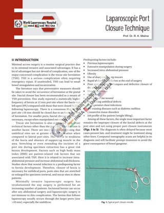
  • 1. INTRODUCTION Minimal access surgery is a routine surgical practice due to its minimal invasive and associated advantages. It has a lot of advantages but not devoid of complication, one of the major concerned complication is the trocar site herniation (TSH). TSH is a serious complication often requiring emergency repair. If unattended, TSH can lead to small bowel strangulation and incarceration. The literature says that preventative measures should be taken to avoid the occurrence of herniation at the portal site. Fascial closure has been recommended as a means of TSH prevention. One study reported a statistically higher frequency of hernia at 12 mm port site where the fascia was left open (8%) compared with those that were closed (0.22%) following laparoscopy. There is a consensus that all the port site ≥10 mm should be closed due to an increased risk of herniation. For smaller ports, fascial closure may not be necessary, except when manipulated extensively. Trocar site herniation is also associated with other technical factors other than the port site. Port location is another factor. There are many reports suggesting that umbilical sites are at greater risk of herniation when compared to lateral port sites. This is due to weakness of the fascia and absence of supporting muscle in the area. Stretching or even extending the incision of a port site during specimen extraction has a great risk hernia development. Factors such as high body mass index (BMI) are patient-related risk factors that are associated with TSH. Here it is related to increase intra- abdominal pressure and increase abdominal wall thickness. Studies show that wound infection is a predisposing factor to hernia development. Therefore, closure of fascia is necessary for umbilical ports, ports sites that are stretched or enlarged for specimen retrieval, and trocar sites in obese patients. Minimally invasive laparoscopic surgery has revolutionized the way surgery is performed for an increasing number of patients. Incisional hernia can occur after any abdominal surgery and laparoscopic surgery is not immune to this complication. The hernia that follows laparoscopy usually occurs through the larger ports (size >10 mm), especially the umbilicus. Fig. 1: Typical port closure needle. Laparoscopic Port ClosureTechnique Predisposing factors include: ■ Previous laparoscopies ■ Extensive manipulation during surgery ■ Increased intra-abdominal pressure ■ Obesity ■ Use of sharp cutting-tip trocars ■ Rapid abdominal deflation at the end of surgery ■ Poor port removal techniques and defective closure of the abdominal fascia ■ Wound extension ■ Male sex ■ Infection of the wound ■ Pre-existing umbilical defects ■ Postoperative chest infections ■ Pre-existing diseases such as diabetes mellitus ■ Connective tissue disorders ■ Job profile of the patient (weight lifting). Among all these factors, the single most important factor remains the improper closure of the fascial defects at the port sites and not using proper port closure instruments (Figs. 1 to 3). The diagnosis is often delayed because most cases present late, and treatment might be instituted along other lines. Computed tomography scans are helpful in its diagnosis and will facilitate prompt treatment to avoid the grave consequence of bowel gangrene. Prof. Dr. R. K. Mishra
  • 2. 166 SECTION 1: Essentials of Laparoscopy Fig. 2: Laparoscopic port closure Cobbler’s needle. Fig. 3: Incisional hernia development due to improper closure of port should be repaired later by mesh. Fig. 4: The tip of telescope should be introduced in and cannula is pulled over telescope to prevent suction of omentum or bowel. While surgical techniques and instrumentation have made significant advances, it is usual that the surgical incision is closed using invasive suturing techniques or by the use of tapes or by the use of topical cyanoacrylate skin adhesives (TCAs) for closure of surgical wounds. The incidence of incisional hernia occurring at the port sites after laparoscopic surgery lies between 0.02 and 3.6% and usually remainsunreported,untilthedevelopmentofcomplications. Any port closure technique should have following characteristics: ■ Effective (strong and secure) surgical wound closure ■ Faster wound closure ■ Better scar cosmesis ■ Occlusive microbial wound dressing ■ Less tissue trauma, reduced inflammatory reaction ■ No requirement for suture/staple removal ■ Easy to use/simple learning curve ■ Reduced risk of needlestick injury—safety and costs ■ Cost effective. WITHDRAWAL OF INSTRUMENTS AND PORTS Once the surgery is finished, all the instruments should be removed carefully under vision. All the accessory ports should be removed and the gas is removed by releasing the valve of 10 mm cannulas. The primary port should be taken out in the end (Fig. 4). If last port is suddenly withdrawn, sudden suction effect of cannula can pull the omentum or bowel inside the port wound, the chance of port-site hernia and adhesion is much higher in this case. It is a good practice to insert some blunt instrument or telescope inside the abdomen while removing the last cannula out over that instrument, to prevent inadvertent entrapment of omentum or bowel. The access technique will result in breach in continuity of abdominal wall which need to be repaired at the end of surgery. All the 10 mm or >10 mm port should be repaired properly to prevent any future possibility of hernia. The rectus sheath should be sutured with Vicryl. Only one stitch is required in middle which will convert 10 mm wound into 5 mm. The 5 mm port wounds are not necessary to repair. Laparoscopic Port Closure Instruments Various types of port closure instruments are available. The suture passer is a convenient instrument for port closure. It is used to pass the thread on the side of cannula and then it is tied externally (Figs. 5A to D). Port Closure Needle This is a simple instrument just like cobblers and it can be effectively used for closing the port. The tip of the instrument isbluntandtheneedlefacestowardthefascia,sothechances of injury to the bowel are less with the use of this instrument (Fig. 6).
  • 3. 167 CHAPTER 12: Laparoscopic Port Closure Technique C A B D Figs. 5A to D: Port closure with the help of suture passer. Fig. 6: Port closure needle. Fig. 7: Aneurysm needle. Aneurysm needle can also be used for closing fascia. The advantage of this needle is that eye is at the tip and due to rigid structure there is no risk of bending or rotation of needle (Fig. 7). After closing the rectus sheath, the skin can be closed by intradermal, skin stapler or any of the surgical skin glues TCAs available (Fig. 8). New Laparoscopic Port Closure Instruments Weck® EFx Shield Fascial Closure System (Figs. 9A and B) The Weck® EFx Shield Fascial Closure System from Teleflex is the only shielded port closure device, providing enhanced sharps protection for uniform and consistent performance.
  • 4. 168 SECTION 1: Essentials of Laparoscopy Fig. 8: Closure of skin wound by skin stapler. A B Figs. 9A and B: Weck® EFx Shield Fascial Closure System. Figs. 10A and B: NeatClose automated port closure device. B A The EFx Shield® System is designed with speed and safety in mind. An array of enhanced features includes: ■ Unique shielded wing design for enhanced sharps protection ■ Intuitive wing deployment ■ Innovative suture retrieval system for unassisted fascial closure. NeatClose Automated Port Closure Device (Figs. 10A and B) NeatStitch of Israel has come up with an automated port closure device known as NeatClose, where it also picked up both the Food and Drug Administration (FDA) and European approvals in the process. It is marketed to be an alternative to manual port closure, making it a speedy and efficient manner to help laparoscopic surgeon save time and money by lowering intraoperative costs. This system lets surgeons produce a watertight seal quickly, and it goes without saying that this would go a long way in aiding the recovery of a patient, never mind that one does not have Wolverine’s healing factor. When inside the operating cavity, the surgeon can squeeze the handle leavers in order to release a couple of blunt needle guides, where said guides are specially positioned in a perpendicular manner to the
  • 5. 169 CHAPTER 12: Laparoscopic Port Closure Technique port plane. With the activation button pressed, it will release the needles from the guide, via the tissue and back to the NeatClose cartridge. Once the system is pulled outside the port, you can be sure that a safe and efficient airtight seal is created, hence aiding the recovery of a patient for surgery quickly. Now we are still waiting for a painless method without the need for anesthesia. Carter-Thomason CloseSure System—Port-site Closure (Figs. 11A and B) Closing any trocar site is a simple, fast and safe procedure with the Carter-Thomason CloseSure System. The cone- shaped Pilot® Guide correctly angles the suture passer to achieve full-thickness closure. It closes the port including fascia and peritoneum (preventing Richter’s hernias)— while maintaining pneumoperitoneum. The guide’s unique design ensures precise placement of the suture passer for consistent, reproducible results on any body type. VersaOne™ Fascial Closure System (Figs. 12 and 13) Port-site hernias are serious complications across procedures and are a burden on patients, clinicians, and health systems. Appropriate port-site closure is considered to be one of the most critical factors for the prevention of port-site herniation. The VersaOne™ Fascial Closure System is a novel all-in-one device that serves as a trocar and fascial closure device to deliver consistent port-site closure and suture placement. The unique system features a special cannula that allows for defect closure without the need of additional devices. As a result, the VersaOne™ Fascial Closure System: ■ Provides procedure efficiency ■ Eliminates the need to remove the trocar before closing ■ Makes reinsufflation unnecessary—pneumoperitoneum can be maintained throughout the procedure ■ Enables tissue layers to remain aligned. B A Figs. 11A and B: Carter-Thomason CloseSure System—port-site closure. Fig. 12: VersaOne™ Fascial Closure System with its trocar. Fig. 13: VersaOne™ Fascial Closure System demonstrating insertion of suture.
  • 6. 170 SECTION 1: Essentials of Laparoscopy Fig. 14: Remove the stylet from the cannula. Fig. 15: Pass a suture material through the cannula from the tip. Fig. 16: Take suture out from the other end. Fig. 17: Tie the loop and hide the knot in the cannula. There are a number of methods of post site closure but there is no gold standard. Use of traditional suturing techniques are difficult due to blind closure of the fascial defect. Varying degrees of success are achieved by modified hand suturing techniques. Finding the rectus sheath and suturing through the layers of a thicker abdominal wall through a relatively small hole is challenging particularly in the obese. In such cases, we need special instrument for efficientclosureoftheportsite.Veressneedleisaninstrument that is commonly used for creating pneumoperitoneum but it has been used to close the port site efficiently under vision. VERESS NEEDLE TECHNIQUE OF PORT CLOSURE In 1983, Janos Veress of Hungary developed a specially designed spring-loaded needle. Interestingly, Veress did not promote the use of his needle for laparoscopy purposes. He used Veress needle for the induction of pneumothorax. But now Veress needle is the most important instrument today to create pneumoperitoneum. Veress needle consists of an outercannulawithabeveledneedlepointforcuttingthrough tissues. Inside the cannula of Veress needle is an inner stylet, stylet is loaded with a spring forward in response to the sudden decrease in pressure encountered upon crossing the abdominal wall and entering the peritoneal cavity. TECHNIQUE OF PORT CLOSURE BY VERESS NEEDLE (FIGS. 14 TO 25) Occlude the port site with a finger so that the pneumo- peritoneum is maintained and pass the Veress beside the fingerthroughallthelayersexcepttheskinandsubcutaneous tissue under vision. Maintenance of pneumoperitoneum is important as it is very difficult to close the port if vision is compromised. Minimal access surgeries are the present and future of surgical procedures and no surgery is complete without port site closure. There are a lot of methods to close the port-site but no gold standard. This procedure with the Veress needle is safe, efficacious, and cost-effective. One of the preventable complications is port-site incisional hernia (PIH), which could develop at any port site, most frequently at the midline, possibly because of the absence of supporting muscle. The incidence of PIH is variable from center to center, depending on several factors including surgical technique and of course surgical experience.
  • 7. 171 CHAPTER 12: Laparoscopic Port Closure Technique Fig. 18: Insert the suture material (that should close the port site) into the cannula tip about 2 cm deep and bend it so that it stays in place. Fig. 19: Now Veress needle is ready for port closure. Fig. 20: Veress needle inserted with loop and tying suture on one side. Fig. 21: Retract the Veress and the suture is automatically retained inside. Fig. 22: Insert the Veress from the other side of the defect. Fig. 23: Entangle the suture in the loop of the Veress. The trocar diameter, trocar design, pre-existing fascial defects, tissue retrieval from the port site, and some operation and patient-related factors, direction of the port insertion, use of drain are the risk factors for development of PIH. In obese and bariatric patients because of larger preperitonealspaceandelevatedintra-abdominalpressure, the risk of formation of trocar site hernia is greater. Size of the port is another major risk factor and it is advisable to close the hole >5 mm at the fascia level. The meticulous closure of the laparoscopic ports is important to prevent and reduce the chances of formation of PIH. Port-site closure by Veress needle is an efficient
  • 8. 172 SECTION 1: Essentials of Laparoscopy Fig. 24: Tighten the loop and retract the Veress along with the suture and tie the knot outside. Fig. 25: Thus, the port site closed under vision and is safe procedure. and safe technique done under vision and there is no need to buy additional equipment to close the port site thus cost effective. The hernia may become evident at any time following laparoscopic surgery and the patient may either have an uncomplicated hernia, or may be afflicted with a variety of complications such as evisceration of the bowel or omentum and it may become a cause of significant morbidity. Meticulous closure of the fascia, avoidance of unnecessary wound extension, the use of nonabsorbable sutures when faced with defects >2 cm in size, completely defining the extent of any pre-existing hernia and repairing this at the time of port-site closure, are recommended to minimize the incidence of port-site hernia after laparoscopic surgery. BIBLIOGRAPHY 1. Ahmad SA, Schuricht AL, Azurin DJ, Arroyo LR, Paskin DL, Bar AH, et al. Complications of laparoscopic cholecystectomy: the experience of a university-affiliated teaching hospital. J Laparoendosc Adv Surg Tech A. 1997;7:29-35. 2. Al-Haijar N, Duca S, Molnar G, Vasilescu A, Nicolescu N. Incidents and postoperative complications of laparoscopic cholecystectomies for acute cholecystitis. Rom J Gastroenterol. 2002;11:115-9. 3. Azurin DJ, Go LS, Arroyo LR, Kirkland ML. Trocar site herniation followinglaparoscopiccholecystectomyandthesignificanceofan incidental preexisting umbilical hernia. Am Surg. 1995;61:718-20. 4. Baird DR, Wilson JP, Mason EM, Duncan TD, Evans JS, Luke JP, et al. An early review of 800 laparoscopic cholecystectomies at a university-affiliated community teaching hospital. Am Surg. 1992;58:206-10. 5. Bender E, Sell H. Small bowel obstruction after laparoscopic cholecystectomy as a result of a Maydl’s herniation of the small bowel through a trocar site. Surgery. 1996;119:480. 6. Berthou JC, Charbonneau P. Elective laparoscopic management of sigmoid diverticulitis: results in a series of 110 patients. Surg Endosc. 1999;13:457-60. 7. Bhoyrul S, Payne J, Steffes B, Swanstrom L, Way LW. A randomized prospective study of radially expanding trocars in laparoscopic surgery. J Gastrointest Surg. 2000;4:392-7. 8. Boughey JC, Nottingham JM, Walls AC. Richter’s hernia in the laparoscopic era: four case reports and review of the literature. Surg Laparosc Endosc Percutan Tech. 2003;13:55-8. 9. Bowrey DJ, Blom D, Crookes PF, Bremner CG, Johansson JL, Lord RV, et al. Risk factors and the prevalence of trocar site herniation after laparoscopic fundoplication. Surg Endosc. 2001;15:663-6. 10. Cadeddu MO, Schlachta CM, Mamazza J, Seshadri PA, Poulin EC. Soft-tissue images: trocar-site hernia after laparoscopic procedures. Can J Surg. 2002;45:9-10. 11. CalleryMP,StrasbergSM,SoperNJ.Complicationsoflaparoscopic general surgery. Gastrointest Endosc Clin N Am. 1996;6:423-44. 12. Chevallier JM, Zinzindohoue F, Elian N, Cherrak A, Blanche JP, Berta JL, et al. Adjustable gastric banding in a public university hospital: prospective analysis of 400 patients. Obes Surg. 2002;12:93-9. 13. Coda A, Bossotti M, Ferri F, Mattio R, Ramellini G, Poma A, et al. Incisional hernia and fascial defect following laparo- scopic surgery. Surg Laparosc Endosc Percutan Tech. 2000;10: 34-8. 14. CottamDR,GoreckiPJ,CurveloM,WeltmanD,AngusLD,Shaftan G. Preperitoneal herniation into a laparoscopic port site without a fascial defect. Obes Surg. 2002;12:121-3. 15. Crist DW, Gadacz TR. Complications of laparoscopic surgery. Surg Clin North Am. 1993;73:265-89. 16. De Giuli M, Festa V, Denoye GC, Morino M. Large postoperative umbilical hernia following laparoscopic cholecystectomy: a case report. Surg Endosc. 1994;8:904-5. 17. Di Lorenzo N, Coscarella G, Lirosi F, Gaspari A. Port-site closure: a new problem, an old device. JSLS. 2002;6:181-3. 18. Dresel A, Kuhn JA, Westmoreland MV, Talaasen LJ, McCarty TM. Establishing a laparoscopic gastric bypass program. Am J Surg. 2002;184:617-20. 19. Duron JJ, Hay JM, Msika S, Gaschard D, Domergue J, Gainant A, et al. Prevalence and mechanisms of small intestinal obstruction following laparoscopic abdominal surgery: a retrospective multicenter study. Arch Surg. 2000; 135:208-12. 20. Fear RE. Laparoscopy: a valuable aid in gynecologic diagnosis. Obstet Gynecol. 1968;31:297-309. 21. Fitzgibbons RJ Jr, Annibali R, Litke BS. Gallbladder and gallstone removal, open versus closed laparoscopy, and pneumoperitoneum. Am J Surg. 1993;165:497-504. 22. Freedman AN, Sigman HH. Incarcerated paraumbilical incisional hernia and abscess: complications of a spilled gallstone. J Laparoendosc Surg. 1995;5:189-91.
  • 9. 173 CHAPTER 12: Laparoscopic Port Closure Technique 23. Hass BE, Schrager RE. Small bowel obstruction due to Richter’s hernia after laparoscopic procedures. J Laparoendosc Surg. 1993;3:421-3. 24. Horgan PG, O’Connell PR. Subumbilical hernia following laparoscopic cholecystectomy. Br J Surg. 1993;80:1595. 25. Kadar N, Reich H, Liu CY, Manko GF, Gimpelson R. Incisional hernias after major laparoscopic gynecologic procedures. Am J Obstet Gynecol. 1993;168:1493-5. 26. KomutaK,HaraguchiM,InoueK,FuruiJ,KanematsuT.Herniation of the small bowel through the port site following removal of drains during laparoscopic surgery. Dig Surg. 2000;17:544-6. 27. Kopelman D, Schein M, Assalia A, Hashmonai M. Small bowel obstruction following laparoscopic cholecystectomy: diagnosis of incisional hernia by computed tomography. Surg Laparosc Endosc. 1994;4:325-6. 28. Kulacoglu IH. Regarding: small bowel obstruction and incisional hernia after laparoscopic surgery: should 5-mm trocar sites be sutured? J Laparoendosc Adv Surg Tech A. 2000;10:227-8. 29. Lafullarde T, Van Hee R, Gys T. A safe and simple method for routine open access in laparoscopic procedures. Surg Endosc. 1999;13:769-72. 30. Larson GM, Vitale GC, Casey J, Voyles CR. Multipractice analysis of laparoscopic cholecystectomy in 1,983 patients. Am J Surg. 1992;163:221-6. 31. Leibl BJ, Schmedt CG, Schwarz J, Kraft K, Bittner R. Laparoscopic surgery complications associated with trocar tip design: review of literature and own results. J Laparoendosc Adv Surg Tech A. 1999;9:135-40. 32. Li P, Chung RS. Closure of trocar wounds using a suture carrier. Surg Laparosc Endosc. 1996;6:469-71. 33. Liu CD, McFadden DW. Laparoscopic port sites do not require fascial closure when nonbladed trocars are used. Am Surg. 2000;66:853-4. 34. Lumley J, Stitz R, Stevenson A, Fielding G, Luck A. Laparoscopic colorectal surgery for cancer: intermediate to long-term outcomes. Dis Colon Rectum. 2002;45:867-72. 35. Maio A, Ruchman RB. CT diagnosis of post laparoscopic hernia. J Comput Assist Tomogr. 1991;15:1054-5. 36. Matter I, Nash E, Abrahamson J, Eldar S. Incisional hernia via a lateral 5-mm trocar port following laparoscopic cholecystectomy. Isr J Med Sci. 1996;32:790-1. 37. Matthews BD, Heniford BT, Sing RF. Preperitoneal Richter hernia after a laparoscopic gastric bypass. Surg Laparosc Endosc Percutan Tech. 2001;11:47-9. 38. Mayol J, Garcia-Aguilar J, Ortiz-Oshiro E, De-Diego Carmona JA, Fernandez-Represa JA. Risks of the minimal access approach for laparoscopic surgery: multivariate analysis of morbidity related to umbilical trocar insertion. World J Surg. 1997;21:529-33. 39. McMillan J, Watt I. Herniation at the site of cannula insertion after laparoscopic cholecystectomy. Br J Surg. 1993;80:915. 40. McMurrick PJ, Polglase AL. Early incisional hernia after use of the 12-mm port for laparoscopic surgery. Aust N Z J Surg. 1993;63:574-5. 41. Montz FJ, Holschneider CH, Munro MG. Incisional hernia following laparoscopy: a survey of the American Association of Gynecologic Laparoscopists. Obstet Gynecol. 1994;84: 881-4. 42. Morrison CP, Wemyss-Holden SA, Iswariah H, Maddern GJ. Lateral laparoscopic port sites should all be closed: the incisional “spigelian” hernia. Surg Endosc. 2002;16:1364. 43. Nakajima K, Wasa M, Kawahara H, Hasegawa T, Soh H, Taniguchi E, et al. Revision laparoscopy for incarcerated hernia at a 5-mm trocar site following pediatric laparoscopic surgery. Surg Laparosc Endosc Percutan Tech. 1999;9:294-5. 44. Nassar AH, Ashkar KA, Rashed AA, Abdulmoneum MG. Laparoscopic cholecystectomy and the umbilicus. Br J Surg. 1997;84:630-3. 45. Ok E, Sozuer E. Intra-abdominal gallstone spillage detected during umbilical trocar site hernia repair after laparoscopic cholecystectomy: report of a case. Surg Today. 2000;30:1046-8. 46. Patterson M, Walters D, Browder W. Postoperative bowel obstruction following laparoscopic surgery. Am Surg. 1993; 59:656-7. 47. Petrakis I, Sciacca V, Chalkiadakis G, Vassilakis SI, Xynos E. A simple technique for trocar site closure after laparoscopic surgery. Surg Endosc. 1999;13:1249-51. 48. Plaus WJ. Laparoscopic trocar site hernias. J Laparoendosc Surg. 1993;3:567-70. 49. Rabinerson D, Avrech O, Neri A, Schoenfeld A. Incisional hernias after laparoscopy. Obstet Gynecol Surv. 1997;52: 701-3. 50. Ramachandran CS. Umbilical hernial defects encountered before and after abdominal laparoscopic procedures. Int Surg. 1998;83:171-3. 51. Reardon PR, McKinney G, Craig ES. The 2-mm trocar: a safe and effective way of closing trocar sites using existing equipment. J Am Coll Surg. 2003;196:333-6. 52. Reardon PR, Preciado A, Scarborough T, Matthews B, Marti JL. Hernia at 5-mm laparoscopic port site presenting as early postoperative small bowel obstruction. J Laparoendosc Adv Surg Tech A. 1999;9:523-5. 53. Rosen M, Ponsky J. Minimally invasive surgery. Endoscopy. 2001;33:358-66. 54. Sanz-Lopez R, Martinez-Ramos C, Nunez-Pena JR, Ruiz de Gopegui M, Pastor-Sirera L, Tamames-Escobar S. Incisional hernias after laparoscopic vs open cholecystectomy. Surg Endosc. 1999;13:922-4. 55. SchauerPR,IkramuddinS,GourashW,RamanathanR,LuketichJ. Outcomes after laparoscopic Roux-en-Y gastric bypass for morbid obesity. Ann Surg. 2000;232:515-29. 56. Schiller VL, Joyce PW, Sarti DA. Small-bowel obstruction due to hernia through the primary laparoscopic port: a complication of laparoscopic cholecystectomy. Am J Roentgenol. 1994;163: 480-1. 57. Susmallian S, Ezri T, Charuzi I. Laparoscopic repair of access port site hernia after Lap-Band system implantation. Obes Surg. 2002;12:682-4. 58. Velasco JM, Vallina VL, Bonomo SR, Hieken TJ. Post laparoscopic small bowel obstruction: rethinking its management. Surg Endosc. 1998;12:1043-5. 59. Voyles CR, Petro AB, Meena AL, Haick AJ, Koury AM. A practical approach to laparoscopic cholecystectomy. Am J Surg. 1991;161:365-70. 60. Wagner M, Farley GE. Incarcerated hernia with intestinal obstruction after laparoscopic cholecystectomy. Wis Med J. 1994;93:169-71. 61. Waldhaussen JH. Incisional hernia in a 5-mm trocar site following pediatric laparoscopy. J Laparoendosc Surg. 1996;6 (Suppl 1):S89-90. 62. Wallace DH, O’Dwyer PJ. Clinical experience with open laparoscopy. J Laparoendosc Adv Surg Tech A. 1997;7:285-8. 63. Williams MD, Flowers SS, Fenoglio ME, Brown TR. Richter hernia: a rare complication of laparoscopy. Surg Laparosc Endosc. 1995;5:419-21.