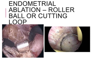
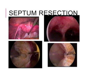
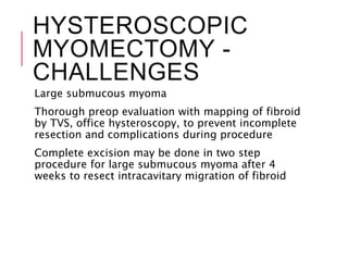
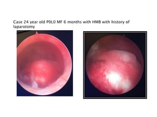
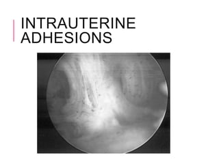
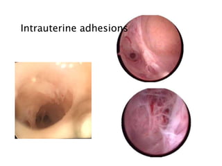
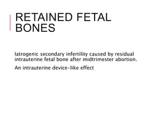
OPERATIVE
HYSTEROS
COPY
Dr Meenakshi
Sharma
MD (Obs & Gynae) (AIIMS),
FICMCH
Senior Consultant Obs &
Gynae
Yashoda Superspeciality
Hospitals, Kaushambi
Shanti Mukund Hospital,
Delhi
BASIC
PRINCIPLES OF
OPERATIVE
HYSTEROSCOPY
PREREQUISITES FOR
OPERATIVE
HYSTEROSCOPY
Should be performed as in patient or day care
hospital
Minor surgical procedures can be done as office
hysteroscopy procedures but advisable to perform
in well equiped OT where anaesthesia facility
available
A thorough physical exam is essential prerequisite
for surgical hysteroscopy.
Improper patient selection and poor technique are
the most frequent causes of complications during
surgery
CONTRAINDICATIONS
FOR SURGICAL
HYSTEROSCOPY
Pelvic inflammatory Disease
Acute cervicovaginitis
Intense Metrorrhagia
Pregnancy
INSTRUMENTATION
The complete set of resectoscopic
surgery instruments consists of
Resectoscope
Camera equipment
Light source
Endomat / Hysteromat
Unipolar high frequency electrosurgical
generator with automatically controlled
output and acoustic control
INSTRUMENTATION
Rigid Hysteroscope with a diameter of 7 mm
and eqipped with 2 channels – one for
distension media, the other for introduction of
ancillary instruments including probes,
catheters, miniature rigid or semi rigid scissors
and various biopsy forceps
Procedures done with operative hysteroscope
are adhesions, thin septum, small polyp and
guided biopsy and removal of foreign body
RESECTOSCOPE
90% of all hysteroscopic surgery is performed using
resectoscope
26 Fr resectoscope with ancillary instruments
It has provisions for connection to hysteromat,
connection to electrosurgical generator, connection
to light source and camera equipment
The resectoscope with continuous flow sheath for
distension of uterine cavity increases the options
available to the surgeon in the removal of
submucous and intramural myomas
ANAESTHESIA
Local
Paracervical block
Epidural block
GA
DISTENSION MEDIA
Carbon dioxide and high viscosity
dextran solution not used in
hysteroscopic surgery
Saline for nonelectrosurgical procedures
Sorbitol or mannitol
Glycine – most commonly used
distension media where electrosurgery
used
INDICATIONS OF
SURGICAL
HYSTEROSCOPY
•Endometrial ablation
•Surgery of uterine malformations –
septum
•Treatment of intrauterine adhesions
•Myomectomy – submucous fibroid
•Fallopian tube catheterisation
•Removal of foreign bodies and IUD
HYSTEROSCOPIC
ENDOMETRIAL
ABLATION
Indications –Drug resistant HMB with
malignancy ruled out
limited with use of LNG IUS, and GnRH
Performed under GA and even LA
Preoperative thinning of endometrium
by Danazol or GnRH for 2-3 months or
therapeutic D&C- better success
HYSTEROSCOPIC
ENDOMETRIAL
ABLATION
Technique
Entire endometrium must be ablated without
leaving islands of normal endometrium
Ablation deep into myometrium to be avoided to
prevent adhesion formation
Endometrium in isthmic region is spared to prevent
asherman syndrome
3-5 mm deep endometrial slices are resected with
resectoscope loop using cutting current and
resection should stop at level of fasciculated
myometrium
ENDOMETRIAL
ABLATION – ROLLER
BALL OR CUTTING
LOOP
HYSTEROSCOPY FOR
UTERINE
MALFORMATION
Arcuate Uterus
Septate Uterus
Bicornuate Uterus
Uterus Didelphys
Septate uterus presents with reproductive difficulty and
need corrective surgery
Diagnosis – Two hemicavities with clear central division
or y shaped cavity on HSG
Concomitant laparoscopy should be done to distinguish
bicornuate uterus and septate uterus
UTERINE SEPTUM
HYSTEROSCOPIC
UTERINE SEPTUM
RESECTIONSemi rigid scissors 5-7Fr
 Semi rigid scissors perfect for thin uterine septum as they
produce required force and small enough to pass through
operating sheath of the hysteroscope and along cervical canal
without any difficulty or risk. Blades can be opened wide
enough for resection of even thick septa.
 Continuous flow irrigation to be used
 Vision clear if only no bleeding
 Resection till level of ostia
Resectoscope with collens knife
 Cutting current 30-40W/s
(KTP/532), (Nd:YAG), or Argon Lasers
 Laser beam with glass fibers of 0.6 micron diameter
 Septum vaporised as a result of the fibers contacting the
target tissue
HYSTEROSCOPIC
UTERINE SEPTUM
RESECTION
Goal of Septum Resection -Satisfactory uterine cavity
Resection to be stopped at the level of osita... Both ostia
will be visible in panoramic view to prevent perforation
and adhesion formation
Cautery near ostia must be avoided
Preoperative treatment with Danazol or GnRH can be
given for better vision
If Uterocervical septum –complete resection with
inclusion of cervical part is mandatory
Follow up after 2 months with HSG or hysteroscopy
Full term pregnancy rate 70-80%, no need of LSCS
SEPTUM RESECTION
HYSTEROSCOPIC
CORRECTION OF
ARCUATE UTERUS
PRIOR TO IVF?Measurement by sonohysterography
 Fm (fundal myometrial thickness)
 Cm (cornual myometrial thickness)
Incision of the incomplete septum.
 Fm >11 mm
 Fm-Cm >5 mm,
Meta-analyses of five studies
 Improving result in subsequent IVF cycle
 (relative risk = 1.75, 95% CI 1.51-2.03).
HYSTERSCOPIC
MYOMECTOMYSubmucous myoma - 5.5-16.6% of all
fibroids
AAGL in 2012 -submucous fibroid
contribute to infertility and its removal
improves fertility rates
European Society of Hysteroscopy (ESH)
 Type 0 - completely within the cavity
 Type I - extend < 50 % into the myometrium
 Type II - extend >50 % within the
myometrium
Indications
 AUB
HYSTEROSCOPIC
MYOMECTOMY
HYSTEROSCOPIC
MYOMECTOMY –
SURGICAL
TECHNIQUESelection of Case
 Myoma within uterine cavity _ pedunculated or limited
implant base
 Myoma with partial intramural development , endocavitary
component >50%. Angle of protrusion between myoma and
uterine wall <900
Operative hysteroscope, resectoscope 26Fr
 Working element with electrosurgergical instrument – thermal
loops or vaporising electrode and mechanical instruments –
cold loops or intrauterine morcellator device can be attached
 Monopolar electrodes require non conducting distension
media glycine 1.5%. Bipolar electrodes can be used with saline
distension media
 Cold loops used mechanically to enucleate intramural portion
of myoma
Excision of only intracavitary component of fibroid
Preoperative GnRH therapy if submucous fibroid >2
HYSTEROSCOPIC
MYOMECTOMY -
CHALLENGES
Large submucous myoma
Thorough preop evaluation with mapping of fibroid
by TVS, office hysteroscopy, to prevent incomplete
resection and complications during procedure
Complete excision may be done in two step
procedure for large submucous myoma after 4
weeks to resect intracavitary migration of fibroid
HYSTEROSCOPIC
MYOMECTOMY –
SURGICAL TECHNIQUE
FOR LARGE FIBROIDOne step procedure – first resect intracavitary portion of
fibroid by usual slicing, then using cold loop
mechanically intramural portion of fibroid is resected by
enucleation and blunt dissection. Enucleation is followed
by excision and resection of intramural component of the
fibroid
Litta’s technique –elliptical incision given at junction of
endometrium and its reflectiom on uterine wall till
cleavage zone of fibroid is reached. Connecting bridges
between fibroid and surrounding mycytes is slowly
resected
Lasmar’s technique –used collins L shaped knife to
dissect endometrium around fibroid followed by direct
mobilization of fibroid in all directions coagulating only
bleeding vessels
Hydromassage
Manual massage
Lasmar Classification for hysteroscopic operability
HYSTEROSCOPIC
MYOMECTOMY -
PREVENTING
COMPLICATIONSGenital tract burns due to monopolar current minimised
by
 Maintain contact of external sheath of resectoscopewith cervix
 Avoid activation of electrosurgical unit when electrode is not in
contact with tissue
 Ensure integrity of insulation of the electrode
 Minimize use of high voltage (coagulation) current during
myomectomy
 Use concomitant laparoscopy in case of type 2 deep seated
myomas
Fluid overload
 Careful selection of fibroid large fibroid –two stage procedure
Uterine perforation
 Detect early and stop procedure
Minimise adhesion formation by opposing tissue should
not be resected during single surgery
 Second look hyateroscopy effective for postoperative adhesions
HYSTEREOSCOPIC
MYOMECTOMY -
POSTOP CARE
Postoperative GnRH analogs can be continued for
2-3 months if the intramural portion of fibroid was
not fully removed and a two step procedure can be
planned
Intraoperative antibiotics are administered to all
patients.
Patients discharged same day
Very few patient require 24 hour observation for
fluid overload
Follow up hysteroscopy planned after 2-3 months
Case 24 year old P0L0 MF 6 months with HMB with history of
laparotomy
Case 50 year lady with postmenopausal increased ET on scan, no PM
HYSTEROSCOPIC
MORCELLATOR
HYSTEROSCOPIC
MORCELLATORS
ADVANTAGES
 Operate in Saline
 Decreased risk of fluid
overload
 Mechanical
 No thermal injury
 Remove Tissue Pieces
 Clear visual field
Decreases risks of
multiple instrument
placement
Uterine perforation,
false passageway and air
embolus
Are Easy to Use
 Facilitate Removal Type
0 and I Myomas
 Decreased operative
time and fluid deficit
 Small Diameter Can Be
Used in the Office
HYSTEROSCOPIC
MORCELLATORS
DISADVANTAGES
 No electrosurgery for hemostasis
IOGYN Mistral has electrosurgery
 Type 2 myomas are difficult
 Fundal pathology is difficult
 Cost of fluid management system
 Mistral is incorporated into device
COMPLICATIONS OF
HYSTEROSCOPIC
MYOMECTOMY
Electrolyte abnormalities with non-electrolyte
media
Excessive bleeding
Incomplete resection
Need for additional procedure
Increased operative time
Wamsteker K, 1993
INTRAUTERINE
ADHESIONS
INTRAUTERINE
ADHESIONS
Trauma
 Missed or incomplete abortion, postpartum hemorrhage, or
retained placental remnants
Genital tuberculosis
Classifications
 Endometrial adhesions - filmsy adhesions
 Myometrial adhesions – dense adhesions
 Connective fiber synechiae
Goal – Restore normal anatomy
 Scissors
 Resectoscope collins knife for dense adhesions
Intrauterine adhesions
RETAINED FETAL
BONES
Iatrogenic secondary infertility caused by residual
intrauterine fetal bone after midtrimester abortion.
An intrauterine device-like effect
FORGOTTON IUCD
TUBERCULAR
ENDOMETRITIS
TUBERCULAR
ENDOMETRITIS
HYSTEROSCOPIC
CANNULATION
HYSTEROSCOPIC
CANNULATION
Intramural portion of tube initial 1 cm
rectilinear, later 1.5 cm irregular and
sometimes difficult to cannulate
Indications
 For proximal tubal occlusion
 Transfer of gametes or embryos in some ART
 Placement of intratubal devices for reversible
sterilization GIFT
PTO account for 10-25% of tubal factor
infertility
Cooks cannulation set 9 Fr outer catheter and 3
Fr inner catheter with guide wire
"CHROMOHYSTEROS
COPY"
5 ml of 1% methylene blue dye
Group I: 19 patients focal dark staining
 10 cases of endometritis
Group II: 15 patients diffuse light blue staining
 normal histopathology
HYSTEROSCOPIC
STERILISATION
•Hysteroscopic placement of
radiopaque inserts in the proximal
portion of the fallopian tube
•Tissue ingrowth occurs through
the insert creating natural barrier
Device Length: ~3.85 cm
• PET Fiber Length: ~1.75 cm
• Expanded Outer Diameter: 1.5 –
2.0 mm
• Inserts are visible by X-Ray,
Ultrasound, MRI and CT Scan
CONCLUSION
Diagnostic hysteroscopy should be a routine
procedure during diagnostic laparoscopy in infertile
women
Office mini-hysteroscopy should be incorporated in
infertility work up
Operative hysteroscopy

Operative hysteroscopy

  • 1.
    OPERATIVE HYSTEROS COPY Dr Meenakshi Sharma MD (Obs& Gynae) (AIIMS), FICMCH Senior Consultant Obs & Gynae Yashoda Superspeciality Hospitals, Kaushambi Shanti Mukund Hospital, Delhi
  • 2.
  • 3.
    PREREQUISITES FOR OPERATIVE HYSTEROSCOPY Should beperformed as in patient or day care hospital Minor surgical procedures can be done as office hysteroscopy procedures but advisable to perform in well equiped OT where anaesthesia facility available A thorough physical exam is essential prerequisite for surgical hysteroscopy. Improper patient selection and poor technique are the most frequent causes of complications during surgery
  • 4.
    CONTRAINDICATIONS FOR SURGICAL HYSTEROSCOPY Pelvic inflammatoryDisease Acute cervicovaginitis Intense Metrorrhagia Pregnancy
  • 5.
    INSTRUMENTATION The complete setof resectoscopic surgery instruments consists of Resectoscope Camera equipment Light source Endomat / Hysteromat Unipolar high frequency electrosurgical generator with automatically controlled output and acoustic control
  • 6.
    INSTRUMENTATION Rigid Hysteroscope witha diameter of 7 mm and eqipped with 2 channels – one for distension media, the other for introduction of ancillary instruments including probes, catheters, miniature rigid or semi rigid scissors and various biopsy forceps Procedures done with operative hysteroscope are adhesions, thin septum, small polyp and guided biopsy and removal of foreign body
  • 7.
    RESECTOSCOPE 90% of allhysteroscopic surgery is performed using resectoscope 26 Fr resectoscope with ancillary instruments It has provisions for connection to hysteromat, connection to electrosurgical generator, connection to light source and camera equipment The resectoscope with continuous flow sheath for distension of uterine cavity increases the options available to the surgeon in the removal of submucous and intramural myomas
  • 8.
  • 9.
    DISTENSION MEDIA Carbon dioxideand high viscosity dextran solution not used in hysteroscopic surgery Saline for nonelectrosurgical procedures Sorbitol or mannitol Glycine – most commonly used distension media where electrosurgery used
  • 10.
    INDICATIONS OF SURGICAL HYSTEROSCOPY •Endometrial ablation •Surgeryof uterine malformations – septum •Treatment of intrauterine adhesions •Myomectomy – submucous fibroid •Fallopian tube catheterisation •Removal of foreign bodies and IUD
  • 11.
    HYSTEROSCOPIC ENDOMETRIAL ABLATION Indications –Drug resistantHMB with malignancy ruled out limited with use of LNG IUS, and GnRH Performed under GA and even LA Preoperative thinning of endometrium by Danazol or GnRH for 2-3 months or therapeutic D&C- better success
  • 12.
    HYSTEROSCOPIC ENDOMETRIAL ABLATION Technique Entire endometrium mustbe ablated without leaving islands of normal endometrium Ablation deep into myometrium to be avoided to prevent adhesion formation Endometrium in isthmic region is spared to prevent asherman syndrome 3-5 mm deep endometrial slices are resected with resectoscope loop using cutting current and resection should stop at level of fasciculated myometrium
  • 13.
  • 14.
    HYSTEROSCOPY FOR UTERINE MALFORMATION Arcuate Uterus SeptateUterus Bicornuate Uterus Uterus Didelphys Septate uterus presents with reproductive difficulty and need corrective surgery Diagnosis – Two hemicavities with clear central division or y shaped cavity on HSG Concomitant laparoscopy should be done to distinguish bicornuate uterus and septate uterus
  • 15.
  • 16.
    HYSTEROSCOPIC UTERINE SEPTUM RESECTIONSemi rigidscissors 5-7Fr  Semi rigid scissors perfect for thin uterine septum as they produce required force and small enough to pass through operating sheath of the hysteroscope and along cervical canal without any difficulty or risk. Blades can be opened wide enough for resection of even thick septa.  Continuous flow irrigation to be used  Vision clear if only no bleeding  Resection till level of ostia Resectoscope with collens knife  Cutting current 30-40W/s (KTP/532), (Nd:YAG), or Argon Lasers  Laser beam with glass fibers of 0.6 micron diameter  Septum vaporised as a result of the fibers contacting the target tissue
  • 17.
    HYSTEROSCOPIC UTERINE SEPTUM RESECTION Goal ofSeptum Resection -Satisfactory uterine cavity Resection to be stopped at the level of osita... Both ostia will be visible in panoramic view to prevent perforation and adhesion formation Cautery near ostia must be avoided Preoperative treatment with Danazol or GnRH can be given for better vision If Uterocervical septum –complete resection with inclusion of cervical part is mandatory Follow up after 2 months with HSG or hysteroscopy Full term pregnancy rate 70-80%, no need of LSCS
  • 18.
  • 19.
    HYSTEROSCOPIC CORRECTION OF ARCUATE UTERUS PRIORTO IVF?Measurement by sonohysterography  Fm (fundal myometrial thickness)  Cm (cornual myometrial thickness) Incision of the incomplete septum.  Fm >11 mm  Fm-Cm >5 mm, Meta-analyses of five studies  Improving result in subsequent IVF cycle  (relative risk = 1.75, 95% CI 1.51-2.03).
  • 20.
    HYSTERSCOPIC MYOMECTOMYSubmucous myoma -5.5-16.6% of all fibroids AAGL in 2012 -submucous fibroid contribute to infertility and its removal improves fertility rates European Society of Hysteroscopy (ESH)  Type 0 - completely within the cavity  Type I - extend < 50 % into the myometrium  Type II - extend >50 % within the myometrium Indications  AUB
  • 21.
  • 23.
    HYSTEROSCOPIC MYOMECTOMY – SURGICAL TECHNIQUESelection ofCase  Myoma within uterine cavity _ pedunculated or limited implant base  Myoma with partial intramural development , endocavitary component >50%. Angle of protrusion between myoma and uterine wall <900 Operative hysteroscope, resectoscope 26Fr  Working element with electrosurgergical instrument – thermal loops or vaporising electrode and mechanical instruments – cold loops or intrauterine morcellator device can be attached  Monopolar electrodes require non conducting distension media glycine 1.5%. Bipolar electrodes can be used with saline distension media  Cold loops used mechanically to enucleate intramural portion of myoma Excision of only intracavitary component of fibroid Preoperative GnRH therapy if submucous fibroid >2
  • 24.
    HYSTEROSCOPIC MYOMECTOMY - CHALLENGES Large submucousmyoma Thorough preop evaluation with mapping of fibroid by TVS, office hysteroscopy, to prevent incomplete resection and complications during procedure Complete excision may be done in two step procedure for large submucous myoma after 4 weeks to resect intracavitary migration of fibroid
  • 25.
    HYSTEROSCOPIC MYOMECTOMY – SURGICAL TECHNIQUE FORLARGE FIBROIDOne step procedure – first resect intracavitary portion of fibroid by usual slicing, then using cold loop mechanically intramural portion of fibroid is resected by enucleation and blunt dissection. Enucleation is followed by excision and resection of intramural component of the fibroid Litta’s technique –elliptical incision given at junction of endometrium and its reflectiom on uterine wall till cleavage zone of fibroid is reached. Connecting bridges between fibroid and surrounding mycytes is slowly resected Lasmar’s technique –used collins L shaped knife to dissect endometrium around fibroid followed by direct mobilization of fibroid in all directions coagulating only bleeding vessels Hydromassage Manual massage
  • 26.
    Lasmar Classification forhysteroscopic operability
  • 28.
    HYSTEROSCOPIC MYOMECTOMY - PREVENTING COMPLICATIONSGenital tractburns due to monopolar current minimised by  Maintain contact of external sheath of resectoscopewith cervix  Avoid activation of electrosurgical unit when electrode is not in contact with tissue  Ensure integrity of insulation of the electrode  Minimize use of high voltage (coagulation) current during myomectomy  Use concomitant laparoscopy in case of type 2 deep seated myomas Fluid overload  Careful selection of fibroid large fibroid –two stage procedure Uterine perforation  Detect early and stop procedure Minimise adhesion formation by opposing tissue should not be resected during single surgery  Second look hyateroscopy effective for postoperative adhesions
  • 29.
    HYSTEREOSCOPIC MYOMECTOMY - POSTOP CARE PostoperativeGnRH analogs can be continued for 2-3 months if the intramural portion of fibroid was not fully removed and a two step procedure can be planned Intraoperative antibiotics are administered to all patients. Patients discharged same day Very few patient require 24 hour observation for fluid overload Follow up hysteroscopy planned after 2-3 months
  • 30.
    Case 24 yearold P0L0 MF 6 months with HMB with history of laparotomy
  • 31.
    Case 50 yearlady with postmenopausal increased ET on scan, no PM
  • 32.
  • 33.
    HYSTEROSCOPIC MORCELLATORS ADVANTAGES  Operate inSaline  Decreased risk of fluid overload  Mechanical  No thermal injury  Remove Tissue Pieces  Clear visual field Decreases risks of multiple instrument placement Uterine perforation, false passageway and air embolus Are Easy to Use  Facilitate Removal Type 0 and I Myomas  Decreased operative time and fluid deficit  Small Diameter Can Be Used in the Office
  • 34.
    HYSTEROSCOPIC MORCELLATORS DISADVANTAGES  No electrosurgeryfor hemostasis IOGYN Mistral has electrosurgery  Type 2 myomas are difficult  Fundal pathology is difficult  Cost of fluid management system  Mistral is incorporated into device
  • 36.
    COMPLICATIONS OF HYSTEROSCOPIC MYOMECTOMY Electrolyte abnormalitieswith non-electrolyte media Excessive bleeding Incomplete resection Need for additional procedure Increased operative time Wamsteker K, 1993
  • 37.
  • 38.
    INTRAUTERINE ADHESIONS Trauma  Missed orincomplete abortion, postpartum hemorrhage, or retained placental remnants Genital tuberculosis Classifications  Endometrial adhesions - filmsy adhesions  Myometrial adhesions – dense adhesions  Connective fiber synechiae Goal – Restore normal anatomy  Scissors  Resectoscope collins knife for dense adhesions
  • 39.
  • 40.
    RETAINED FETAL BONES Iatrogenic secondaryinfertility caused by residual intrauterine fetal bone after midtrimester abortion. An intrauterine device-like effect
  • 42.
  • 43.
  • 44.
  • 45.
  • 46.
    HYSTEROSCOPIC CANNULATION Intramural portion oftube initial 1 cm rectilinear, later 1.5 cm irregular and sometimes difficult to cannulate Indications  For proximal tubal occlusion  Transfer of gametes or embryos in some ART  Placement of intratubal devices for reversible sterilization GIFT PTO account for 10-25% of tubal factor infertility Cooks cannulation set 9 Fr outer catheter and 3 Fr inner catheter with guide wire
  • 47.
    "CHROMOHYSTEROS COPY" 5 ml of1% methylene blue dye Group I: 19 patients focal dark staining  10 cases of endometritis Group II: 15 patients diffuse light blue staining  normal histopathology
  • 48.
    HYSTEROSCOPIC STERILISATION •Hysteroscopic placement of radiopaqueinserts in the proximal portion of the fallopian tube •Tissue ingrowth occurs through the insert creating natural barrier Device Length: ~3.85 cm • PET Fiber Length: ~1.75 cm • Expanded Outer Diameter: 1.5 – 2.0 mm • Inserts are visible by X-Ray, Ultrasound, MRI and CT Scan
  • 49.
    CONCLUSION Diagnostic hysteroscopy shouldbe a routine procedure during diagnostic laparoscopy in infertile women Office mini-hysteroscopy should be incorporated in infertility work up