SlideShare a Scribd company logo
1 of 23
Download to read offline
Other Minimal Access
Surgical Procedures
TWO-PORT CHOLECYSTECTOMY
Laparoscopic cholecystectomy is the gold standard for the
treatment of gallstone disease. The operation is routinely
performed using four or three ports of entry into the
abdomen. At laparoscopy hospital, we frequently perform
cholecystectomy by two-port method using modified
extracorporeal knot (Fig. 1).
With this technique, we can give traction over the
gallbladder in any direction for proper exposure. This new
innovative two-port method of gallbladder removal can be
used only for simple uncomplicated cholelithiasis cases
by experienced surgeon, but it has definite advantage
over conventional three- or four-port cholecystectomy. In
two-port cholecystectomy, fundus is retracted by help of
strategically passed suture (Figs. 2 and 3).
Once the proper exposure of cystic pedicle is achieved,
Maryland is used for dissection (Figs. 4A to D).
Extracorporeal knot can be applied for cystic duct
without any problem after nice dissection of cystic pedicle
(Figs. 5A to D).
The knot, which is tied over the cystic pedicle, is used to
pull the neck of the gallbladder up and with the help of hook,
gallbladder is separated from the liver (Figs. 6 and 7).
Patients undergoing cholecystectomy by two-port
methodhadabetterresumptionofdietandlesspostoperative
pain. Two-port cholecystectomy is technically feasible
and may further improve the surgical outcomes in terms
of postoperative pain and better cosmetic value. The two-
port cholecystectomy should be performed by experienced
laparoscopic surgeon because skilled choreographic hand
movement is very important in this surgery. Bimanual
skill and correct interpretation of anatomy are must before
proceeding for this technique. We do not recommend two-
port cholecystectomy as a routine procedure.
Ending of the Operation
The instruments and ports are removed. Telescope should
be removed leaving gas valve of umbilical port open to let out
all the gas. At the time of removing umbilical port, telescope
should be again inserted and umbilical port should be
removed over the telescope to prevent any entrapment of
omentum. The wound is then closed with suture. Vicryl
should be used for rectus and unabsorbable intradermal or
stapler for skin. A single suture is used to close the umbilicus
and upper midline fascial opening. Many laparoscopic
surgeons routinely leave this fascial defect without ill effect.
Some surgeon likes to inject local anesthetic agent over port
site to avoid postoperative pain. Sterile dressing over the
wound should be applied.
TWO-PORT REPAIR OF VENTRAL HERNIA
Two-port ventral hernia is one of the options in case of small
uncomplicated ventral hernia surgery. Patient should be
under general anesthesia, nasogastric tube is introduced,
and there should not be any organomegaly, if surgeon is
planning two-port laparoscopic repair of ventral hernia.
Access is performed through Palmer’s point. Veress needle
or open technique both can be used for access from the
Palmer’s point. All the safety indicators should be used
and checked at the time of access. A 10-mm port should
be introduced carefully through Palmer’s point. It should
be introduced perpendicular not oblique toward anus to
avoid injury of splenic flexor of colon. Telescope should be
introduced and the size, extent, and content of hernia are
assessed.
Fig. 1: Port position for two-port cholecystectomy.
Prof. Dr. R. K. Mishra
534 SECTION 6: Miscellaneous
Figs. 2A to D: Fundus is retracted up with the help of needle and thread is passed through intercostals space under vision.
Figs. 3A to D: Another Vicryl is applied over Hartmann’s pouch to provide anterolateral traction. Any leak from the
gallbladder is irrigated and sucked nicely with the help of suction irrigation instrument.
A
C
B
D
A
C
B
D
535
CHAPTER 43: Other Minimal Access Surgical Procedures
Figs. 4A to D: Dissection of cystic pedicle is performed by Maryland.
Figs. 5A to D: Clip or extracorporeal Meltzer’s knot is applied over cystic artery and duct.
A
C
B
D
A
C
B
D
536 SECTION 6: Miscellaneous
Figs. 6A to D: The extracorporeal knot of cystic duct is used to pull the neck up and to expose bed of the gallbladder.
Figs. 7A to D: Any leak should be sucked and gallbladder is separated with the help of hook.
A
C
B
D
A
C
B
D
537
CHAPTER 43: Other Minimal Access Surgical Procedures
Fig. 8: Access is done through the Palmer’s point. Fig. 9: 45° azimuth angle is kept for second port.
Fig. 10: 12 cm long Prolene is tied at three corner of the mesh keeping
6 cm long pair of free suture at each end.
Fig. 11: Each pair of suture is pulled out through the same skin incision
but different rectus and muscle layer.
After initial assessment of ventral hernia, one more port
is introduced according to baseball diamond concept but
keeping the azimuth angle (angle between telescope and
instrument) 45° (Figs. 8 and 9).
Content of hernia is reduced and adhesiolysis should be
performed for any possible omental or bowel adhesion. Size
of the mesh is selected in such a way that at least it should
cover 4 cm all around beyond the healthy margin of defect. A
1-mm wide, just skin, deep stab incision should be given at
all the four corner of mesh.
A 12-cm long Prolene is tied around three corner of
the mesh and one of the remaining corners should be tied
through the needle and thread introduced through one
of the stab wound of skin (Fig. 10). The thread which was
introduced percutaneously will help to stabilize the mesh
and it will act as the third port (Fig. 11).
Both the end of Prolene is pulled out through the same
skin puncture side but keeping rectus and peritoneum in
between (Fig. 12).
Fig. 12: Both end of suture is tied outside the skin. Skin is lifted to slip
the knot subcutaneously.
The end of both the thread should be ligated using
Tumble Square knot and it should be slipped inside skin
538 SECTION 6: Miscellaneous
depth before locking to avoid loosening. This two-port
technique can be accomplished with the help of Anchor or
ProTack or Tacker, if patient can afford. Two-port technique
using Prolene is safe and economical method of performing
laparoscopic repair of ventral hernia. Although using
strategically placed knot, we have performed one-port repair
of ventral hernia also with the help of suture passer but if
adhesions are present, one-port technique is not possible.
Two-port techniques should be included in the practice of
repair of ventral hernia surgery laparoscopically because in
case of any difficulty, the third port can be introduced any
time without any difficulty.
SINGLE-INCISION LAPAROSCOPIC SURGERY
Usually, when a new surgical technique is introduced, the
focus will be on the feasibility, safety, and clinical advantage
of the method. On the other hand, safety is highly dependent
on how easily the new technique can be learned by average
surgeons. It is a well-known fact that the implementation
phase of new techniques is associated with an increased risk
of complications emphasizing the importance of thorough
training and education for the operating surgeon. The first
report of single-incision laparoscopic surgery (SILS) was by
Navarra et al. who performed an SILS cholecystectomy in
1997. Since then, there have been many reports regarding the
use of SILS for appendectomy, splenectomy, nephrectomy,
prostatectomy, colectomy, sleeve gastrectomy, adrena-
lectomy, and adjustable gastric band. However, there have
been no reported randomized clinical trials with direct
comparison between SILS to conventional laparoscopic
surgery.
Despite the lack of evidence demonstrating any
superiority of SILS, it is being increasingly performed
unfortunatelyinalargelyunregulatedfashionwithoutformal
training. Concern has been raised that this new procedure is
more technically challenging and is likely associated with a
significant learning curve and also its own disadvantage and
complication.
Single-incision laparoscopic surgery is a new technique
that has now been utilized in many centers for minimal
access surgery (MAS) (Figs. 13A and B). The major difficulty
with this new technique is the sacrifice that has to be made in
terms of comfort and ergonomics. One of the great advantage
of SILS is patients are very satisfied with their single scar and
particularly enthusiastic in regard to the cosmetic outcome
of single incision approach.
Single-incision laparoscopic surgery was first performed
for the treatment of appendicitis at Department of Pediatric
Surgery, Dokuz Eylül Medical School University, Izmir,
Turkey and first presented at—The Annual Congress of
Turkish Association of Pediatric Surgeons, October 2005.
Synonym
All over the world, SILS is called by following names:
	
■ SPL: Single Port Laparoscopy
	
■ SPA: Single Port Access Surgery
	
■ SILS: Single-incision Laparoscopic Surgery
	
■ LESS: Laparoendoscopic Single-site Surgery
	
■ OPUS: One-port Umbilical Surgery
	
■ NOTUS: Natural Orifice Transumbilical Surgery.
Equipment
Many companies are making SILS equipment (Figs. 14
and 15). Few of the famous brands are:
	
■ SILS device from: Covidien
	
■ GelPOINT system from: Applied Medical
	
■ R-Port and TriPort from: Advanced Surgical Concepts
	
■ Uni-X from: Pnavel.
Figs. 13A and B: Single-incision laparoscopic surgery.
Fig. 14: Articulating instruments used in single-incision
laparoscopic surgery.
A B
539
CHAPTER 43: Other Minimal Access Surgical Procedures
Fig. 15: Some of the popular single-incision laparoscopic surgery (SILS) equipment.
Case selection is paramount for the success of SILS and
at world laparoscopy hospital, we performed SILS for the
cases, which is not complicated and has minimum risk.
Good ergonomics in SILS need articulating instrument and
high end energy sources such as tissue response generator
(LigaSure) and ultrasonic dissector (Harmonic).
Advantages of Single-incision
Laparoscopic Surgery
	
■ Cosmesis +++ (Figs. 16 and 17)
	
■ Ease of tissue retrieval ++++
	
■ Combination procedure +++
	
■ Patient acceptance ++++
	
■ Quality of life analysis ++
	
■ Standard equipment ++
	
■ Do not violate natural orifices ++++
	
■ Surgeon’s domain +++.
Disadvantages of Single-incision
Laparoscopic Surgery
	
■ More pain compared to laparoscopic surgery
	
■ Violating principles of ergonomics
Figs. 16A and B: Comparison of scar of single-incision laparoscopic
surgery (SILS) and conventional laparoscopic surgery.
	
■ Incisional hernia chances are more compared to
laparoscopic surgery
	
■ More wound infection compared to laparoscopic surgery
	
■ Bigger learning curve compared to laparoscopic surgery
	
■ Not cost-effective compared to laparoscopic surgery.
The evolution of surgery toward less invasive approaches
has act as stimulant effect toward the development of
new less invasive techniques in entering the abdominal
cavity. An example of such technique is the use of a single
skin incision through which multiple instruments can be
A B
540 SECTION 6: Miscellaneous
Figs. 17A to C: Comparison of scar of single-incision laparoscopic surgery (SILS), laparoscopic surgery, and open surgery.
Figs. 18A and B: Position of surgical team in single-incision laparoscopic surgery (SILS).
inserted into the abdomen. This single-incision laparoscopic
technique has been described by a variety of names as we
have discussed earlier. With this single incision of entry,
SILS is theoretically less invasive approach compared to the
standard multiport laparoscopic surgery. However, SILS may
not allow the same level of manual dexterity and technical
performance compared to conventional laparoscopic
surgery that in certain aspect, it even violates the principle of
laparoscopic surgery (Figs. 18A and B).
Laparoscopic cholecystectomy (LC) has become the
standard procedure for treating gallstones, cholecystitis, or
gallbladder polyps. Traditionally, LC has involved four ports.
Many laparoscopic techniques happen to be developed
using this four-port LC and it has become possible to do
these techniques safely. Now, having established the security
of LC, our interest centered on lowering the invasiveness and
scarring brought on by the procedure. Cuesta et al. reported
single-incision laparoscopic cholecystectomy (SILC),
in which two 5 mm ports were introduced through the
umbilicus, along with a Kirschner wire hook was introduced
with the right subcostal area to pull in an upright direction in
order to visualize Calot’s triangle.
Several surgeons have described performing SILC using
three 5 mm ports in the umbilicus. Meanwhile, Merchant
et al. also performed SILS by inserting a GelPort (Applied
Medical, Rancho Santa Margarita, CA, USA) to stretch the
umbilical fascia incision for simple access with instruments
into the abdominal cavity. Furthermore, a method involving
several transumbilical-placed ports for SILS was newly
developed and SILS by way of the ASC TriPort (Advanced
Surgical Concepts, Wicklow, Ireland) continues to be
described successively. On the other hand, a fascinating new
instrument named SPIDER (TransEnterix, Inc., Research
Triangle Park, NC) to be used in single-incision surgery was
created and its use in SILS in an animal experiment has been
reported. As a result of these clinical studies, using SILC has
spread rapidly. Various ports and instruments are available
and various surgical methods utilized in performing SILS
can be found in many institutions; however, it is necessary
to develop a great method that can be performed safely
such as the conventional four-port LC and it is also essential
to balance safety, operability, and economy in this new
technique.
Under general anesthesia, an approximately 25 mm
vertical skin incision is made through the center of the
umbilicus, the peritoneal cavity should be entered with
the open method, and then the SILS port inserted (Figs. 19A
to E). Three exclusive 5 mm ports were inserted through the
SILS port and one 5 mm port changeable for an exclusive
12mmport.Thepneumoperitoneumshouldsetat12–15mm
and flexible scope (Olympus, Tokyo, Japan) should be used,
if possible for that intra-abdominal visualization. A couple
of loop-type retractor (Mini-loop retractor II; Covidien)
should inserted directly within the right subcostal area.
A B
A B C
541
CHAPTER 43: Other Minimal Access Surgical Procedures
Figs. 19A to E: Single-incision laparoscopic surgery (SILS) port and its access.
Following the patient should be put into the reverse
Trendelenburg position and slightly rotated to the left,
the fundus of the gallbladder should tightened by way of
this loop-type retractor and also the gallbladder thereafter
suspended. In dissecting the gallbladder, a curved grasper,
bipolar forceps, or monopolar hooks should be utilized in
the two remaining apertures. The cystic duct and artery
should be exposed and clipped having a 5-mm clip applier
(Endo Clip; Covidien) after which divided with laparoscopic
scissors. The gallbladder should be extracted by having an
endoscopic retrieval bag (Endo Catch Gold; Covidien).
Easy replacing a 5-mm port having a 12-mm port is
one of the advantages of this port. Actually, SILS, while
using SILS port, was proven as safe as conventional four-
port LC and complications, for example, bile duct injury
or uncontrolled bleeding did not occur. However, the issue
areas where improvements are needed would be that the
umbilical scar through the SILS port is larger than that of
conventional four-port LC. Concretely, the umbilical scar
length when it comes to conventional four-port LC involved
15 mm; however, while using SILS port, it was approximately
25 mm and furthermore in instances where the umbilicus
bottom was shallow, the scar may be unexpectedly large.
Conflict between your operative instruments and also the
scope is inevitable and also the procedure seemed to be
inconvenient to do since the surgeon and also the assistant
needed to stand in the same side from the patient.
The fundus from the gallbladder may be tightened with
the Roeder’s or Meltzer’s knot and so the straight needle
was inserted from the abdominal cavity right subcostal
abdominal wall. The gallbladder should be elevated by
raising this nylon suture and a good surgical field obtained.
The surgeon operated both one instrument and also the
5 mm flexible scope by hand and the assistant designed a
good surgical field such as Calot’s triangle through the
traction of the gallbladder utilizing a fine loop retractor
A
C
B
D
E
542 SECTION 6: Miscellaneous
and nylon suture. This technique relieved the interference
between your surgeon and also the assistant and between
yourforcepsthemselves.Toextracttheexfoliatedgallbladder,
one 5 mm port should be removed and an endoscopic
retrieval bag should be inserted directly with an original hole
and the gallbladder ended up being extracted.
The fascial defect of the umbilicus incision should be
repaired with approximately two stitches and an intradermal
suture should be performed on the skin (Figs. 20A and B).
It should be noted that during MAS, generally, we do not
shave the hair of skin around the port site. If it is required,
just trimming of hair should be done on the operation table
itself. Shaving of hair many hours before the surgery is
liable to bring the bacteria on the surface and may increase
the chances of skin infection. This method represents
noninvasive surgery that mixes low invasiveness with a
scarless outcome. The surgeon operates one instrument
along with a 5-mm flexible scope and the assistant pulls
or pushes the fine loop retractor and the nylon suture.
This straightforward transition is also an advantage of our
two-port technique, since it can be created in any case of
cholecystitis or intraperitoneal adhesion.
With the global growth of using SILS, large series of
cases happen to be reported in many institutes. Curcillo et
al. reported that within their multi-institutional 297 case
series, the utilization of one more port away from umbilicus
occurred in only 34 cases plus they concluded that SILS
was safe and can serve as an alternative choice to multiport
therapy with fewer scars and cosmesis. Erbella and Bunch
surprisingly reported that their mean operative time was
30 minutes (from 22 to 75 minutes) in 100 consecutive SILS
cases. Rivas et al. reported that they observed surgeons in
training and located that experienced laparoscopic surgeons
may not have to undergo a steep learning curve plus they
concluded that SILS was becoming the conventional
process of most elective patients with gallbladder disease.
Other reports also concluded that SILS was safe; however,
Hernandez et al. reported that biliary complication (cystic
duct stump leak) took place among 100 SILS cases and
Edwards et al. described that biliary complications occurred
in 3.7% of the SILS patients (cystic duct stump leak; 1 and
accessory duct leak; 2). Moreover, iatrogenic combined bile
duct and right hepatic artery injury during SILS has already
been reported and the authors recommended that surgeons
should have a low threshold to add additional ports at the
appropriate interval to make sure that procedures were
completed safely, especially in their early stages.
As described, SILS is really a useful technique; however,
it is important to make sure that the procedure is as safe as
conventional four-port LC. In our department, to secure the
safety, acute cholecystitis is excluded in the indication for
SILS for that present. Comparative studies between SILS
and conventional four-port LC regarding operating time,
operative cost, complications, postoperative pain, cosmetic
result, and time for you to go back to normal activity have
been performed gradually with time. Fronza et al. reported
that the operating time was significantly longer in SILS
and 12% of SILS patients were readmitted within 24 hours
after the operation, although these readmissions were due
to complications similar to those present in four-port LC.
Similarly, Chang et al. figured that there is a substantial
difference in operative time (SILS was approximately 1.6
times longer) as well as in operative cost (SILS was 1.29
times more expensive), but no difference in postoperative
discomfort was observed. However, they result that patients
who underwent SILS returned to normal activity 1.8 days
earlier than four-port LC patients seem to demonstrate the
usefulness of SILS. Furthermore, two randomized controlled
trials (RCTs) that compared SILS with conventional four-port
LC have already been published. One of these trials included
70 patients and the other included 40 patients. Inside a
result present with both trials, the operating amount of time
in SILS was more than that in four-port LC, although it was
discovered that the two methods differed with regard to the
patients’ postoperative pain. According to the conventional
reports, the benefit of SILS has not yet become clear;
therefore, well-designed RCTs are essential to judge the
corrective operative outcomes and also the necessity of SILS.
Figs. 20A and B: Port closure technique in single-incision laparoscopic surgery (SILS).
A B
543
CHAPTER 43: Other Minimal Access Surgical Procedures
RETROPERITONEOSCOPY
Traditionally, laparoscopic surgeries are performed by
transperitoneal approach following establishment of
pneumoperitoneum by closed technique using the Veress
needle or by the open mini-laparotomy. The parietal
peritoneum is then secondarily incised and dissected to
obtain access to the retroperitoneal target organs such as
kidneys,ureters,adrenals,andlymphnodes.Transperitoneal
laparoscopy, although seemingly expeditious, invites the
potential calamities of possible vascular and bowel injuries.
On the other hand, retroperitoneal laparoscopy has its
own difficulty due to working in a contained limited space.
However, with technical refinements, several recent reports
of successful retroperitoneoscopic surgeries have proved the
feasibility and distinct advantages of this approach.
Historical Perspective
Retroperitoneoscopy has experienced a delayed develop-
ment and acceptance compared with peritoneoscopy.
Difficulties in providing adequate visualization and room for
surgical maneuvering as well as concerns about deleterious
effects of insufflating the retroperitoneal space account for
thisretardedprogression.Althoughpneumoretroperitoneum
has been used safely for >50 years to aid in radiographic
visualization of the kidneys and adrenals, the conjunctive
use of endoscopes has only recently been attempted.
Retroperitoneoscopy through the flank in a human was
pioneered by Wickham, who performed an extraperitoneal
laparoscopic ureterolithotomy in 1978.
Earliest techniques, termed pelviscopy, utilized a
telescopetobluntlydissectwithinthepelvicretroperitoneum
to sample pelvic lymph nodes. The obvious disadvantage
of such technique has been the difficulty in exposing the
obturator nodes, precluding the performance of an adequate
staging lymphadenectomy. In the initial series reported by
Fig. 21: Finger of glove over sheath of cannula. Fig. 22: Place for primary trocar insertion in retroperitoneoscopy.
Hald and Rasmussen, many of the patients had no lymph
nodes found in their surgical specimen. With refinements
of surgical techniques, subsequently more complete
pelviscopic node dissections have been reported.
Surgical Technique
Probably, the most important initial step in retro-
peritoneoscopy is the expansion and distention of the
retroperitoneal space by an expanding balloon device. A
balloon device modified fingers of a latex rubber glove is tied
off and the glove is secured over the distal end of the sheath
of cannula (Fig. 21).
The device is placed in the retroperitoneum and
expanded by injecting saline. Special trocars are available
with transparent balloons at their inner end that can be
inflated with air or fluid to allow laparoscopic visibility of
retroperitoneum through the clear distended balloon.
Retroperitoneoscopy through the Flank
This approach is applicable to surgery on adrenals, kidneys,
and upper ureters. The patient is placed in a lateral decubitus
position with slight forward tilt. A small incision is made
about 2 cm below the 12th rib, just lateral to the sacrospinalis.
The incision is deepened through the fused lamellae of
lumbar fascia to enter the perinephric space (Fig. 22).
Thespaceisfurtherdissectedbybluntdigitalexploration.
The expanding device is introduced into the retroperitoneal
space and about 800–1,000 mL saline are injected to inflate
the balloon. The balloon is then deflated and removed
(Figs. 23A and B).
Laparoscope in 11 mm trocar is introduced into the
retroperitoneal space and carbon dioxide (CO2) insufflation
is continued to maintain a pressure of about 14 mm Hg.
With the posterior parietal peritoneum pushed away by the
expandingdevice,theretroperitonealspaceiswidelyopened
544 SECTION 6: Miscellaneous
Figs. 23A and B: Finger dissection of retroperitoneal space.
Fig. 24: Laparoscope with balloon cannula system. Fig. 25: Ureter as seen during retroperitoneoscopy.
by continued insufflation and subsequent working ports are
established under camera vision. The number and location
of accessory ports are determined by the surgical procedure
to be undertaken. However, it is usually advisable to keep the
ports posterior to the anterior axillary line to avoid puncture
of the lateral peritoneal reflection. During laparoscopic
dissection, it is often helpful to move the laparoscope to the
anterior ports for better visibility as the situation may dictate
(Fig. 24).
Retroperitoneoscopy: Anterolateral
For surgery on mid and lower ureters and for internal
spermaticveinligation,theretroperitoneoscopyisperformed
by a small incision at McBurney’s point. The external oblique
aponeurosis is incised and underlying fibers of the internal
oblique and transverse muscles of the abdomen are split to
reach the extraperitoneal space. Following careful digital
dissection, the expanding device is introduced and inflated
in the retroperitoneum (Figs. 25 and 26).
Retroperitoneoscopy for Pelvic Lymph Node
Pelvic retroperitoneoscopy is ideal for bilateral staging
pelvic lymph node dissection and bladder neck suspension
procedures. A small midline incision is made about 2 cm
belowtheumbilicus.Thelineaalbaisopenedandunderlying
extraperitoneal space is developed by digital dissection
behind the rectus muscle of the abdomen. The expanding
device is introduced and expanded by injecting about
1,200 mL saline solution. The balloon is decompressed and
removed. The laparoscope is introduced with high-flow
CO2 insufflation. Anatomic landmarks of the symphysis
pubis, superior pubic rami, bladder neck, and external
iliac vein pulsations in the pelvis are easily identified.
Complete bilateral staging lymphadenectomy is
accomplished by en bloc dissection of the fibrofatty
lymphatic tissue from the triangular area bounded laterally
by the external iliac vein, proximally by the hypogastric
artery, and inferiorly by the endopelvic fascia (Figs. 27
and 28).
A B
545
CHAPTER 43: Other Minimal Access Surgical Procedures
Figs. 26A and B: Two clips are applied over ureter and cut in between.
Fig. 27: Posterior dissection of left kidney.
Conclusion
The pioneering concept of retroperitoneal expansion
by Gaur and coworkers has led to the resurgence of
retroperitoneoscopy. Artificial balloon expansion creates
the necessary space and retraction of neighboring viscera, so
that subsequent insufflation can maintain the open space for
surgical maneuvering. In a way, this technique simulates the
steps of dissection and retraction traditionally used during
open surgery.
Aside from preventing the potential hazards of
transperitoneal access and intraperitoneal dissection, there
are certain distinct advantages of retroperitoneoscopy in
patient positioning, intestinal retraction, anatomic approach
to the renal hilum, and postoperative wound drainage. For
retroperitoneal approaches to the adrenals, kidneys, and
ureters, the patient is placed in the lateral decubitus position,
as opposed to the supine position for laparoscopy. The
intestines contained within the intact peritoneal envelope
remain displaced during retroperitoneal dissection, thereby
avoiding extensive colonic mobilization and constant
retraction of bowel. During nephrectomy, the renal hilum is
approached from the posterior aspect, allowing easier initial
control of the renal arteries. Similarly, exposure of other
retroperitoneal structures such as the adrenal gland in the
right side is relatively easier than with the anterior approach.
The closed extraperitoneal space allows more effective
postoperative drainage, especially following reconstructive
and reparative surgery such as pyeloplasty, partial
nephrectomy, pyelolithotomy, and ureterolithotomy.
The perceived difficulties of surgical dissection of kidneys
and adrenal glands in a restricted environment during
retroperitoneoscopy have not proved true with the present
technique or initial balloon expansion. However, organ
entrapment, especially of large specimens, in the limited
space is difficult.
The anterolateral extraperitoneal approach allows access
for ureterolithotomy on the lower ureter at and above the
pelvicinlet.Internalspermaticveinligationextraperitoneally
is done for treatment of varicocele. The extraperitoneal
dissection appears, however, to be more extensive and
such an approach will not be suitable for bilateral variocele
surgery. In the pelvis, the excellent anatomic appearance of
the bladder, bladder neck, and symphysial structures makes
the extraperitoneal approach ideally suited for procedures
such as laparoscopic bladder-neck suspension and surgery
for urachal pathologies. The majority of bladder diverticula
are located posterolaterally and are, therefore, not often
amenable to extraperitoneal excision.
For the bilateral staging of pelvic lymphadenectomy, the
extraperitonealapproachhasproveditssafetyandfeasibility.
The lymph node dissection is anatomically precise,
simulating the standards of open pelvic lymphadenectomy.
The advantage of retroperitoneal CO2 insufflation is that
by avoiding CO2 contact with the peritoneal membrane,
there is less hypothermia and reduced postoperative
pain from diaphragmatic irritation. In conclusion,
retroperitoneoscopy offers another viable option in the
A B
546 SECTION 6: Miscellaneous
Figs. 28A and B: Dissection of hilum of kidney and application of clips before cutting.
developing field of laparoscopy. The surgical indications that
warrant an extraperitoneal approach in open surgery hold
true for retroperitoneoscopy as well. As a minimally invasive
technique, retroperitoneoscopy emulates the established
standards and principles of open urologic procedures
without compromising surgical efficiency and patient safety.
MINIMAL ACCESS LINX PROCEDURE
Northwestern Medicine has launched LINX®
, a new
laparoscopic procedure to treat gastroesophageal reflux
disease (GERD). This procedure involves a string of metal
beads that are drawn together by a magnet. When this string
is placed around the lower esophagus, the magnet pulls
the beads together and closes off the esophagus from the
stomach. When the patient swallows, the beads separate to
allow food to pass into the stomach. The magnet then draws
the beads back together to keep acid from going up into the
esophagus (Fig. 29).
The success rate for LINX®
is very high. Of the patients
who undergo this procedure, 90–95% are able to stop taking
medication completely and the remaining 5–10% rarely
take medication. There are also several advantages to this
Fig. 29: LINX® procedure for gastroesophageal reflux disease (GERD).
procedure over the previous approaches used to treat GERD.
Recovery time is typically quick, with patients commonly
going home the same day. Patients are also encouraged to
eat right away after the LINX®
procedure, whereas previous
surgical approaches have required patients to follow a liquid
diet for up to 2 weeks.
MINIMAL ACCESS TRANSORAL
INCISIONLESS FUNDOPLICATION
PROCEDURE
The transoral incisionless fundoplication (TIF) is a minimally
invasivetreatmentforGERD.TheTIFprocedureisperformed
from inside the patient’s stomach without incisions. This
procedure delivers patient outcomes similar to those
provided by conventional antireflux surgical procedures,
but is less invasive, has fewer adverse effects, and does not
limit future treatment options. Following the principles of
antireflux surgery for GERD, the TIF procedure repairs the
antireflux barrier by reducing a hiatal hernia (≤2 cm) and
creating a valve 2–4 cm in length and >270º circumferential
wrap, thus restoring the dynamics of the angle of His
(Fig. 30).
A B
547
CHAPTER 43: Other Minimal Access Surgical Procedures
MINIMAL ACCESS NECK SURGERY
One of the newest frontiers is in minimally invasive soft-
tissue surgery performed outside an established body
cavity. The neck has been one of the soft tissue spaces
of considerable interest and endoscopic or endoscopic-
assisted techniques have recently been used to perform both
thyroidectomy and parathyroidectomy. Several technical
advances have facilitated the development of these new
procedures, including the availability of balloon dilator for
making artificial space, external lifts, ultrasonic coagulators,
and smaller 2–3 mm diameter endoscopic instrumentation.
Background
In whole world, thyroidectomy and parathyroidectomy
are the two most commonly performed endocrine surgical
procedures. The most common indication for thyroidectomy
is a solitary nodule that is not clearly benign on fine-needle
biopsy. Parathyroidectomy is most commonly performed for
primary hyperparathyroidism, in which a single enlarged
gland or adenoma accounts for maximum number of cases.
The principles of neck exploration for these two disorders
are well established and the morbidity of operation is low
when carried out by an experienced minimal access surgeon.
Unlike many open abdominal operations, recovery is also
rapid and most patients are discharged from the hospital the
day after surgery and return to unlimited physical activity
within fortnight.
Already many surgeons are attempting to perform
thyroidectomy and parathyroidectomy through smaller
and smaller open incisions to achieve better cosmetic
results. However, as open incisions become smaller, surgical
exposure, access, ease of dissection, and even safety may be
Fig. 30: Transoral incisionless fundoplication.
compromised. Further evidence is that parathyroidectomy,
for example, is viewed as an invasive procedure by patients
and by referring endocrinologists is the reluctance of many
individuals with asymptomatic or minimally symptomatic
disease to undergo a definitive and curative operation,
despite the cumulative risks of hyperparathyroidism over
time, including osteoporosis and other metabolic sequelae.
In parathyroid surgery, there has also been interest in a
focused, unilateral exploration of the neck rather than the
accepted gold standard of bilateral neck exploration with
identificationandbiopsyofallfourparathyroids.Exploration
of both sides of the neck avoids the problem of missed
multiple adenomas or asymmetric hyperplasia, which can
occur in up to 5–15% of cases and eliminates the need for
preoperative localization studies. However, the advantages
of unilateral neck exploration are that it results in less
dissection and operative times are shorter. There may also
be fewer injuries to the recurrent laryngeal nerve and the
other parathyroid glands from leaving the contralateral neck
undisturbed. Improvements in the accuracy of parathyroid
imaging, such as 99m
Tc sestamibi scanning and intraoperative
assessment of curative resection with the quick parathyroid
hormone assay, have led to better outcomes from and wider
application of the unilateral approach. These considerations
become increasingly important in the current economic
environment in health care.
Under these circumstances, minimal access approach
to neck exploration may offer certain possible benefits,
includingimprovedvisualizationduetomagnification,better
cosmesis, less trauma to the neck musculature, less pain,
and a more rapid recovery. Disadvantages of this approach
might include longer operative times, increased hospital
costs, possible risk of injury to the recurrent laryngeal nerve,
potentialtumorspillage,inabilitytolocalizetheparathyroids,
and adverse effects of neck insufflation. Consideration of
an endoscopic approach to neck exploration, at the least,
presents several challenges from an anatomic standpoint.
Unlike the abdominal cavity, in which there is an easily
distensible space for laparoscopy, the area that must be
expanded and maintained to allow endoscopic access in
the neck is composed of only potential spaces between soft
tissue and muscle planes and the trachea. The thyroid and
parathyroid glands are situated within the pretracheal space
and are covered by the strap muscles anteriorly and laterally,
which also limits exposure and access.
The absence of a discrete anatomic compartmental
boundary in the neck adds further problems, if insufflation
is used because of the potential for gaseous diffusion
subcutaneously and into the mediastinum. The thyroid
and parathyroids are also highly vascular structures and
are intimately related to the recurrent laryngeal nerve
and inferior thyroid artery. Further, the location of the
parathyroids, especially the inferior glands, is often variable.
548 SECTION 6: Miscellaneous
Several technologic advances have been necessary to
facilitate the development of endoscopic neck exploration.
Miniature 2–3 mm endoscopic instruments have been
constructed suitable for smaller working space and the more
delicate structures in the neck. Many balloon space maker
devices have been invented, just like used in laparoscopic
hernia repair, could be adapted to create a working space.
Gasless laparoscopy has been used with mechanical lifts
and retractors to maintain the working space and, thus,
eliminate the need for insufflation of the neck. Ultrasonic
coagulators and small clip appliers may be more appropriate
for obtaining hemostasis in the neck rather than monopolar
cautery. Endoscopic ultrasound also aids in intraoperative
localization of the parathyroid adenoma, which is localized
preoperatively by sestamibi scanning. These considerations
led our group to first explore the possibility of an endoscopic
approach to neck exploration in an experimental animal
model.
Endoscopic Parathyroidectomy
Endoscopic parathyroidectomy in humans was first
performed successfully by Gagner in 1995. The patient
had familial hyperparathyroidism and initially presented
with acute pancreatitis for which he required laparoscopic
pancreaticojejunostomy with stone extraction as well as LC.
A preoperative sestamibi scan showed four-gland uptake
consistent with generalized parathyroid hyperplasia and a
subtotal parathyroidectomy was performed endoscopically.
Access to the neck was obtained with four 5 mm ports placed
1 cm above the clavicle and sternal notch. Exposure was
achieved by insufflation of the subplatysmal space with 15
mm Hg2+
pressure, which was maintained throughout the
operation. Operative time was 5 hours and intraoperatively,
the patient experienced tachycardia and hypercarbia.
Postoperatively, he had subcutaneous emphysema from the
eyelids to the scrotum. He recovered uneventfully, however,
and was discharged on the fourth postoperative day with a
normal serum calcium level.
Since this initial report, endoscopic parathyroidectomy
has been carried out by a small number of surgeons using
either low-level gas insufflation of the neck or external
retractors without CO2 gas. Gagner has excised parathyroid
adenomas in several cases, but uses a lower CO2 insufflation
pressure (7–10 mm Hg2+
) to reduce the adverse effects of
this technique. Duluq has also successfully performed
endoscopic parathyroidectomy in several patients with
low-level (7 mm Hg2+
) CO2 insulation for exposure. Norman
and Albrink attempted parathyroidectomy in four patients
after preoperative localization with sestamibi imaging.
Initial access to the pretracheal space was achieved via a
1.5-cm incision, but CO2 at a low insulation pressure (8 mm
Hg2+
) was used to maintain a working space. Although the
parathyroidadenomawasvisualizedinthreeofthefourcases,
endoscopic excision was successful in only two patients and
only one normal parathyroid was identified out of these four
explorations. At the conclusion of the endoscopic procedure,
all patients were converted to open exploration via a
3.5-cm incision, through which the ipsilateral remaining
parathyroids,bothnormalandadenomatous,wereidentified
and either biopsied or removed. Postoperatively, there was
subcutaneous air in the anterior neck, but no other sequela
of CO2 insulation was noted.
We recently performed endoscopic parathyroidectomy
in two patients with primary hyperparathyroidism using
a gasless technique. Preoperative localization of the
parathyroid adenoma was carried out with 99m
Tc sestamibi
scanning, which identified abnormal uptake in the left
neck of both patients. Following the induction of general
anesthesia, the parathyroid adenoma was more precisely
localized with transcutaneous ultrasound and in each case
was posterior to the thyroid lobe. A 1.5-cm incision was then
made at the sternal notch and the strap muscles were divided
in the midline to enter the pretracheal space under direct
vision. In the first patient, a modified space maker balloon
was inserted into this space and inflated to 60 mL volume.
Afterremovaloftheballoon,aworkingspacewasmaintained
with a handheld S-shaped retractor. The strap muscles were
further separated from the left lobe of the thyroid and the
thyroid was retracted medially with a Babcock clamp placed
through the open insertion site. Endoscopic visualization
was achieved with a 3-mm 30° arthroscope. Two 4-mm ports
were placed in the neck anterior to the sternocleidomastoid
muscle.
A normal inferior parathyroid was identified and biopsied
and the adenoma was localized to the superior position
with the aid of laparoscopic ultrasound. The enlarged gland
was posterior to the thyroid lobe and wedged between two
branches of the inferior thyroid artery and the recurrent
laryngealnerve,whichledtoalengthyandtediousdissection.
Excision was accomplished by blunt dissection with 3 mm
endoscopic instruments and the ultrasonic scalpel. Small
Ligaclips placed through the open insertion site were
used to ligate the vascular pedicle. The second patient was
approached in a similar fashion, but a small lift ring attached
to a mechanical retractor was used to maintain exposure.
A left superior adenoma was removed that weighed 1.7 g.
The recurrent laryngeal nerve and inferior thyroid artery
were identified during the dissection, but it is difficult to
locate the inferior parathyroid despite careful examination
of the region of the thyrothymic ligament. Total operative
time in our two patients has averaged approximately
4 hours. Exposure was suboptimal at times due to the small
space and there was difficulty in retracting the strap muscles
laterally and the thyroid gland medially. Very small amounts
of bleeding or fluid accumulation obscured the operative
field and required frequent sponging through the open
549
CHAPTER 43: Other Minimal Access Surgical Procedures
insertionsite.Manipulationandretractionoftheparathyroid
with the small instruments was sometimes difficult as well.
Parathyroid tissue was confirmed in all specimens and
serum calcium levels have been normal postoperatively.
Miccoli used an endoscopic-assisted approach in
approximately 20 patients. Handheld retractors are used to
maintain exposure and the dissection has been carried out
with one or two lateral ports. A brief period of insufflation is
used initially to aid in expanding the pretracheal space, but
the remainder of the operation is carried out, with gasless
retraction. Preliminary results have been favorable, but
not all patients have had a normal ipsilateral parathyroid
identified. Confirmation of successful excision of the
parathyroid adenoma was made intraoperatively with use of
the quick parathyroid hormone assay.
Alternatives to Endoscopic Parathyroidectomy
Minimally invasive or less invasive approaches to
parathyroidectomy have been described recently that do
not require endoscopic techniques or instrumentation.
Norman and Chheda performed parathyroidectomy
through a minimal 2–3 cm open incision after precise
preoperative localization of the adenoma with sestamibi
imaging. The technique used for parathyroid localization
is analogous to that used for sentinel node mapping with
radiolymphoscintigraphy. The 99m
Tc sestamibi scanning
is carried out 3 hours prior to surgical exploration. The
operation is then directed with an 11-mm Neoprobe, which
is used to scan and quantitate radioactivity in all four
quadrants of the neck. A 2–3 cm incision is made over the
site of maximal gamma activity and the adenoma is excised
through this minimal incision. The authors have used this
technique in 14 patients, 13 of whom had adenomas and one
who was correctly predicted to have parathyroid hyperplasia.
The adenomas were located operatively on average in just 19
minutes. Nine cases were carried out under local anesthesia
and 11 (79%) patients were discharged the same day as
surgery. Serum calcium levels were normal postoperatively
and there were no operative complications. This approach
is potentially very attractive because it requires minimal
dissection and can be carried out under local anesthesia
as strictly as outpatient procedure. Both operative and
recovery times should be short, which may result in lower
hospital costs, despite the use of preoperative scintigraphic
localization. Frozen section examination by pathology may
also become unnecessary if, after excision, all radioactivity
is confined to the resected specimen. The limitations of
this approach currently are that neither the ipsilateral
parathyroid nor the recurrent laryngeal nerves have been
routinely identified in these dissections. Further, the
accuracy of “sentinel” mapping of the parathyroid adenoma
must be confirmed by other investigators.
Thoracoscopic Parathyroidectomy
Video-assisted thoracoscopy should be considered as
an alternative to median sternotomy in patients with
ectopic mediastinal parathyroid adenomas. Prim and
coworkers reported the use of thoracoscopic techniques to
successfully excise mediastinal parathyroids in four patients
with persistent hyperparathyroidism after failed cervical
exploration. All glands were localized preoperatively by a
combination of radionuclide scintigraphy and CT scan. The
location of the abnormal glands in these four cases included
the aortopulmonary window, near the ascending aorta, the
aortic arch, and the region of the main pulmonary artery.
Three thoracoscopic ports were used, including a 10-mm
initial access port placed in the midaxillary line at the 6th
intercostal space. Operative times averaged 3.25 hours
and all patients became normocalcemic postoperatively,
although one patient with secondary hyperparathyroidism
developed recurrent hypercalcemia 9 months after surgery.
A subxiphoid laparoscopic approach has also been used
to excise a mediastinal parathyroid adenoma, but this
technique would appear to provide access to glands in the
anterior mediastinum only.
Endoscopic Thyroidectomy
Endoscopic excision of the thyroid is more technically
demanding because of the more complex blood supply and
the intimate relationship of the thyroid gland to the recurrent
laryngeal nerve. A lateral approach is used in which three
laparoscopic trocars are placed in the subplatysmal space
along the anterior border of the sternocleidomastoid muscle
from the jugular notch to the angle of the mandible. Both low
pressure CO2 and a wall lifter inserted at the jugular trocar
site are used to maintain a working space. Division of the
strap muscles is necessary to access the thyroid. The thyroid
vessels are divided with clips and an ultrasonic dissector
is used to dissect the thyroid from the recurrent laryngeal
nerve. In addition, both parathyroids are identified and
preserved as is the external branch of the superior laryngeal
nerve.
Conclusion
Early experience with endoscopic neck exploration prevents
any definitive conclusions about its role in the management
of patients with either hyperparathyroidism or thyroid
disorders. Published experiences have to date been limited
to small case reports and results and outcomes have not been
reported in detail. The minimally invasive open approach
of “sentinel” parathyroidectomy reported by Norman
and Chheda has much to commend it, including accurate
localization, rapid operative times, and improved cosmesis,
and it is an outpatient operation that can be performed
under local anesthesia.
550 SECTION 6: Miscellaneous
Although the laparoscope provides optical magnification
of important neurovascular structures, including the
recurrent laryngeal nerve, better methods for exposure, and
retraction of the strap muscles and thyroid, it would greatly
facilitate visualization and dissection. Improved instruments
are needed that allow safe manipulation of the parathyroid
to lower the risk of parathyroid rupture as well as to speed
the operative dissection. Suction and irrigation devices
designed specifically for small spaces such as the neck would
help maintain a dry operative field. Surgeons will also need
flexibility in the exposure and operative approach to deal
successfully with variations in parathyroid anatomy.
Patient selection should be careful for endoscopic
approach until there is further experience and improved
operative technique. Individuals, who are obese, have a
nodular goiter, have had previous neck surgery, or who are
likely to have generalized parathyroid hyperplasia, should
not be considered as a good candidate for an endoscopic
exploration. Despite these limitations and challenges,
the search for less invasive means for performing neck
exploration will undoubtedly continue and has already led
to renewed interest in a unilateral operative approach in
patients with primary hyperparathyroidism.
MINIMAL ACCESS SURGERY IN
ORTHOPEDIC SURGERY
Introduction
Conventional (open) methods result in high amount of
morbidity. To reduce the morbidity during the secondary
injury, i.e., the surgical procedures while opening to reach
the site of pathology, encourage the clinician to use the MAS
or endoscopic techniques in orthopedic surgery. MAS with
endoscope in orthopedic practice is useful in the following
fields:
	
■ Arthroscopic surgery in sports-related injuries and other
pathologies in shoulder, elbow, wrist, hip, knee, foot, and
ankle
	
■ Arthroscopic-assisted surgery in orthopedic trauma
	
■ Spine surgery
	
■ Benign bone tumors.
Arthroscopy is a minimally invasive surgical procedure
in which a physical examination of the interior of a joint
is performed using an arthroscope, a type of endoscope
that is inserted into the joint through a small incision. The
advantage of arthroscopy over traditional open procedures
is that the joint does not have to be opened up fully and
surgery is performed with two small incisions—one for the
arthroscope and other for the surgical instruments. This
reduces the recovery time for the patient and may increase
the rate of surgical success due to lesser trauma to the
connective tissue. It is especially useful for professional
athletes, who frequently injure joints and require faster
healing. There is also less scarring because of the smaller
incisions. In procedures where endoscope or arthroscope
is used, the advantage increases manifold by providing
magnified view. The advantages of magnification and
minimal scarring are extended also to the management of
fracture fixation, carpal tunnel release at wrist joint, and
spinal surgeries. As technology becomes more and more
advanced, a greater number of minimally invasive surgical
interventions have evolved. With the increase in proficiency
of arthroscopic or endoscopic surgery, surgeons are now
using the same technique for intramedullary lesion and
tumor surgery also.
Clinical effectiveness of MAS procedures over open
procedures was proven beyond doubt. Hundreds of
controlled randomized trials of procedures using the MAS
techniques were published in the 1970s and 1980s. The
advantages of MAS over conventional open surgery are listed
in Box 1.
In an era of rising healthcare costs, MAS offers a
significant economic advantage over conventional open
surgery. Decreased hospital time, decreased rehabilitation
time, and a rapid return to normal activities all add up to
a significant “savings” in economic and social costs. Many
surgical procedures require a combination of both minimal
invasive and open techniques. Therefore, the use of MAS
must be tempered with knowledge of its limitations.
History
Medical endoscopy for internal organs has begun in the
early 1800s by Bozzini. In 1918, Prof Kenji Takagi reported
the arthroscopic examination in cadaveric knee at Tokyo
University with the cystoscope. Dr Eugen Bircher was
the first to perform and publish the first arthroscopy on
live patients to diagnose tuberculosis. Initially, internal
examination was done by direct visualization through the
eyepiece till the advent of fiberoptic light source and camera.
On the other hand, the surgical skills in arthroscopy surgery
have improved with fine instrumentation. Since, then the
developments in arthroscopy have become manifold.
Aswithanyothersurgicaltechnique,arthroscopicsurgery
continues to evolve, improvements in fiberoptics, video
reproduction,andminiaturization,itwillenhanceandwiden
its application. During the past two decades, arthroscopic
procedures have been replacing traditional, more invasive
orthopedic surgical procedures. Today arthroscopy is being
BOX 1: The benefits of minimal access surgery (MAS) in
orthopedics.
• Less painful
• Faster rehabilitation
• Better visualization of the pathology
• Shorter hospital stay
• Cheaper (long term)
• Esthetic
• More precise
551
CHAPTER 43: Other Minimal Access Surgical Procedures
done in almost all joints. High performance athletes need a
minimal surgical exposure for a faster recovery and quick
return to the field with very minimal morbidity. Recently,
training simulators (virtual reality) have come into vogue
to teach the skills necessary for arthroscopy, especially the
knee.
ARTHROSCOPIC SURGERY IN SPORTS-
RELATED INJURIES AND OTHER PATHOLOGY
Knee Joint
Diagnosis
Kneearthroscopy,withtheadvantageofdirectandmagnified
view inside joint, makes it an excellent diagnostic tool. Its
diagnostic accuracy rate of 95% has considerable advantages
ascomparedwiththe75%accuracyrateofclinicalevaluation
alone. The high sensitivity of MRI for arthroscopically
remediable lesions in cases of internal derangement of the
knee indicates that it could be used as a screening test before
arthroscopy. Comparison of magnetic resonance imaging
and arthroscopy confirmed the higher accuracy of magnetic
resonance imaging in the diagnosis of internal derangement,
but the results for articular cartilage lesions were much less
good. Intra-articular fracture, chondral injury, and meniscal
and ligamentous injuries (partial or complete) can be
diagnosed and treated simultaneously.
Trauma
Anatomic reduction, typically obtained by direct visualization
through an arthrotomy and internal fixation (open reduction
and internal fixation), is the traditional treatment method
for displaced intra-articular condylar fractures of the distal
femur and proximal tibia. Arthroscopic-assisted reduction
and internal fixation, of a displaced, malrotated intra-
articular fracture fragment involving the tibia or femur, have
benefits of decreased blood loss, shortened operative time,
excellent intra-articular visualization, decreased soft-tissue
dissection, and shortened postoperative recovery.
Ligamentous Injury
In acute ligamentous injuries, arthroscopy has limited or
no role for repair of these ligamentous structures. Once
the acute stage subsides, the ligamentous structures can
be reinforced or reconstructed. Arthroscopic-assisted
ligamentous reconstruction is the gold standard treatment
for ruptured ligament.
Meniscal Injury
Meniscal injury is caused due to meniscal tear.
Chondral Injury
According to recent research, up to 10–12% of individuals
present with chondral injuries. When symptomatic,
chondral lesions manifest as swelling and knee pain.
The loss of cartilage may be partial or complete and
it may affect one or multiple locations. Nonsurgical
treatment modalities include analgesics, knee brace, and
physiotherapy. Surgical treatment varies from arthroscopic
debridement to implantation of autologous chondrocytes
beneath a periosteal patch covering the lesion. Autologous
chondrocyte transplantation has a durable outcome for as
long as 11 years.
Osteoarthritis
Arthroscopic debridement in early osteoarthritic patients
may provide early symptomatic relief to pain. The long-term
results are comparable with conservative management.
Hip Joint
Hip arthroscopy is technically demanding, with a steep
learning curve, and requires special distraction tools and
operating equipment. Access to the hip joint is difficult
because of the resistance to distraction resulting from the
large muscular envelope, the strength of the iliofemoral
ligament, and the negative intra-articular pressure. This
operation should not be done without specific education in
its methods. Hip arthroscopy allows thorough visualization
of the acetabular labrum, femoral head, and acetabular
chondral surfaces as well as of the fovea, ligamentum teres,
and adjacent synovium. Microsurgical tools developed
specifically for arthroscopic hip surgery can be used to
provide the least intrusive means of diagnosis and treatment
of conditions involving the above mentioned structures
(Box 2).
No radiographic study, including high-contrast
gadolinium-enhanced arthrography and magnetic
resonance imaging, is entirely sensitive or specific for the
diagnosis of labral tears or chondral lesions. Thus, a high
level of clinical suspicion based on the patient’s symptoms
and positive physical findings is paramount for the clinician
to recognize subtle abnormalities in the hip joint.
BOX 2: Indications for hip arthroscopy.
• Labral tears
• Loose bodies
• Acetabular and femoral head chondral flap lesions
• Foreign body removal
• Synovial chondromatosis
• Collagen diseases with impinging synovitis
• Crystalline hip arthropathies
• Ruptured or impinging ligamentum teres
• Capsular shrinkage (Ehlers–Danlos syndrome)
• Posttraumatic conditions (e.g., Pipkin fracture)
• After total hip arthroplasty
• Osteonecrosis (early stages prior to collapse)
• Extra-articular conditions
552 SECTION 6: Miscellaneous
Ankle Joint
The advantages and experiences of arthroscopy in large
joints were extended to the small joints such as ankle and
wrist. Arthroscopy of the ankle is a relatively new discipline
but has in recent years been increasingly applied to the
diagnostic and therapeutic treatment of ankle disorders.
Indications for arthroscopy in ankle joint are given in Box 3.
About 30° wide angles, 2.7 mm arthroscope with a
3.5-mm shaver is used for ankle joint. Ankle joint is also
distended, maximum up to 50 mm Hg pressure with the help
of pump. To distract the ankle joint, ankle strap can also be
used for manual traction. Standard portals are anteromedial,
medial to the tibialis and anterior tendon, and located about
5 mm proximal to the medial malleolus and anterolateral,
just lateral to the peroneus tertius tendon. Initial arthroscopy
is performed with the scope in the anteromedial portal,
but for the majority of the case, this portal will be used for
instrumentation. Possible complications with anterior
approach are injury to greater saphenous nerve and vein and
injury to the dorsal lateral branch of the peroneal nerve.
Recent studies suggest that, with the patient in the prone
position, arthroscopic equipment may be introduced into
the posterior aspect of the ankle without gross injury to the
posterior neurovascular structures.
Shoulder Joint
The shoulder joint is well encapsulated with muscular
covering throughout its circumference. Open surgical
procedures lead to bleeding and high morbidity; hence,
minimal access procedures are preferred with the use
of arthroscope. Indications for shoulder arthroscopy are
enumerated in Box 4.
Beach chair position is comfortable for both the patient
and the surgeon as it allows free access to shoulder joint and
the option of converting to an open procedure. Standard
portals for shoulder joint are posterior, anterior, and lateral.
Complication for shoulder arthroscopy and its position are
brachial plexus strain and hypoglossal nerve injury.
Elbow Joint
Arthroscopic surgery for elbow joint is still in primitive stage
and limited to arthroscopic synovectomy. Arthroscopic
synovectomy is a reliable procedure to alleviate pain in
early grades of rheumatoid arthritis. The fundamental of
arthroscopy is visualization and access. Visualization and
access to the ulnohumeral and radiocapitellar articulation
are rather difficult. Recent study has come out with a joint
jack to widen the ulnohumeral joint space to work better
posteriorly.
Wrist Joint
Wrist arthroscopy is the third most common joint after knee
and shoulder joint to be examined by arthroscope.
MINIMAL ACCESS SURGERY IN
ORTHOPEDIC TRAUMA
Opening of the fracture site during exposure further
jeopardizethevascularityatthefracturesite,whichadversely
effects the healing at the fracture site. The involvement of
intra-articular fracture needs minimal tissue stripping to
further jeopardize the vascularity. This principle leads to
the foundation of the minimal access surgical principle in
orthopedic trauma surgery. This principle helps to maintain
the biology around the fracture site, so this fixation is also
known as biological fixation. Biological fixation or MAS is
extremely useful at the site of fractures with comminution or
areas with doubtful vascularity.
SPINE SURGERY AND ARTHROSCOPY
Spinal surgeries are thought to be risky due to the vicinity
of the important structures and high vascularity (venous
plexus) around the spine. The conventional (open) exposure
to reach the site of pathology needs wide exposure, which,
in turn, leads to large amount of morbidity and prolongs
the period of recovery, especially in the thoracic spine
surgeries. The advantage of endoscopy such as precision,
magnification, and small incision for exposure has lead to
the endoscopic spinal surgery to the great advantage than
conventional open procedure.
The endoscope allows the surgeon to use a “keyhole”
incision to access the herniated disk. Muscle and tissue
are dilated rather than being cut when accessing the disk.
This leads to less tissue destruction, less postoperative
pain, quicker recovery times, earlier rehabilitation, and
avoidance of general anesthesia. Thermal annuloplasty is
an adjunctive procedure that uses bipolar electrothermal
energy (radiofrequency and/or laser) to ablate or depopulate
the sensitized pain nociceptors in the annulus, ablate any
BOX 3: Indications for ankle arthroscopy.
• Soft tissue:
– Soft-tissue impingement
– Synovitis (diagnosis and biopsy)
– Arthrofibrosis
• Osteochondral defect of the talus
• Intra-articular fracture and occult intra-articular injury
• Arthrodesis of ankle joint
BOX 4: Indications for shoulder arthroscopy.
• Soft tissue:
– Soft-tissue impingement
– Synovitis (diagnosis and biopsy)
– Arthroscopic-assisted lysis of adhesion
• Arthroscopic subacromial decompression (ASAD) and
acromioplasty
• Arthroscopic reconstruction (Bankart lesion repair and slap lesions)
553
CHAPTER 43: Other Minimal Access Surgical Procedures
inflammatory/granulation tissue that has grown into the
annulus, and to shrink and tighten the stretched or torn
collagen fibers of the annulus.
Scoliosis is a three-dimensional problem. The aim of
surgery is to try to restore the normal contour of the back
from both the front view and the side view. A technique to
assist in getting a maximum of correction with a minimum of
scar and morbidity by releasing the contacted anterior tissue
with the use of the endoscope to go into the chest (similar to
the way surgeon stakes out gallbladders now) in front where
the actual vertebra is and take out the disks in front, thus
relaxing up the spine so we can get better correction and the
fusion in back. This method goes in through the chest using
three or four small incisions to reach the front of the spine.
Once inside the chest, the spine is clearly visible and “soft”
tissues can be cleaned off exposing the spine. The disks are
easily seen and can be removed.
BONE ENDOSCOPY AND TUMORS
With the increasing experience of seeing inside the soft
cavitiessuchasjoint,thesamearthroscopeisnowusedtosee
inside the bony cavities. The initial results are equal than the
open surgical procedure with added advantages of minimal
invasive, no immobilization, quick hospital discharge,
minimal chances of pathological fracture, and very small
surgical scar. This is especially useful for benign cystic
lesion of bone such as giant cell tumor, simple bone cyst, or
enchondroma. With the endoscopic technique, curettage
of the cystic lesion and filling of the cavity by morselized
autologous bone or bone cement can be done effectively.
This minimal invasive technique allows the lesion to heal at
much faster rate and with minimal scaring. This technique is
being in practice for the management of these cystic lesions
in soft bones or in cancellous bones.
BIBLIOGRAPHY
1. Abbou CC, Cicco A, Gasman D, Hoznek A, Antiphon P,
Chopin DK, et al. Retroperitoneal laparoscopic versus open
radical nephrectomy. J Urol. 1999;161(6):1776-80.
2. Abolyosr A. Laparoscopic versus open orchidopexy in the
management of abdominal testis: a descriptive study. Int J Urol.
2006;13(11):1421-4.
3. American Society of Anesthesiologists. New classification of
physical status. Anesthesiology. 1963;24(1):111.
4. Ashton RC, McGinnis KM, Connery CP, Swistel DG, Ewing DR,
DeRoseJJ.Totallyendoscopicroboticthymectomyformyasthenia
gravis. Ann Thorac Surg. 2003;75(2):569-71.
5. Azagra JS, Goergen M, Gilbart E, Jacobs D. Laparoscopic
anatomical (hepatic) left lateral segmentectomy—technical
aspects. Surg Endosc. 1996;10(7):758-61.
6. Baba S, Ito K, Yanaihara H, Nagata H, Murai M, Iwamura M.
Retroperitoneoscopic adrenalectomy by a lumbodorsal
approach: clinical experience with solo surgery. Word J Urol.
1999;17(4):54-8.
7. Berkmen F, Alagol H. Germinal cell tumors of the testis in
cryptorchids. J Exp Clin Cancer Res. 1998;17(4):409-12.
8. Bisgaard T, Klarskov B, Trap R, Kehlet H, Rosenberg J. Pain after
microlaparoscopic cholecystectomy. A randomized double-blind
controlled study. Surg Endosc. 2000;14(6):340-4.
9. Bode CO, Nwawolo CC, Giwa-Osagie OF. Surgical education at the
WestAfricanCollegeofSurgeons.WorldJSurg.2008;32(6):2162-6.
10. Carmignani L, Morabito A, Gadda F, Bozzini G, Rocco F,
Colpi GM. Prognostic parameters in adult impalpable ultra-
sonographic lesions of the testicle. J Urol. 2005;174(30):1035-8.
11. Chandrasekharam VVSS. Laparoscopy vs inguinal exploration
for the nonpalpable undescended testis. Indian J Pediatr.
2005;72(12):1021-3.
12. Charny CK, Jarnagin WR, Schwartz LH, Frommeyer HS,
DeMatteo RP, Fong Y, et al. Management of 155 patients with
benign liver tumors. Br J Surg. 2001;88(6):808-13.
13. Cheah WK, Lenzi JE, So JB, Kum CK, Goh PM. Randomized trial
of needlescopic versus laparoscopic cholecystectomy. Br J Surg.
2001;88(5):45-7.
14. Cherqui D, Husson E, Hammoud R, Malassagne B, Stéphan D,
Bensaid S, et al. Laparoscopic liver resections: a feasibility study
in 30 patients. Ann Surg. 2000;232(6):753-62.
15. Cherqui D, Rahmouni A, Charlotte F, Boulahdour H, Métreau JM,
Meignan M, et al. Management of focal nodular hyperplasia
and hepatocellular adenoma in young women: a series of 41
patients with clinical, radiological, and pathological correlations.
Hepatology. 1995;22(6):1674-81.
16. Cherqui D. Laparoscopic liver resection. Br J Surg. 2003;90(6):
644-6.
17. Cleary K, Nguyen C. State of the art in surgical robotics: clinical
applications and technology challenges. Comput Aided Surg.
2001;6(6):312-28.
18. Cortes D, Thorup J, Visfeldt J. Multinucleated spermatogonia in
cryptorchid boys: a possible association with an increased risk of
testicular malignancy later in life? APMIS. 2003;111(1):25-30.
19. Cortez D, Thorup J, Peterson BL. Testicular neoplasia in
undescended testes of cryptorchid boys—does surgical strategy
have an impact on the risk of invasive testicular neoplasia? Turk J
Pediatr. 2004;46 Suppl:35-42.
20. Corvin S, Sturm W, Anastasiadis A, Kuczyk M, Stenzl A.
Laparoscopic management of adult nonpalpable testicle. Urol
Int. 2005;75(4):337-9.
21. Cuschieri A, Shimi S, Banting S, Nathanson LK, Pietrabissa A.
Intraoperative cholangiography during laparoscopic chole-
cystectomy. Routine vs selective policy. Surg Endosc. 1994;
8(4):302-5.
22. Delaunay L, Bonnet F, Cherqui D, Rimaniol JM, Dahan E,
Atlan G. Laparoscopic cholecystectomy minimally impairs
postoperative cardiorespiratory and muscle performance. Br J
Surg. 1995;82(3):373-6.
23. DeRose JJ, Swistel DG, Safavi A, Connery CP, Ashton RC.
Mediastinal mass evaluation using advanced robotic techniques.
Ann Thorac Surg. 2003;75(6):571-3.
24. Desai CS, Prabhu RY, Supe AN. Laparoscopic orchidectomy for
undescended testis in adults. J Postgrad Med. 2002;48(5):25-6.
25. Descottes B, Glineur D, Lachachi F, Valleix D, Paineau J, Hamy A,
et al. Laparoscopic liver resection of benign liver tumors. Surg
Endosc. 2003;17(1):23-30.
26. Descottes B, Lachachi F, Sodji M, Valleix D, Durand-Fontanier S,
deLaclauseBP,etal.Earlyexperiencewithlaparoscopicapproach
forsolidlivertumors:initial16cases.AnnSurg.2000;232(5):641-5.
27. Dunlap KD, Wanzer L. Is the robotic arm a cost-effective surgical
tool? Assoc Oper Room Nurs J. 1998;68(4):265-72.
28. El-Anany F, Gad El-Moula M, Abdel Moneim A, AbdallahA,
Takahashi M, Kanayama H, et al. Laparoscopy for impalpable
testis: classification-based management. Surg Endosc. 2007;
21(3):449-545.
29. Elder JS. Ultrasonography is unnecessary in evaluating boys with
a nonpalpable testis. Pediatrics. 2002;110(4):748-51.
554 SECTION 6: Miscellaneous
30. Farges O, Jagot P, Kirstetter P, Marty J, Belghiti J. Prospective
assessment of the safety and benefit of laparoscopic liver
resections. J Hepatobiliary Pancreat Surg. 2002;9(2):242-8.
31. Farrer JH, Walker AH, Rajfer J. Management of the postpubertal
cryptorchid testis: a statistical review. J Urol. 1985;134(4):1071-6.
32. Fong Y, Jarnagin W, Conlon KC, Dematteo R, Dougherty E,
Blumgart LH. Hand-assisted laparoscopic liver resection. Arch
Surg. 2000;135(7):854-9.
33. Ford TF, Parkinson MC, Pryor JP. The undescended testis in adult
life. Br J Urol. 1985;57(2):181-4.
34. Gagner M, Rheault M, Dubuc J. Laparoscopic partial hepatectomy
for liver tumor [abstract]. Surg Endosc. 1992;6(2):99.
35. Hamdorf JM, Hall JC. Acquiring surgical skills. Br J Surg.
2000;87(5):28-37.
36. HrebinkoRL,BellingerMF.Thelimitedroleofimagingtechniques
in managing children with undescended testes. J Urol. 1993;
150(2 Pt 1):458-60.
37. Hüscher CG, Lirici MM, Chiodini S. Laparoscopic liver resections.
Semin Laparosc Surg. 1998;5(3):204-10.
38. IHPBA Brisbane 2000 terminology of liver anatomy and
resections. HPB Surg. 2000;2(3):333-9.
39. Ivanov A, Dewey C, Fahlenkamp D, Luning M. MRT in
nonpalpable testes. Rofo. 1994;160(3):249-53.
40. JarnaginWR,GonenM,FongY,DeMatteoRP,Ben-PoratL,LittleS,
et al. Improvement in perioperative outcome after hepatic
resection: analysis of 1,803 consecutive cases over the past
decade. Ann Surg. 2002;236(4):397-406.
41. Kammula US, Buell JF, Labow DM, Rosen S, Millis JM, Posner MC.
Surgical management of benign tumors of the liver. Int J
Gastrointest Cancer. 2001;30(3):141-6.
42. Kaneko H, Takagi S, Shiba T. Laparoscopic partial hepatectomy
and left lateral segmentectomy: technique and results of a clinical
series. Surgery. 1996;120(3):468-75.
43. Katkhouda N, Hurwitz M, Gugenheim J, Mavor E, Mason RJ,
Waldrep DJ, et al. Laparoscopic management of benign solid and
cystic lesions of the liver. Ann Surg. 1999;229(4):460-6.
44. Kavoussi LR, Moore RG, Adams JB, Partin AW. Comparison
of robotic versus human laparoscopic camera control. J Urol.
1995;154(6):2134-6.
45. Kneebone R, ApSimon D. Surgical skills training: simulation and
multimedia combined. Med Educ. 2001;35(9):909-15.
46. Kondraske GV, Hamilton EC, Scott DJ, Fischer CA, Tesfay ST,
Taneja R, et al. Surgeon workload and motion efficiency with
robot and human laparoscopic camera control. Surg Endosc.
2002;16(6):1523-7.
47. Kucheria R, Sahai A, Sami TA, Challacombe B, Godbole H, Khan
MS, et al. Laparoscopic management of cryptorchidism in adults.
Eur Urol. 2005;48(3):453-7.
48. Laurent A, Cherqui D, Lesurtel M, Brunetti F, Tayar C, Fagniez PL.
Laparoscopic liver resection for subcapsular hepatocellular
carcinoma complicating chronic liver disease. Arch Surg.
2003;138(7):763-9.
49. Le Bartz G, Petit T, Ravasse P. Is there any interest to perform
ultrasonography in boys with undescended testis? Arch Pediatr.
2006;13(5):426-8.
50. Leggett PL, Churchman-Winn R, Miller R. Minimizing ports
to improve laparoscopic cholecystectomy. Surg Endosc.
2000;14(7):32-6.
51. Lesurtel M, Cherqui D, Laurent A, Tayar C, Fagniez PL.
Laparoscopic versus open left lateral hepatic lobectomy: a case-
control study. J Am Coll Surg. 2003;196(2):236-42.
52. Leung KF, Lee KW, Cheung TY, Leung LC, Lau KW. Laparo-
scopic cholecystectomy: two-port technique. Endoscopy.
1996;6(4):505-7.
53. Lomanto D, De Angelis L, Ceci V, Dalsasso G, So J, Frattaroli
FM, et al. Two-trocar laparoscopic cholecystectomy: a
reproducible technique. Surg Laparosc Endosc Percutan Tech.
2001;11(4):248-51.
54. Mcheik JN, Levard G. Laparoscopic treatment of the nonpalpable
testis. Results. Prog Urol. 2002;12(2):294-7.
55. Menconi GF, Melfi FAM, Givigliano F, Angeletti CA. Robotic
pulmonary lobectomy: early operative experience and
preliminary clinical results. Eur Surg. 2002;34(5):173-6.
56. Merola S, Weber P, Wasielewski A, Ballantyne GH. Comparison
of laparoscopic colectomy with and without the aid of a
robotic camera holder. Surg Laparosc Endosc Percutan Tech.
2002;12(6):46-51.
57. Moher D, Schulz KF, Altman DG. The CONSORT statement:
revised recommendations for improving the quality of reports of
parallel-group randomised trials. Lancet. 2001;357(6):1191-4.
58. Morgan JA, Thornton BA, Peacock JC, Hollingsworth KW, Smith
CR,OzMC,etal.Doesrobotictechnologymakeminimallyinvasive
cardiac surgery too expensive? A hospital cost analysis of robotic
and conventional techniques. J Card Surg. 2005;20(4):246-51.
59. Morino M, Morra I, Rosso E, Miglietta C, Garrone C. Laparoscopic
vs open hepatic resection: a comparative study. Surg Endosc.
2003;17(12):1914-8.
60. Nijs SM, Eijsbouts SW, Madern GC, Leyman PM, Lequin MH,
Hazebroek FW. Nonpalpable testes: is there a relationship
between ultrasonographic and operative findings? Pediatr Radiol.
2007;37(4):374-9.
61. Panait L, Doarn CR, Merrell RC. Applications of robotics in
surgery. Chirurgia (Bucur). 2002;97(5):549-55.
63. Perniceni T, Slim K. What are the validated indications for
laparoscopy in digestive surgery? Gastroenterol Clin Biol.
2001;25(4 Suppl):B57-70.
63. Poon CM, Chan KW, Ko CW, Chan KC, Lee DW, Cheung HY,
et al. Two-port laparoscopic cholecystectomy: initial results
of a modified technique. J Laparoendosc Adv Surg Tech A.
2002;12(5):259-62.
64. PoonRT,FanST,LoCM,LiuCL,LamCM,YuenWK,etal.Improving
perioperative outcome expands the role of hepatectomy in
management of benign and malignant hepatobiliary diseases:
analysis of 1222 consecutive patients from a prospective database.
Ann Surg. 2004;240(4):698-710.
65. Raina V, Shukla NK, Gupta NP, Deo S, Rath GK. Germ cell tumours
in uncorrected cryptorchid testis at Institute Rotary Cancer
Hospital, New Delhi. Br J Cancer. 1995;71(2):380-2.
66. Ramachandran CS, Arora V. Two-port laparoscopic cholecys-
tectomy: an innovative new method for gallbladder removal.
J Laparoendosc Adv Surg Tech A. 1998;8(3):303-8.
67. Rangarajan M, Jayakar SM. Laparoscopic orchidectomy for the
adult impalpable testis—experiences in a rural teaching hospital.
Surg Endosc. 2007;21(1):66-9.
68. Rodriguez MDA, Freire AU, Orpez AR, Lorenzo CG. Diagnostic
and therapeutic laparoscopy for nonpalpable testis. Surg Endosc.
2003;17(11):1756-8.
69. Rau HG, Buttler E, Meyer G, Schardey HM, Schildberg FW.
Laparoscopic liver resection compared with conventional partial
hepatectomy—a prospective analysis. Hepatogastroenterology.
1998;45(24):2333-8.
70. Ruurda JP, Hanlo PW, Hennipman A, Broeders IA. Robot-assisted
thoracoscopic resection of a benign mediastinal neurogenic
tumor: technical note. Neurosurgery. 2003;52(4):462-4.
71. Satar N, Bayazit Y, Doran S. Laparoscopy in the management of
impalpable testicle. Acta Chir Belg. 2005;105(6):662-6.
72. Shin D, Lemack GE, Goldstein M. Induction of spermatogenesis
and pregnancy after adult orchiopexy. J Urol. 1997;158(6):2242.
73. Siemer S, Humke U, Hildebrandt U, Karadiakos N, Ziegler M.
Diagnosis of nonpalpable testes in childhood: comparison of
magnetic resonance imaging and laparoscopy in a prospective
study. Eur J Pediatr Surg. 2000;10(2):114-8.
555
CHAPTER 43: Other Minimal Access Surgical Procedures
74. Soper NJ, Brunt LM, Kerbl K. Laparoscopic general surgery. N
Engl J Med. 1994;330(6):409-19.
75. Soper NJ, Dunnegan DL. Routine versus selective intra-operative
cholangiography during laparoscopic cholecystectomy. World J
Surg. 1992;16(6):1133-40.
76. Terkivatan T, de Wilt JH, de Man RA, van Rijn RR, Zondervan PE,
Tilanus HW, et al. Indications and long-term outcome of
treatment for benign hepatic tumors: a critical appraisal. Arch
Surg. 2001;136(9):1033-8.
77. Vibert E, Perniceni T, Levard H, Denet C, Shahri NK, Gayet B.
Laparoscopic liver resection. Br J Surg. 2006;93(1):67-72.
78. Waseem T. Technologic advances in robotic surgery. J Coll
Physicians Surg Pak. 2005;15(9):559-61.
79. Weimann A, Ringe B, Klempnauer J, Lamesch P, Gratz KF,
Prokop M, et al. Benign liver tumors: differential diagnosis and
indications for surgery. World J Surg. 1997;21(9):983-90.
80. Welvaart K, Tijssen JGP. Management of the undescended
testis in relation to the development of cancer. J Surg Oncol.
2006;173(4):219-23.
81. Wolverson MK, Houttuin E, Heiberg E, Sundaram M, Shields JB.
Comparison of computed tomography with high-resolution
real-time ultrasound in the localization of the impalpable
undescended testis. Radiology. 1983;146(1):133-6.
82. Yoshino I, Hashizume M, Shimada M, Tomikawa M, Tomiyasu M,
Suemitsu R, et al. Thoracoscopic thymomectomy with the da
Vinci computer-enhanced surgical system. J Thorac Cardiovasc
Surg. 2001;122(5):783-5.

More Related Content

What's hot

Endoscopy in gynaecology rabi
Endoscopy in gynaecology rabiEndoscopy in gynaecology rabi
Endoscopy in gynaecology rabiRabi Satpathy
 
Misadventures in endoscopic surgery dr rabi
Misadventures in endoscopic surgery dr rabiMisadventures in endoscopic surgery dr rabi
Misadventures in endoscopic surgery dr rabiRabi Satpathy
 
Basics of laparoscopy by Dr.Mohsin Khan
Basics of laparoscopy by Dr.Mohsin KhanBasics of laparoscopy by Dr.Mohsin Khan
Basics of laparoscopy by Dr.Mohsin KhanDr.Mohsin Khan
 
Laparoscopy: Historic, Present and Emerging Trends
Laparoscopy: Historic, Present and Emerging TrendsLaparoscopy: Historic, Present and Emerging Trends
Laparoscopy: Historic, Present and Emerging TrendsGeorge S. Ferzli
 
BASICS OF LAPAROSCOPY IN GYNAECOLGY
BASICS OF LAPAROSCOPY IN GYNAECOLGYBASICS OF LAPAROSCOPY IN GYNAECOLGY
BASICS OF LAPAROSCOPY IN GYNAECOLGYNeha Sharma
 
Dr Ayman Ewies - Principles of Safe Laparoscopic Entry
Dr Ayman Ewies - Principles of Safe Laparoscopic EntryDr Ayman Ewies - Principles of Safe Laparoscopic Entry
Dr Ayman Ewies - Principles of Safe Laparoscopic EntryAymanEwies
 
Minimal access surgery
Minimal access surgeryMinimal access surgery
Minimal access surgeryAjayKumar4497
 
Laparoscopy: The impact on the future
Laparoscopy: The impact on the futureLaparoscopy: The impact on the future
Laparoscopy: The impact on the futureGeorge S. Ferzli
 
Natural Orifice Transluminal Endoscopic Surgery"NOTES"
Natural Orifice Transluminal Endoscopic Surgery"NOTES"Natural Orifice Transluminal Endoscopic Surgery"NOTES"
Natural Orifice Transluminal Endoscopic Surgery"NOTES"Hisham Ahmed,M.D,PhD,MRCS
 
Safe entry and how to avoid complications
Safe entry and how to avoid complicationsSafe entry and how to avoid complications
Safe entry and how to avoid complicationsMahmoud zakherah
 

What's hot (20)

Laparoscopic Port Closure Technique
Laparoscopic Port Closure TechniqueLaparoscopic Port Closure Technique
Laparoscopic Port Closure Technique
 
Laparoscopic Ovarian Surgery
Laparoscopic Ovarian SurgeryLaparoscopic Ovarian Surgery
Laparoscopic Ovarian Surgery
 
Minimal Access Robotic Surgery
Minimal Access Robotic SurgeryMinimal Access Robotic Surgery
Minimal Access Robotic Surgery
 
Laparoscopic Hysterectomy
Laparoscopic HysterectomyLaparoscopic Hysterectomy
Laparoscopic Hysterectomy
 
Laparoscopic Repair of Hiatus Hernia
Laparoscopic Repair of Hiatus HerniaLaparoscopic Repair of Hiatus Hernia
Laparoscopic Repair of Hiatus Hernia
 
Abdominal access-techniques
Abdominal access-techniquesAbdominal access-techniques
Abdominal access-techniques
 
Essentials of Hysteroscopy
Essentials of HysteroscopyEssentials of Hysteroscopy
Essentials of Hysteroscopy
 
Endoscopy in gynaecology rabi
Endoscopy in gynaecology rabiEndoscopy in gynaecology rabi
Endoscopy in gynaecology rabi
 
Laparoscopic Dissection Techniques
Laparoscopic Dissection TechniquesLaparoscopic Dissection Techniques
Laparoscopic Dissection Techniques
 
Diagnostic Laparoscopy
Diagnostic LaparoscopyDiagnostic Laparoscopy
Diagnostic Laparoscopy
 
Misadventures in endoscopic surgery dr rabi
Misadventures in endoscopic surgery dr rabiMisadventures in endoscopic surgery dr rabi
Misadventures in endoscopic surgery dr rabi
 
Basics of laparoscopy by Dr.Mohsin Khan
Basics of laparoscopy by Dr.Mohsin KhanBasics of laparoscopy by Dr.Mohsin Khan
Basics of laparoscopy by Dr.Mohsin Khan
 
Laparoscopy: Historic, Present and Emerging Trends
Laparoscopy: Historic, Present and Emerging TrendsLaparoscopy: Historic, Present and Emerging Trends
Laparoscopy: Historic, Present and Emerging Trends
 
BASICS OF LAPAROSCOPY IN GYNAECOLGY
BASICS OF LAPAROSCOPY IN GYNAECOLGYBASICS OF LAPAROSCOPY IN GYNAECOLGY
BASICS OF LAPAROSCOPY IN GYNAECOLGY
 
Dr Ayman Ewies - Principles of Safe Laparoscopic Entry
Dr Ayman Ewies - Principles of Safe Laparoscopic EntryDr Ayman Ewies - Principles of Safe Laparoscopic Entry
Dr Ayman Ewies - Principles of Safe Laparoscopic Entry
 
Minimal access surgery
Minimal access surgeryMinimal access surgery
Minimal access surgery
 
Laparoscopy: The impact on the future
Laparoscopy: The impact on the futureLaparoscopy: The impact on the future
Laparoscopy: The impact on the future
 
Notes
Notes Notes
Notes
 
Natural Orifice Transluminal Endoscopic Surgery"NOTES"
Natural Orifice Transluminal Endoscopic Surgery"NOTES"Natural Orifice Transluminal Endoscopic Surgery"NOTES"
Natural Orifice Transluminal Endoscopic Surgery"NOTES"
 
Safe entry and how to avoid complications
Safe entry and how to avoid complicationsSafe entry and how to avoid complications
Safe entry and how to avoid complications
 

Similar to Other Minimal Access Surgical Procedures

World's Most Popular Hands-On Laparoscopic Training Institute
World's Most Popular Hands-On Laparoscopic Training InstituteWorld's Most Popular Hands-On Laparoscopic Training Institute
World's Most Popular Hands-On Laparoscopic Training Instituteraja766604
 
Episiotomy slideshare by dr alka mukherjee & dr apurva mukherjee ms
Episiotomy slideshare by dr alka mukherjee & dr apurva mukherjee msEpisiotomy slideshare by dr alka mukherjee & dr apurva mukherjee ms
Episiotomy slideshare by dr alka mukherjee & dr apurva mukherjee msalka mukherjee
 
Hydrocelectomy
HydrocelectomyHydrocelectomy
Hydrocelectomycimomoci
 
The Obstetric Gynaecologis - 2013 - Stavroulis - Methods for specimen remov...
The Obstetric   Gynaecologis - 2013 - Stavroulis - Methods for specimen remov...The Obstetric   Gynaecologis - 2013 - Stavroulis - Methods for specimen remov...
The Obstetric Gynaecologis - 2013 - Stavroulis - Methods for specimen remov...Amer Raza
 
Surgical drains & its recent advancements (by dr. hakeem ullah)
Surgical drains & its recent advancements (by dr. hakeem ullah)Surgical drains & its recent advancements (by dr. hakeem ullah)
Surgical drains & its recent advancements (by dr. hakeem ullah)Saleemullah Khan Yousafzai
 
Surgical drains & its recent advancements
Surgical drains & its recent advancementsSurgical drains & its recent advancements
Surgical drains & its recent advancementsHakeemullahkhan2
 
Art 3 a10.1007-2fs00464-011-2009-2
Art 3 a10.1007-2fs00464-011-2009-2Art 3 a10.1007-2fs00464-011-2009-2
Art 3 a10.1007-2fs00464-011-2009-2Sameh Naguib
 
Video assisted thoracic surgery (vats)
Video assisted thoracic surgery (vats)Video assisted thoracic surgery (vats)
Video assisted thoracic surgery (vats)Vijay Verma
 
Surgical management of the failed airway a guide to percutaneous cricothyrotomy
Surgical management of the failed airway  a guide to percutaneous cricothyrotomySurgical management of the failed airway  a guide to percutaneous cricothyrotomy
Surgical management of the failed airway a guide to percutaneous cricothyrotomyEmergency Live
 
Laparoscopic Management of Hepaticopancreatic Diseases
Laparoscopic Management of Hepaticopancreatic DiseasesLaparoscopic Management of Hepaticopancreatic Diseases
Laparoscopic Management of Hepaticopancreatic DiseasesWorld Laparoscopy Hospital
 
Basics of laparoscopic surgery.pptx
Basics of laparoscopic surgery.pptxBasics of laparoscopic surgery.pptx
Basics of laparoscopic surgery.pptxnavdeepsingh375470
 
Drain technique in elective total joint arthroplasty
Drain technique in elective total joint arthroplastyDrain technique in elective total joint arthroplasty
Drain technique in elective total joint arthroplastyAdriana Jg
 
Blocked epidural catheter
Blocked epidural catheterBlocked epidural catheter
Blocked epidural catheterAshok Jadon
 
Principles of Laparoscopic Surgery and SAFE Cholecystectomy.pptx
Principles of Laparoscopic Surgery and SAFE Cholecystectomy.pptxPrinciples of Laparoscopic Surgery and SAFE Cholecystectomy.pptx
Principles of Laparoscopic Surgery and SAFE Cholecystectomy.pptxAkshaySarraf1
 
Trocar issues in laparoscopy
Trocar issues in laparoscopyTrocar issues in laparoscopy
Trocar issues in laparoscopyDrVarun Raju
 

Similar to Other Minimal Access Surgical Procedures (20)

Abdominal-Access-Techniques.pdf
Abdominal-Access-Techniques.pdfAbdominal-Access-Techniques.pdf
Abdominal-Access-Techniques.pdf
 
World's Most Popular Hands-On Laparoscopic Training Institute
World's Most Popular Hands-On Laparoscopic Training InstituteWorld's Most Popular Hands-On Laparoscopic Training Institute
World's Most Popular Hands-On Laparoscopic Training Institute
 
Final pdf
Final pdfFinal pdf
Final pdf
 
Episiotomy slideshare by dr alka mukherjee & dr apurva mukherjee ms
Episiotomy slideshare by dr alka mukherjee & dr apurva mukherjee msEpisiotomy slideshare by dr alka mukherjee & dr apurva mukherjee ms
Episiotomy slideshare by dr alka mukherjee & dr apurva mukherjee ms
 
V10p0073
V10p0073V10p0073
V10p0073
 
Hydrocelectomy
HydrocelectomyHydrocelectomy
Hydrocelectomy
 
The Obstetric Gynaecologis - 2013 - Stavroulis - Methods for specimen remov...
The Obstetric   Gynaecologis - 2013 - Stavroulis - Methods for specimen remov...The Obstetric   Gynaecologis - 2013 - Stavroulis - Methods for specimen remov...
The Obstetric Gynaecologis - 2013 - Stavroulis - Methods for specimen remov...
 
Surgical drains & its recent advancements (by dr. hakeem ullah)
Surgical drains & its recent advancements (by dr. hakeem ullah)Surgical drains & its recent advancements (by dr. hakeem ullah)
Surgical drains & its recent advancements (by dr. hakeem ullah)
 
Surgical drains & its recent advancements
Surgical drains & its recent advancementsSurgical drains & its recent advancements
Surgical drains & its recent advancements
 
Art 3 a10.1007-2fs00464-011-2009-2
Art 3 a10.1007-2fs00464-011-2009-2Art 3 a10.1007-2fs00464-011-2009-2
Art 3 a10.1007-2fs00464-011-2009-2
 
Video assisted thoracic surgery (vats)
Video assisted thoracic surgery (vats)Video assisted thoracic surgery (vats)
Video assisted thoracic surgery (vats)
 
RECTAL_PROLAPSE.pdf
RECTAL_PROLAPSE.pdfRECTAL_PROLAPSE.pdf
RECTAL_PROLAPSE.pdf
 
Surgical management of the failed airway a guide to percutaneous cricothyrotomy
Surgical management of the failed airway  a guide to percutaneous cricothyrotomySurgical management of the failed airway  a guide to percutaneous cricothyrotomy
Surgical management of the failed airway a guide to percutaneous cricothyrotomy
 
Laparoscopic Management of Hepaticopancreatic Diseases
Laparoscopic Management of Hepaticopancreatic DiseasesLaparoscopic Management of Hepaticopancreatic Diseases
Laparoscopic Management of Hepaticopancreatic Diseases
 
Basics of laparoscopic surgery.pptx
Basics of laparoscopic surgery.pptxBasics of laparoscopic surgery.pptx
Basics of laparoscopic surgery.pptx
 
Abdominal incisions
Abdominal incisionsAbdominal incisions
Abdominal incisions
 
Drain technique in elective total joint arthroplasty
Drain technique in elective total joint arthroplastyDrain technique in elective total joint arthroplasty
Drain technique in elective total joint arthroplasty
 
Blocked epidural catheter
Blocked epidural catheterBlocked epidural catheter
Blocked epidural catheter
 
Principles of Laparoscopic Surgery and SAFE Cholecystectomy.pptx
Principles of Laparoscopic Surgery and SAFE Cholecystectomy.pptxPrinciples of Laparoscopic Surgery and SAFE Cholecystectomy.pptx
Principles of Laparoscopic Surgery and SAFE Cholecystectomy.pptx
 
Trocar issues in laparoscopy
Trocar issues in laparoscopyTrocar issues in laparoscopy
Trocar issues in laparoscopy
 

More from World Laparoscopy Hospital (11)

Minimal Access Bariatric Surgery
Minimal Access Bariatric SurgeryMinimal Access Bariatric Surgery
Minimal Access Bariatric Surgery
 
Laparoscopic Surgery in Pregnancy: Precautions and Complications
Laparoscopic Surgery in Pregnancy: Precautions and ComplicationsLaparoscopic Surgery in Pregnancy: Precautions and Complications
Laparoscopic Surgery in Pregnancy: Precautions and Complications
 
Laparoscopic Urological Procedures
Laparoscopic Urological ProceduresLaparoscopic Urological Procedures
Laparoscopic Urological Procedures
 
Laparoscopic Fundoplication
Laparoscopic FundoplicationLaparoscopic Fundoplication
Laparoscopic Fundoplication
 
Laparoscopic Imaging Systems
Laparoscopic Imaging SystemsLaparoscopic Imaging Systems
Laparoscopic Imaging Systems
 
Laparoscopic Equipment and Instruments
Laparoscopic Equipment and InstrumentsLaparoscopic Equipment and Instruments
Laparoscopic Equipment and Instruments
 
Laparoscopic CBD Exploration
Laparoscopic CBD ExplorationLaparoscopic CBD Exploration
Laparoscopic CBD Exploration
 
Laparoscopic Bariatric Surgery
Laparoscopic Bariatric SurgeryLaparoscopic Bariatric Surgery
Laparoscopic Bariatric Surgery
 
Laparoscopic Cholecystectomy
Laparoscopic CholecystectomyLaparoscopic Cholecystectomy
Laparoscopic Cholecystectomy
 
Complications of Minimal Access Surgery
Complications of Minimal Access SurgeryComplications of Minimal Access Surgery
Complications of Minimal Access Surgery
 
Anesthesia in Laparoscopic Surgery
Anesthesia in Laparoscopic SurgeryAnesthesia in Laparoscopic Surgery
Anesthesia in Laparoscopic Surgery
 

Recently uploaded

Call Girls Horamavu WhatsApp Number 7001035870 Meeting With Bangalore Escorts
Call Girls Horamavu WhatsApp Number 7001035870 Meeting With Bangalore EscortsCall Girls Horamavu WhatsApp Number 7001035870 Meeting With Bangalore Escorts
Call Girls Horamavu WhatsApp Number 7001035870 Meeting With Bangalore Escortsvidya singh
 
Russian Call Girls in Pune Riya 9907093804 Short 1500 Night 6000 Best call gi...
Russian Call Girls in Pune Riya 9907093804 Short 1500 Night 6000 Best call gi...Russian Call Girls in Pune Riya 9907093804 Short 1500 Night 6000 Best call gi...
Russian Call Girls in Pune Riya 9907093804 Short 1500 Night 6000 Best call gi...Miss joya
 
Russian Call Girls Chickpet - 7001305949 Booking and charges genuine rate for...
Russian Call Girls Chickpet - 7001305949 Booking and charges genuine rate for...Russian Call Girls Chickpet - 7001305949 Booking and charges genuine rate for...
Russian Call Girls Chickpet - 7001305949 Booking and charges genuine rate for...narwatsonia7
 
Call Girls Colaba Mumbai ❤️ 9920874524 👈 Cash on Delivery
Call Girls Colaba Mumbai ❤️ 9920874524 👈 Cash on DeliveryCall Girls Colaba Mumbai ❤️ 9920874524 👈 Cash on Delivery
Call Girls Colaba Mumbai ❤️ 9920874524 👈 Cash on Deliverynehamumbai
 
Call Girl Service Bidadi - For 7001305949 Cheap & Best with original Photos
Call Girl Service Bidadi - For 7001305949 Cheap & Best with original PhotosCall Girl Service Bidadi - For 7001305949 Cheap & Best with original Photos
Call Girl Service Bidadi - For 7001305949 Cheap & Best with original Photosnarwatsonia7
 
VIP Mumbai Call Girls Hiranandani Gardens Just Call 9920874524 with A/C Room ...
VIP Mumbai Call Girls Hiranandani Gardens Just Call 9920874524 with A/C Room ...VIP Mumbai Call Girls Hiranandani Gardens Just Call 9920874524 with A/C Room ...
VIP Mumbai Call Girls Hiranandani Gardens Just Call 9920874524 with A/C Room ...Garima Khatri
 
Russian Call Girls Chennai Madhuri 9907093804 Independent Call Girls Service ...
Russian Call Girls Chennai Madhuri 9907093804 Independent Call Girls Service ...Russian Call Girls Chennai Madhuri 9907093804 Independent Call Girls Service ...
Russian Call Girls Chennai Madhuri 9907093804 Independent Call Girls Service ...Nehru place Escorts
 
CALL ON ➥9907093804 🔝 Call Girls Baramati ( Pune) Girls Service
CALL ON ➥9907093804 🔝 Call Girls Baramati ( Pune)  Girls ServiceCALL ON ➥9907093804 🔝 Call Girls Baramati ( Pune)  Girls Service
CALL ON ➥9907093804 🔝 Call Girls Baramati ( Pune) Girls ServiceMiss joya
 
Ahmedabad Call Girls CG Road 🔝9907093804 Short 1500 💋 Night 6000
Ahmedabad Call Girls CG Road 🔝9907093804  Short 1500  💋 Night 6000Ahmedabad Call Girls CG Road 🔝9907093804  Short 1500  💋 Night 6000
Ahmedabad Call Girls CG Road 🔝9907093804 Short 1500 💋 Night 6000aliya bhat
 
CALL ON ➥9907093804 🔝 Call Girls Hadapsar ( Pune) Girls Service
CALL ON ➥9907093804 🔝 Call Girls Hadapsar ( Pune)  Girls ServiceCALL ON ➥9907093804 🔝 Call Girls Hadapsar ( Pune)  Girls Service
CALL ON ➥9907093804 🔝 Call Girls Hadapsar ( Pune) Girls ServiceMiss joya
 
VIP Call Girls Indore Kirti 💚😋 9256729539 🚀 Indore Escorts
VIP Call Girls Indore Kirti 💚😋  9256729539 🚀 Indore EscortsVIP Call Girls Indore Kirti 💚😋  9256729539 🚀 Indore Escorts
VIP Call Girls Indore Kirti 💚😋 9256729539 🚀 Indore Escortsaditipandeya
 
Russian Call Girl Brookfield - 7001305949 Escorts Service 50% Off with Cash O...
Russian Call Girl Brookfield - 7001305949 Escorts Service 50% Off with Cash O...Russian Call Girl Brookfield - 7001305949 Escorts Service 50% Off with Cash O...
Russian Call Girl Brookfield - 7001305949 Escorts Service 50% Off with Cash O...narwatsonia7
 
Housewife Call Girls Hoskote | 7001305949 At Low Cost Cash Payment Booking
Housewife Call Girls Hoskote | 7001305949 At Low Cost Cash Payment BookingHousewife Call Girls Hoskote | 7001305949 At Low Cost Cash Payment Booking
Housewife Call Girls Hoskote | 7001305949 At Low Cost Cash Payment Bookingnarwatsonia7
 
Call Girls Doddaballapur Road Just Call 7001305949 Top Class Call Girl Servic...
Call Girls Doddaballapur Road Just Call 7001305949 Top Class Call Girl Servic...Call Girls Doddaballapur Road Just Call 7001305949 Top Class Call Girl Servic...
Call Girls Doddaballapur Road Just Call 7001305949 Top Class Call Girl Servic...narwatsonia7
 
Bangalore Call Girls Majestic 📞 9907093804 High Profile Service 100% Safe
Bangalore Call Girls Majestic 📞 9907093804 High Profile Service 100% SafeBangalore Call Girls Majestic 📞 9907093804 High Profile Service 100% Safe
Bangalore Call Girls Majestic 📞 9907093804 High Profile Service 100% Safenarwatsonia7
 
Low Rate Call Girls Pune Esha 9907093804 Short 1500 Night 6000 Best call girl...
Low Rate Call Girls Pune Esha 9907093804 Short 1500 Night 6000 Best call girl...Low Rate Call Girls Pune Esha 9907093804 Short 1500 Night 6000 Best call girl...
Low Rate Call Girls Pune Esha 9907093804 Short 1500 Night 6000 Best call girl...Miss joya
 
Vip Call Girls Anna Salai Chennai 👉 8250192130 ❣️💯 Top Class Girls Available
Vip Call Girls Anna Salai Chennai 👉 8250192130 ❣️💯 Top Class Girls AvailableVip Call Girls Anna Salai Chennai 👉 8250192130 ❣️💯 Top Class Girls Available
Vip Call Girls Anna Salai Chennai 👉 8250192130 ❣️💯 Top Class Girls AvailableNehru place Escorts
 
VIP Call Girls Tirunelveli Aaradhya 8250192130 Independent Escort Service Tir...
VIP Call Girls Tirunelveli Aaradhya 8250192130 Independent Escort Service Tir...VIP Call Girls Tirunelveli Aaradhya 8250192130 Independent Escort Service Tir...
VIP Call Girls Tirunelveli Aaradhya 8250192130 Independent Escort Service Tir...narwatsonia7
 

Recently uploaded (20)

Call Girls Horamavu WhatsApp Number 7001035870 Meeting With Bangalore Escorts
Call Girls Horamavu WhatsApp Number 7001035870 Meeting With Bangalore EscortsCall Girls Horamavu WhatsApp Number 7001035870 Meeting With Bangalore Escorts
Call Girls Horamavu WhatsApp Number 7001035870 Meeting With Bangalore Escorts
 
Russian Call Girls in Pune Riya 9907093804 Short 1500 Night 6000 Best call gi...
Russian Call Girls in Pune Riya 9907093804 Short 1500 Night 6000 Best call gi...Russian Call Girls in Pune Riya 9907093804 Short 1500 Night 6000 Best call gi...
Russian Call Girls in Pune Riya 9907093804 Short 1500 Night 6000 Best call gi...
 
Russian Call Girls Chickpet - 7001305949 Booking and charges genuine rate for...
Russian Call Girls Chickpet - 7001305949 Booking and charges genuine rate for...Russian Call Girls Chickpet - 7001305949 Booking and charges genuine rate for...
Russian Call Girls Chickpet - 7001305949 Booking and charges genuine rate for...
 
Call Girls Colaba Mumbai ❤️ 9920874524 👈 Cash on Delivery
Call Girls Colaba Mumbai ❤️ 9920874524 👈 Cash on DeliveryCall Girls Colaba Mumbai ❤️ 9920874524 👈 Cash on Delivery
Call Girls Colaba Mumbai ❤️ 9920874524 👈 Cash on Delivery
 
Call Girl Service Bidadi - For 7001305949 Cheap & Best with original Photos
Call Girl Service Bidadi - For 7001305949 Cheap & Best with original PhotosCall Girl Service Bidadi - For 7001305949 Cheap & Best with original Photos
Call Girl Service Bidadi - For 7001305949 Cheap & Best with original Photos
 
VIP Mumbai Call Girls Hiranandani Gardens Just Call 9920874524 with A/C Room ...
VIP Mumbai Call Girls Hiranandani Gardens Just Call 9920874524 with A/C Room ...VIP Mumbai Call Girls Hiranandani Gardens Just Call 9920874524 with A/C Room ...
VIP Mumbai Call Girls Hiranandani Gardens Just Call 9920874524 with A/C Room ...
 
Russian Call Girls Chennai Madhuri 9907093804 Independent Call Girls Service ...
Russian Call Girls Chennai Madhuri 9907093804 Independent Call Girls Service ...Russian Call Girls Chennai Madhuri 9907093804 Independent Call Girls Service ...
Russian Call Girls Chennai Madhuri 9907093804 Independent Call Girls Service ...
 
CALL ON ➥9907093804 🔝 Call Girls Baramati ( Pune) Girls Service
CALL ON ➥9907093804 🔝 Call Girls Baramati ( Pune)  Girls ServiceCALL ON ➥9907093804 🔝 Call Girls Baramati ( Pune)  Girls Service
CALL ON ➥9907093804 🔝 Call Girls Baramati ( Pune) Girls Service
 
Ahmedabad Call Girls CG Road 🔝9907093804 Short 1500 💋 Night 6000
Ahmedabad Call Girls CG Road 🔝9907093804  Short 1500  💋 Night 6000Ahmedabad Call Girls CG Road 🔝9907093804  Short 1500  💋 Night 6000
Ahmedabad Call Girls CG Road 🔝9907093804 Short 1500 💋 Night 6000
 
CALL ON ➥9907093804 🔝 Call Girls Hadapsar ( Pune) Girls Service
CALL ON ➥9907093804 🔝 Call Girls Hadapsar ( Pune)  Girls ServiceCALL ON ➥9907093804 🔝 Call Girls Hadapsar ( Pune)  Girls Service
CALL ON ➥9907093804 🔝 Call Girls Hadapsar ( Pune) Girls Service
 
Russian Call Girls in Delhi Tanvi ➡️ 9711199012 💋📞 Independent Escort Service...
Russian Call Girls in Delhi Tanvi ➡️ 9711199012 💋📞 Independent Escort Service...Russian Call Girls in Delhi Tanvi ➡️ 9711199012 💋📞 Independent Escort Service...
Russian Call Girls in Delhi Tanvi ➡️ 9711199012 💋📞 Independent Escort Service...
 
sauth delhi call girls in Bhajanpura 🔝 9953056974 🔝 escort Service
sauth delhi call girls in Bhajanpura 🔝 9953056974 🔝 escort Servicesauth delhi call girls in Bhajanpura 🔝 9953056974 🔝 escort Service
sauth delhi call girls in Bhajanpura 🔝 9953056974 🔝 escort Service
 
VIP Call Girls Indore Kirti 💚😋 9256729539 🚀 Indore Escorts
VIP Call Girls Indore Kirti 💚😋  9256729539 🚀 Indore EscortsVIP Call Girls Indore Kirti 💚😋  9256729539 🚀 Indore Escorts
VIP Call Girls Indore Kirti 💚😋 9256729539 🚀 Indore Escorts
 
Russian Call Girl Brookfield - 7001305949 Escorts Service 50% Off with Cash O...
Russian Call Girl Brookfield - 7001305949 Escorts Service 50% Off with Cash O...Russian Call Girl Brookfield - 7001305949 Escorts Service 50% Off with Cash O...
Russian Call Girl Brookfield - 7001305949 Escorts Service 50% Off with Cash O...
 
Housewife Call Girls Hoskote | 7001305949 At Low Cost Cash Payment Booking
Housewife Call Girls Hoskote | 7001305949 At Low Cost Cash Payment BookingHousewife Call Girls Hoskote | 7001305949 At Low Cost Cash Payment Booking
Housewife Call Girls Hoskote | 7001305949 At Low Cost Cash Payment Booking
 
Call Girls Doddaballapur Road Just Call 7001305949 Top Class Call Girl Servic...
Call Girls Doddaballapur Road Just Call 7001305949 Top Class Call Girl Servic...Call Girls Doddaballapur Road Just Call 7001305949 Top Class Call Girl Servic...
Call Girls Doddaballapur Road Just Call 7001305949 Top Class Call Girl Servic...
 
Bangalore Call Girls Majestic 📞 9907093804 High Profile Service 100% Safe
Bangalore Call Girls Majestic 📞 9907093804 High Profile Service 100% SafeBangalore Call Girls Majestic 📞 9907093804 High Profile Service 100% Safe
Bangalore Call Girls Majestic 📞 9907093804 High Profile Service 100% Safe
 
Low Rate Call Girls Pune Esha 9907093804 Short 1500 Night 6000 Best call girl...
Low Rate Call Girls Pune Esha 9907093804 Short 1500 Night 6000 Best call girl...Low Rate Call Girls Pune Esha 9907093804 Short 1500 Night 6000 Best call girl...
Low Rate Call Girls Pune Esha 9907093804 Short 1500 Night 6000 Best call girl...
 
Vip Call Girls Anna Salai Chennai 👉 8250192130 ❣️💯 Top Class Girls Available
Vip Call Girls Anna Salai Chennai 👉 8250192130 ❣️💯 Top Class Girls AvailableVip Call Girls Anna Salai Chennai 👉 8250192130 ❣️💯 Top Class Girls Available
Vip Call Girls Anna Salai Chennai 👉 8250192130 ❣️💯 Top Class Girls Available
 
VIP Call Girls Tirunelveli Aaradhya 8250192130 Independent Escort Service Tir...
VIP Call Girls Tirunelveli Aaradhya 8250192130 Independent Escort Service Tir...VIP Call Girls Tirunelveli Aaradhya 8250192130 Independent Escort Service Tir...
VIP Call Girls Tirunelveli Aaradhya 8250192130 Independent Escort Service Tir...
 

Other Minimal Access Surgical Procedures

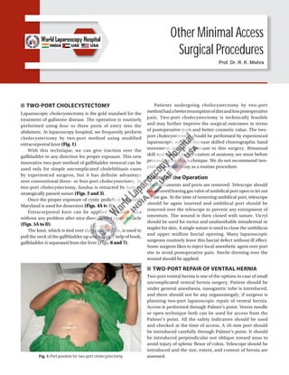
  • 1. Other Minimal Access Surgical Procedures TWO-PORT CHOLECYSTECTOMY Laparoscopic cholecystectomy is the gold standard for the treatment of gallstone disease. The operation is routinely performed using four or three ports of entry into the abdomen. At laparoscopy hospital, we frequently perform cholecystectomy by two-port method using modified extracorporeal knot (Fig. 1). With this technique, we can give traction over the gallbladder in any direction for proper exposure. This new innovative two-port method of gallbladder removal can be used only for simple uncomplicated cholelithiasis cases by experienced surgeon, but it has definite advantage over conventional three- or four-port cholecystectomy. In two-port cholecystectomy, fundus is retracted by help of strategically passed suture (Figs. 2 and 3). Once the proper exposure of cystic pedicle is achieved, Maryland is used for dissection (Figs. 4A to D). Extracorporeal knot can be applied for cystic duct without any problem after nice dissection of cystic pedicle (Figs. 5A to D). The knot, which is tied over the cystic pedicle, is used to pull the neck of the gallbladder up and with the help of hook, gallbladder is separated from the liver (Figs. 6 and 7). Patients undergoing cholecystectomy by two-port methodhadabetterresumptionofdietandlesspostoperative pain. Two-port cholecystectomy is technically feasible and may further improve the surgical outcomes in terms of postoperative pain and better cosmetic value. The two- port cholecystectomy should be performed by experienced laparoscopic surgeon because skilled choreographic hand movement is very important in this surgery. Bimanual skill and correct interpretation of anatomy are must before proceeding for this technique. We do not recommend two- port cholecystectomy as a routine procedure. Ending of the Operation The instruments and ports are removed. Telescope should be removed leaving gas valve of umbilical port open to let out all the gas. At the time of removing umbilical port, telescope should be again inserted and umbilical port should be removed over the telescope to prevent any entrapment of omentum. The wound is then closed with suture. Vicryl should be used for rectus and unabsorbable intradermal or stapler for skin. A single suture is used to close the umbilicus and upper midline fascial opening. Many laparoscopic surgeons routinely leave this fascial defect without ill effect. Some surgeon likes to inject local anesthetic agent over port site to avoid postoperative pain. Sterile dressing over the wound should be applied. TWO-PORT REPAIR OF VENTRAL HERNIA Two-port ventral hernia is one of the options in case of small uncomplicated ventral hernia surgery. Patient should be under general anesthesia, nasogastric tube is introduced, and there should not be any organomegaly, if surgeon is planning two-port laparoscopic repair of ventral hernia. Access is performed through Palmer’s point. Veress needle or open technique both can be used for access from the Palmer’s point. All the safety indicators should be used and checked at the time of access. A 10-mm port should be introduced carefully through Palmer’s point. It should be introduced perpendicular not oblique toward anus to avoid injury of splenic flexor of colon. Telescope should be introduced and the size, extent, and content of hernia are assessed. Fig. 1: Port position for two-port cholecystectomy. Prof. Dr. R. K. Mishra
  • 2. 534 SECTION 6: Miscellaneous Figs. 2A to D: Fundus is retracted up with the help of needle and thread is passed through intercostals space under vision. Figs. 3A to D: Another Vicryl is applied over Hartmann’s pouch to provide anterolateral traction. Any leak from the gallbladder is irrigated and sucked nicely with the help of suction irrigation instrument. A C B D A C B D
  • 3. 535 CHAPTER 43: Other Minimal Access Surgical Procedures Figs. 4A to D: Dissection of cystic pedicle is performed by Maryland. Figs. 5A to D: Clip or extracorporeal Meltzer’s knot is applied over cystic artery and duct. A C B D A C B D
  • 4. 536 SECTION 6: Miscellaneous Figs. 6A to D: The extracorporeal knot of cystic duct is used to pull the neck up and to expose bed of the gallbladder. Figs. 7A to D: Any leak should be sucked and gallbladder is separated with the help of hook. A C B D A C B D
  • 5. 537 CHAPTER 43: Other Minimal Access Surgical Procedures Fig. 8: Access is done through the Palmer’s point. Fig. 9: 45° azimuth angle is kept for second port. Fig. 10: 12 cm long Prolene is tied at three corner of the mesh keeping 6 cm long pair of free suture at each end. Fig. 11: Each pair of suture is pulled out through the same skin incision but different rectus and muscle layer. After initial assessment of ventral hernia, one more port is introduced according to baseball diamond concept but keeping the azimuth angle (angle between telescope and instrument) 45° (Figs. 8 and 9). Content of hernia is reduced and adhesiolysis should be performed for any possible omental or bowel adhesion. Size of the mesh is selected in such a way that at least it should cover 4 cm all around beyond the healthy margin of defect. A 1-mm wide, just skin, deep stab incision should be given at all the four corner of mesh. A 12-cm long Prolene is tied around three corner of the mesh and one of the remaining corners should be tied through the needle and thread introduced through one of the stab wound of skin (Fig. 10). The thread which was introduced percutaneously will help to stabilize the mesh and it will act as the third port (Fig. 11). Both the end of Prolene is pulled out through the same skin puncture side but keeping rectus and peritoneum in between (Fig. 12). Fig. 12: Both end of suture is tied outside the skin. Skin is lifted to slip the knot subcutaneously. The end of both the thread should be ligated using Tumble Square knot and it should be slipped inside skin
  • 6. 538 SECTION 6: Miscellaneous depth before locking to avoid loosening. This two-port technique can be accomplished with the help of Anchor or ProTack or Tacker, if patient can afford. Two-port technique using Prolene is safe and economical method of performing laparoscopic repair of ventral hernia. Although using strategically placed knot, we have performed one-port repair of ventral hernia also with the help of suture passer but if adhesions are present, one-port technique is not possible. Two-port techniques should be included in the practice of repair of ventral hernia surgery laparoscopically because in case of any difficulty, the third port can be introduced any time without any difficulty. SINGLE-INCISION LAPAROSCOPIC SURGERY Usually, when a new surgical technique is introduced, the focus will be on the feasibility, safety, and clinical advantage of the method. On the other hand, safety is highly dependent on how easily the new technique can be learned by average surgeons. It is a well-known fact that the implementation phase of new techniques is associated with an increased risk of complications emphasizing the importance of thorough training and education for the operating surgeon. The first report of single-incision laparoscopic surgery (SILS) was by Navarra et al. who performed an SILS cholecystectomy in 1997. Since then, there have been many reports regarding the use of SILS for appendectomy, splenectomy, nephrectomy, prostatectomy, colectomy, sleeve gastrectomy, adrena- lectomy, and adjustable gastric band. However, there have been no reported randomized clinical trials with direct comparison between SILS to conventional laparoscopic surgery. Despite the lack of evidence demonstrating any superiority of SILS, it is being increasingly performed unfortunatelyinalargelyunregulatedfashionwithoutformal training. Concern has been raised that this new procedure is more technically challenging and is likely associated with a significant learning curve and also its own disadvantage and complication. Single-incision laparoscopic surgery is a new technique that has now been utilized in many centers for minimal access surgery (MAS) (Figs. 13A and B). The major difficulty with this new technique is the sacrifice that has to be made in terms of comfort and ergonomics. One of the great advantage of SILS is patients are very satisfied with their single scar and particularly enthusiastic in regard to the cosmetic outcome of single incision approach. Single-incision laparoscopic surgery was first performed for the treatment of appendicitis at Department of Pediatric Surgery, Dokuz Eylül Medical School University, Izmir, Turkey and first presented at—The Annual Congress of Turkish Association of Pediatric Surgeons, October 2005. Synonym All over the world, SILS is called by following names: ■ SPL: Single Port Laparoscopy ■ SPA: Single Port Access Surgery ■ SILS: Single-incision Laparoscopic Surgery ■ LESS: Laparoendoscopic Single-site Surgery ■ OPUS: One-port Umbilical Surgery ■ NOTUS: Natural Orifice Transumbilical Surgery. Equipment Many companies are making SILS equipment (Figs. 14 and 15). Few of the famous brands are: ■ SILS device from: Covidien ■ GelPOINT system from: Applied Medical ■ R-Port and TriPort from: Advanced Surgical Concepts ■ Uni-X from: Pnavel. Figs. 13A and B: Single-incision laparoscopic surgery. Fig. 14: Articulating instruments used in single-incision laparoscopic surgery. A B
  • 7. 539 CHAPTER 43: Other Minimal Access Surgical Procedures Fig. 15: Some of the popular single-incision laparoscopic surgery (SILS) equipment. Case selection is paramount for the success of SILS and at world laparoscopy hospital, we performed SILS for the cases, which is not complicated and has minimum risk. Good ergonomics in SILS need articulating instrument and high end energy sources such as tissue response generator (LigaSure) and ultrasonic dissector (Harmonic). Advantages of Single-incision Laparoscopic Surgery ■ Cosmesis +++ (Figs. 16 and 17) ■ Ease of tissue retrieval ++++ ■ Combination procedure +++ ■ Patient acceptance ++++ ■ Quality of life analysis ++ ■ Standard equipment ++ ■ Do not violate natural orifices ++++ ■ Surgeon’s domain +++. Disadvantages of Single-incision Laparoscopic Surgery ■ More pain compared to laparoscopic surgery ■ Violating principles of ergonomics Figs. 16A and B: Comparison of scar of single-incision laparoscopic surgery (SILS) and conventional laparoscopic surgery. ■ Incisional hernia chances are more compared to laparoscopic surgery ■ More wound infection compared to laparoscopic surgery ■ Bigger learning curve compared to laparoscopic surgery ■ Not cost-effective compared to laparoscopic surgery. The evolution of surgery toward less invasive approaches has act as stimulant effect toward the development of new less invasive techniques in entering the abdominal cavity. An example of such technique is the use of a single skin incision through which multiple instruments can be A B
  • 8. 540 SECTION 6: Miscellaneous Figs. 17A to C: Comparison of scar of single-incision laparoscopic surgery (SILS), laparoscopic surgery, and open surgery. Figs. 18A and B: Position of surgical team in single-incision laparoscopic surgery (SILS). inserted into the abdomen. This single-incision laparoscopic technique has been described by a variety of names as we have discussed earlier. With this single incision of entry, SILS is theoretically less invasive approach compared to the standard multiport laparoscopic surgery. However, SILS may not allow the same level of manual dexterity and technical performance compared to conventional laparoscopic surgery that in certain aspect, it even violates the principle of laparoscopic surgery (Figs. 18A and B). Laparoscopic cholecystectomy (LC) has become the standard procedure for treating gallstones, cholecystitis, or gallbladder polyps. Traditionally, LC has involved four ports. Many laparoscopic techniques happen to be developed using this four-port LC and it has become possible to do these techniques safely. Now, having established the security of LC, our interest centered on lowering the invasiveness and scarring brought on by the procedure. Cuesta et al. reported single-incision laparoscopic cholecystectomy (SILC), in which two 5 mm ports were introduced through the umbilicus, along with a Kirschner wire hook was introduced with the right subcostal area to pull in an upright direction in order to visualize Calot’s triangle. Several surgeons have described performing SILC using three 5 mm ports in the umbilicus. Meanwhile, Merchant et al. also performed SILS by inserting a GelPort (Applied Medical, Rancho Santa Margarita, CA, USA) to stretch the umbilical fascia incision for simple access with instruments into the abdominal cavity. Furthermore, a method involving several transumbilical-placed ports for SILS was newly developed and SILS by way of the ASC TriPort (Advanced Surgical Concepts, Wicklow, Ireland) continues to be described successively. On the other hand, a fascinating new instrument named SPIDER (TransEnterix, Inc., Research Triangle Park, NC) to be used in single-incision surgery was created and its use in SILS in an animal experiment has been reported. As a result of these clinical studies, using SILC has spread rapidly. Various ports and instruments are available and various surgical methods utilized in performing SILS can be found in many institutions; however, it is necessary to develop a great method that can be performed safely such as the conventional four-port LC and it is also essential to balance safety, operability, and economy in this new technique. Under general anesthesia, an approximately 25 mm vertical skin incision is made through the center of the umbilicus, the peritoneal cavity should be entered with the open method, and then the SILS port inserted (Figs. 19A to E). Three exclusive 5 mm ports were inserted through the SILS port and one 5 mm port changeable for an exclusive 12mmport.Thepneumoperitoneumshouldsetat12–15mm and flexible scope (Olympus, Tokyo, Japan) should be used, if possible for that intra-abdominal visualization. A couple of loop-type retractor (Mini-loop retractor II; Covidien) should inserted directly within the right subcostal area. A B A B C
  • 9. 541 CHAPTER 43: Other Minimal Access Surgical Procedures Figs. 19A to E: Single-incision laparoscopic surgery (SILS) port and its access. Following the patient should be put into the reverse Trendelenburg position and slightly rotated to the left, the fundus of the gallbladder should tightened by way of this loop-type retractor and also the gallbladder thereafter suspended. In dissecting the gallbladder, a curved grasper, bipolar forceps, or monopolar hooks should be utilized in the two remaining apertures. The cystic duct and artery should be exposed and clipped having a 5-mm clip applier (Endo Clip; Covidien) after which divided with laparoscopic scissors. The gallbladder should be extracted by having an endoscopic retrieval bag (Endo Catch Gold; Covidien). Easy replacing a 5-mm port having a 12-mm port is one of the advantages of this port. Actually, SILS, while using SILS port, was proven as safe as conventional four- port LC and complications, for example, bile duct injury or uncontrolled bleeding did not occur. However, the issue areas where improvements are needed would be that the umbilical scar through the SILS port is larger than that of conventional four-port LC. Concretely, the umbilical scar length when it comes to conventional four-port LC involved 15 mm; however, while using SILS port, it was approximately 25 mm and furthermore in instances where the umbilicus bottom was shallow, the scar may be unexpectedly large. Conflict between your operative instruments and also the scope is inevitable and also the procedure seemed to be inconvenient to do since the surgeon and also the assistant needed to stand in the same side from the patient. The fundus from the gallbladder may be tightened with the Roeder’s or Meltzer’s knot and so the straight needle was inserted from the abdominal cavity right subcostal abdominal wall. The gallbladder should be elevated by raising this nylon suture and a good surgical field obtained. The surgeon operated both one instrument and also the 5 mm flexible scope by hand and the assistant designed a good surgical field such as Calot’s triangle through the traction of the gallbladder utilizing a fine loop retractor A C B D E
  • 10. 542 SECTION 6: Miscellaneous and nylon suture. This technique relieved the interference between your surgeon and also the assistant and between yourforcepsthemselves.Toextracttheexfoliatedgallbladder, one 5 mm port should be removed and an endoscopic retrieval bag should be inserted directly with an original hole and the gallbladder ended up being extracted. The fascial defect of the umbilicus incision should be repaired with approximately two stitches and an intradermal suture should be performed on the skin (Figs. 20A and B). It should be noted that during MAS, generally, we do not shave the hair of skin around the port site. If it is required, just trimming of hair should be done on the operation table itself. Shaving of hair many hours before the surgery is liable to bring the bacteria on the surface and may increase the chances of skin infection. This method represents noninvasive surgery that mixes low invasiveness with a scarless outcome. The surgeon operates one instrument along with a 5-mm flexible scope and the assistant pulls or pushes the fine loop retractor and the nylon suture. This straightforward transition is also an advantage of our two-port technique, since it can be created in any case of cholecystitis or intraperitoneal adhesion. With the global growth of using SILS, large series of cases happen to be reported in many institutes. Curcillo et al. reported that within their multi-institutional 297 case series, the utilization of one more port away from umbilicus occurred in only 34 cases plus they concluded that SILS was safe and can serve as an alternative choice to multiport therapy with fewer scars and cosmesis. Erbella and Bunch surprisingly reported that their mean operative time was 30 minutes (from 22 to 75 minutes) in 100 consecutive SILS cases. Rivas et al. reported that they observed surgeons in training and located that experienced laparoscopic surgeons may not have to undergo a steep learning curve plus they concluded that SILS was becoming the conventional process of most elective patients with gallbladder disease. Other reports also concluded that SILS was safe; however, Hernandez et al. reported that biliary complication (cystic duct stump leak) took place among 100 SILS cases and Edwards et al. described that biliary complications occurred in 3.7% of the SILS patients (cystic duct stump leak; 1 and accessory duct leak; 2). Moreover, iatrogenic combined bile duct and right hepatic artery injury during SILS has already been reported and the authors recommended that surgeons should have a low threshold to add additional ports at the appropriate interval to make sure that procedures were completed safely, especially in their early stages. As described, SILS is really a useful technique; however, it is important to make sure that the procedure is as safe as conventional four-port LC. In our department, to secure the safety, acute cholecystitis is excluded in the indication for SILS for that present. Comparative studies between SILS and conventional four-port LC regarding operating time, operative cost, complications, postoperative pain, cosmetic result, and time for you to go back to normal activity have been performed gradually with time. Fronza et al. reported that the operating time was significantly longer in SILS and 12% of SILS patients were readmitted within 24 hours after the operation, although these readmissions were due to complications similar to those present in four-port LC. Similarly, Chang et al. figured that there is a substantial difference in operative time (SILS was approximately 1.6 times longer) as well as in operative cost (SILS was 1.29 times more expensive), but no difference in postoperative discomfort was observed. However, they result that patients who underwent SILS returned to normal activity 1.8 days earlier than four-port LC patients seem to demonstrate the usefulness of SILS. Furthermore, two randomized controlled trials (RCTs) that compared SILS with conventional four-port LC have already been published. One of these trials included 70 patients and the other included 40 patients. Inside a result present with both trials, the operating amount of time in SILS was more than that in four-port LC, although it was discovered that the two methods differed with regard to the patients’ postoperative pain. According to the conventional reports, the benefit of SILS has not yet become clear; therefore, well-designed RCTs are essential to judge the corrective operative outcomes and also the necessity of SILS. Figs. 20A and B: Port closure technique in single-incision laparoscopic surgery (SILS). A B
  • 11. 543 CHAPTER 43: Other Minimal Access Surgical Procedures RETROPERITONEOSCOPY Traditionally, laparoscopic surgeries are performed by transperitoneal approach following establishment of pneumoperitoneum by closed technique using the Veress needle or by the open mini-laparotomy. The parietal peritoneum is then secondarily incised and dissected to obtain access to the retroperitoneal target organs such as kidneys,ureters,adrenals,andlymphnodes.Transperitoneal laparoscopy, although seemingly expeditious, invites the potential calamities of possible vascular and bowel injuries. On the other hand, retroperitoneal laparoscopy has its own difficulty due to working in a contained limited space. However, with technical refinements, several recent reports of successful retroperitoneoscopic surgeries have proved the feasibility and distinct advantages of this approach. Historical Perspective Retroperitoneoscopy has experienced a delayed develop- ment and acceptance compared with peritoneoscopy. Difficulties in providing adequate visualization and room for surgical maneuvering as well as concerns about deleterious effects of insufflating the retroperitoneal space account for thisretardedprogression.Althoughpneumoretroperitoneum has been used safely for >50 years to aid in radiographic visualization of the kidneys and adrenals, the conjunctive use of endoscopes has only recently been attempted. Retroperitoneoscopy through the flank in a human was pioneered by Wickham, who performed an extraperitoneal laparoscopic ureterolithotomy in 1978. Earliest techniques, termed pelviscopy, utilized a telescopetobluntlydissectwithinthepelvicretroperitoneum to sample pelvic lymph nodes. The obvious disadvantage of such technique has been the difficulty in exposing the obturator nodes, precluding the performance of an adequate staging lymphadenectomy. In the initial series reported by Fig. 21: Finger of glove over sheath of cannula. Fig. 22: Place for primary trocar insertion in retroperitoneoscopy. Hald and Rasmussen, many of the patients had no lymph nodes found in their surgical specimen. With refinements of surgical techniques, subsequently more complete pelviscopic node dissections have been reported. Surgical Technique Probably, the most important initial step in retro- peritoneoscopy is the expansion and distention of the retroperitoneal space by an expanding balloon device. A balloon device modified fingers of a latex rubber glove is tied off and the glove is secured over the distal end of the sheath of cannula (Fig. 21). The device is placed in the retroperitoneum and expanded by injecting saline. Special trocars are available with transparent balloons at their inner end that can be inflated with air or fluid to allow laparoscopic visibility of retroperitoneum through the clear distended balloon. Retroperitoneoscopy through the Flank This approach is applicable to surgery on adrenals, kidneys, and upper ureters. The patient is placed in a lateral decubitus position with slight forward tilt. A small incision is made about 2 cm below the 12th rib, just lateral to the sacrospinalis. The incision is deepened through the fused lamellae of lumbar fascia to enter the perinephric space (Fig. 22). Thespaceisfurtherdissectedbybluntdigitalexploration. The expanding device is introduced into the retroperitoneal space and about 800–1,000 mL saline are injected to inflate the balloon. The balloon is then deflated and removed (Figs. 23A and B). Laparoscope in 11 mm trocar is introduced into the retroperitoneal space and carbon dioxide (CO2) insufflation is continued to maintain a pressure of about 14 mm Hg. With the posterior parietal peritoneum pushed away by the expandingdevice,theretroperitonealspaceiswidelyopened
  • 12. 544 SECTION 6: Miscellaneous Figs. 23A and B: Finger dissection of retroperitoneal space. Fig. 24: Laparoscope with balloon cannula system. Fig. 25: Ureter as seen during retroperitoneoscopy. by continued insufflation and subsequent working ports are established under camera vision. The number and location of accessory ports are determined by the surgical procedure to be undertaken. However, it is usually advisable to keep the ports posterior to the anterior axillary line to avoid puncture of the lateral peritoneal reflection. During laparoscopic dissection, it is often helpful to move the laparoscope to the anterior ports for better visibility as the situation may dictate (Fig. 24). Retroperitoneoscopy: Anterolateral For surgery on mid and lower ureters and for internal spermaticveinligation,theretroperitoneoscopyisperformed by a small incision at McBurney’s point. The external oblique aponeurosis is incised and underlying fibers of the internal oblique and transverse muscles of the abdomen are split to reach the extraperitoneal space. Following careful digital dissection, the expanding device is introduced and inflated in the retroperitoneum (Figs. 25 and 26). Retroperitoneoscopy for Pelvic Lymph Node Pelvic retroperitoneoscopy is ideal for bilateral staging pelvic lymph node dissection and bladder neck suspension procedures. A small midline incision is made about 2 cm belowtheumbilicus.Thelineaalbaisopenedandunderlying extraperitoneal space is developed by digital dissection behind the rectus muscle of the abdomen. The expanding device is introduced and expanded by injecting about 1,200 mL saline solution. The balloon is decompressed and removed. The laparoscope is introduced with high-flow CO2 insufflation. Anatomic landmarks of the symphysis pubis, superior pubic rami, bladder neck, and external iliac vein pulsations in the pelvis are easily identified. Complete bilateral staging lymphadenectomy is accomplished by en bloc dissection of the fibrofatty lymphatic tissue from the triangular area bounded laterally by the external iliac vein, proximally by the hypogastric artery, and inferiorly by the endopelvic fascia (Figs. 27 and 28). A B
  • 13. 545 CHAPTER 43: Other Minimal Access Surgical Procedures Figs. 26A and B: Two clips are applied over ureter and cut in between. Fig. 27: Posterior dissection of left kidney. Conclusion The pioneering concept of retroperitoneal expansion by Gaur and coworkers has led to the resurgence of retroperitoneoscopy. Artificial balloon expansion creates the necessary space and retraction of neighboring viscera, so that subsequent insufflation can maintain the open space for surgical maneuvering. In a way, this technique simulates the steps of dissection and retraction traditionally used during open surgery. Aside from preventing the potential hazards of transperitoneal access and intraperitoneal dissection, there are certain distinct advantages of retroperitoneoscopy in patient positioning, intestinal retraction, anatomic approach to the renal hilum, and postoperative wound drainage. For retroperitoneal approaches to the adrenals, kidneys, and ureters, the patient is placed in the lateral decubitus position, as opposed to the supine position for laparoscopy. The intestines contained within the intact peritoneal envelope remain displaced during retroperitoneal dissection, thereby avoiding extensive colonic mobilization and constant retraction of bowel. During nephrectomy, the renal hilum is approached from the posterior aspect, allowing easier initial control of the renal arteries. Similarly, exposure of other retroperitoneal structures such as the adrenal gland in the right side is relatively easier than with the anterior approach. The closed extraperitoneal space allows more effective postoperative drainage, especially following reconstructive and reparative surgery such as pyeloplasty, partial nephrectomy, pyelolithotomy, and ureterolithotomy. The perceived difficulties of surgical dissection of kidneys and adrenal glands in a restricted environment during retroperitoneoscopy have not proved true with the present technique or initial balloon expansion. However, organ entrapment, especially of large specimens, in the limited space is difficult. The anterolateral extraperitoneal approach allows access for ureterolithotomy on the lower ureter at and above the pelvicinlet.Internalspermaticveinligationextraperitoneally is done for treatment of varicocele. The extraperitoneal dissection appears, however, to be more extensive and such an approach will not be suitable for bilateral variocele surgery. In the pelvis, the excellent anatomic appearance of the bladder, bladder neck, and symphysial structures makes the extraperitoneal approach ideally suited for procedures such as laparoscopic bladder-neck suspension and surgery for urachal pathologies. The majority of bladder diverticula are located posterolaterally and are, therefore, not often amenable to extraperitoneal excision. For the bilateral staging of pelvic lymphadenectomy, the extraperitonealapproachhasproveditssafetyandfeasibility. The lymph node dissection is anatomically precise, simulating the standards of open pelvic lymphadenectomy. The advantage of retroperitoneal CO2 insufflation is that by avoiding CO2 contact with the peritoneal membrane, there is less hypothermia and reduced postoperative pain from diaphragmatic irritation. In conclusion, retroperitoneoscopy offers another viable option in the A B
  • 14. 546 SECTION 6: Miscellaneous Figs. 28A and B: Dissection of hilum of kidney and application of clips before cutting. developing field of laparoscopy. The surgical indications that warrant an extraperitoneal approach in open surgery hold true for retroperitoneoscopy as well. As a minimally invasive technique, retroperitoneoscopy emulates the established standards and principles of open urologic procedures without compromising surgical efficiency and patient safety. MINIMAL ACCESS LINX PROCEDURE Northwestern Medicine has launched LINX® , a new laparoscopic procedure to treat gastroesophageal reflux disease (GERD). This procedure involves a string of metal beads that are drawn together by a magnet. When this string is placed around the lower esophagus, the magnet pulls the beads together and closes off the esophagus from the stomach. When the patient swallows, the beads separate to allow food to pass into the stomach. The magnet then draws the beads back together to keep acid from going up into the esophagus (Fig. 29). The success rate for LINX® is very high. Of the patients who undergo this procedure, 90–95% are able to stop taking medication completely and the remaining 5–10% rarely take medication. There are also several advantages to this Fig. 29: LINX® procedure for gastroesophageal reflux disease (GERD). procedure over the previous approaches used to treat GERD. Recovery time is typically quick, with patients commonly going home the same day. Patients are also encouraged to eat right away after the LINX® procedure, whereas previous surgical approaches have required patients to follow a liquid diet for up to 2 weeks. MINIMAL ACCESS TRANSORAL INCISIONLESS FUNDOPLICATION PROCEDURE The transoral incisionless fundoplication (TIF) is a minimally invasivetreatmentforGERD.TheTIFprocedureisperformed from inside the patient’s stomach without incisions. This procedure delivers patient outcomes similar to those provided by conventional antireflux surgical procedures, but is less invasive, has fewer adverse effects, and does not limit future treatment options. Following the principles of antireflux surgery for GERD, the TIF procedure repairs the antireflux barrier by reducing a hiatal hernia (≤2 cm) and creating a valve 2–4 cm in length and >270º circumferential wrap, thus restoring the dynamics of the angle of His (Fig. 30). A B
  • 15. 547 CHAPTER 43: Other Minimal Access Surgical Procedures MINIMAL ACCESS NECK SURGERY One of the newest frontiers is in minimally invasive soft- tissue surgery performed outside an established body cavity. The neck has been one of the soft tissue spaces of considerable interest and endoscopic or endoscopic- assisted techniques have recently been used to perform both thyroidectomy and parathyroidectomy. Several technical advances have facilitated the development of these new procedures, including the availability of balloon dilator for making artificial space, external lifts, ultrasonic coagulators, and smaller 2–3 mm diameter endoscopic instrumentation. Background In whole world, thyroidectomy and parathyroidectomy are the two most commonly performed endocrine surgical procedures. The most common indication for thyroidectomy is a solitary nodule that is not clearly benign on fine-needle biopsy. Parathyroidectomy is most commonly performed for primary hyperparathyroidism, in which a single enlarged gland or adenoma accounts for maximum number of cases. The principles of neck exploration for these two disorders are well established and the morbidity of operation is low when carried out by an experienced minimal access surgeon. Unlike many open abdominal operations, recovery is also rapid and most patients are discharged from the hospital the day after surgery and return to unlimited physical activity within fortnight. Already many surgeons are attempting to perform thyroidectomy and parathyroidectomy through smaller and smaller open incisions to achieve better cosmetic results. However, as open incisions become smaller, surgical exposure, access, ease of dissection, and even safety may be Fig. 30: Transoral incisionless fundoplication. compromised. Further evidence is that parathyroidectomy, for example, is viewed as an invasive procedure by patients and by referring endocrinologists is the reluctance of many individuals with asymptomatic or minimally symptomatic disease to undergo a definitive and curative operation, despite the cumulative risks of hyperparathyroidism over time, including osteoporosis and other metabolic sequelae. In parathyroid surgery, there has also been interest in a focused, unilateral exploration of the neck rather than the accepted gold standard of bilateral neck exploration with identificationandbiopsyofallfourparathyroids.Exploration of both sides of the neck avoids the problem of missed multiple adenomas or asymmetric hyperplasia, which can occur in up to 5–15% of cases and eliminates the need for preoperative localization studies. However, the advantages of unilateral neck exploration are that it results in less dissection and operative times are shorter. There may also be fewer injuries to the recurrent laryngeal nerve and the other parathyroid glands from leaving the contralateral neck undisturbed. Improvements in the accuracy of parathyroid imaging, such as 99m Tc sestamibi scanning and intraoperative assessment of curative resection with the quick parathyroid hormone assay, have led to better outcomes from and wider application of the unilateral approach. These considerations become increasingly important in the current economic environment in health care. Under these circumstances, minimal access approach to neck exploration may offer certain possible benefits, includingimprovedvisualizationduetomagnification,better cosmesis, less trauma to the neck musculature, less pain, and a more rapid recovery. Disadvantages of this approach might include longer operative times, increased hospital costs, possible risk of injury to the recurrent laryngeal nerve, potentialtumorspillage,inabilitytolocalizetheparathyroids, and adverse effects of neck insufflation. Consideration of an endoscopic approach to neck exploration, at the least, presents several challenges from an anatomic standpoint. Unlike the abdominal cavity, in which there is an easily distensible space for laparoscopy, the area that must be expanded and maintained to allow endoscopic access in the neck is composed of only potential spaces between soft tissue and muscle planes and the trachea. The thyroid and parathyroid glands are situated within the pretracheal space and are covered by the strap muscles anteriorly and laterally, which also limits exposure and access. The absence of a discrete anatomic compartmental boundary in the neck adds further problems, if insufflation is used because of the potential for gaseous diffusion subcutaneously and into the mediastinum. The thyroid and parathyroids are also highly vascular structures and are intimately related to the recurrent laryngeal nerve and inferior thyroid artery. Further, the location of the parathyroids, especially the inferior glands, is often variable.
  • 16. 548 SECTION 6: Miscellaneous Several technologic advances have been necessary to facilitate the development of endoscopic neck exploration. Miniature 2–3 mm endoscopic instruments have been constructed suitable for smaller working space and the more delicate structures in the neck. Many balloon space maker devices have been invented, just like used in laparoscopic hernia repair, could be adapted to create a working space. Gasless laparoscopy has been used with mechanical lifts and retractors to maintain the working space and, thus, eliminate the need for insufflation of the neck. Ultrasonic coagulators and small clip appliers may be more appropriate for obtaining hemostasis in the neck rather than monopolar cautery. Endoscopic ultrasound also aids in intraoperative localization of the parathyroid adenoma, which is localized preoperatively by sestamibi scanning. These considerations led our group to first explore the possibility of an endoscopic approach to neck exploration in an experimental animal model. Endoscopic Parathyroidectomy Endoscopic parathyroidectomy in humans was first performed successfully by Gagner in 1995. The patient had familial hyperparathyroidism and initially presented with acute pancreatitis for which he required laparoscopic pancreaticojejunostomy with stone extraction as well as LC. A preoperative sestamibi scan showed four-gland uptake consistent with generalized parathyroid hyperplasia and a subtotal parathyroidectomy was performed endoscopically. Access to the neck was obtained with four 5 mm ports placed 1 cm above the clavicle and sternal notch. Exposure was achieved by insufflation of the subplatysmal space with 15 mm Hg2+ pressure, which was maintained throughout the operation. Operative time was 5 hours and intraoperatively, the patient experienced tachycardia and hypercarbia. Postoperatively, he had subcutaneous emphysema from the eyelids to the scrotum. He recovered uneventfully, however, and was discharged on the fourth postoperative day with a normal serum calcium level. Since this initial report, endoscopic parathyroidectomy has been carried out by a small number of surgeons using either low-level gas insufflation of the neck or external retractors without CO2 gas. Gagner has excised parathyroid adenomas in several cases, but uses a lower CO2 insufflation pressure (7–10 mm Hg2+ ) to reduce the adverse effects of this technique. Duluq has also successfully performed endoscopic parathyroidectomy in several patients with low-level (7 mm Hg2+ ) CO2 insulation for exposure. Norman and Albrink attempted parathyroidectomy in four patients after preoperative localization with sestamibi imaging. Initial access to the pretracheal space was achieved via a 1.5-cm incision, but CO2 at a low insulation pressure (8 mm Hg2+ ) was used to maintain a working space. Although the parathyroidadenomawasvisualizedinthreeofthefourcases, endoscopic excision was successful in only two patients and only one normal parathyroid was identified out of these four explorations. At the conclusion of the endoscopic procedure, all patients were converted to open exploration via a 3.5-cm incision, through which the ipsilateral remaining parathyroids,bothnormalandadenomatous,wereidentified and either biopsied or removed. Postoperatively, there was subcutaneous air in the anterior neck, but no other sequela of CO2 insulation was noted. We recently performed endoscopic parathyroidectomy in two patients with primary hyperparathyroidism using a gasless technique. Preoperative localization of the parathyroid adenoma was carried out with 99m Tc sestamibi scanning, which identified abnormal uptake in the left neck of both patients. Following the induction of general anesthesia, the parathyroid adenoma was more precisely localized with transcutaneous ultrasound and in each case was posterior to the thyroid lobe. A 1.5-cm incision was then made at the sternal notch and the strap muscles were divided in the midline to enter the pretracheal space under direct vision. In the first patient, a modified space maker balloon was inserted into this space and inflated to 60 mL volume. Afterremovaloftheballoon,aworkingspacewasmaintained with a handheld S-shaped retractor. The strap muscles were further separated from the left lobe of the thyroid and the thyroid was retracted medially with a Babcock clamp placed through the open insertion site. Endoscopic visualization was achieved with a 3-mm 30° arthroscope. Two 4-mm ports were placed in the neck anterior to the sternocleidomastoid muscle. A normal inferior parathyroid was identified and biopsied and the adenoma was localized to the superior position with the aid of laparoscopic ultrasound. The enlarged gland was posterior to the thyroid lobe and wedged between two branches of the inferior thyroid artery and the recurrent laryngealnerve,whichledtoalengthyandtediousdissection. Excision was accomplished by blunt dissection with 3 mm endoscopic instruments and the ultrasonic scalpel. Small Ligaclips placed through the open insertion site were used to ligate the vascular pedicle. The second patient was approached in a similar fashion, but a small lift ring attached to a mechanical retractor was used to maintain exposure. A left superior adenoma was removed that weighed 1.7 g. The recurrent laryngeal nerve and inferior thyroid artery were identified during the dissection, but it is difficult to locate the inferior parathyroid despite careful examination of the region of the thyrothymic ligament. Total operative time in our two patients has averaged approximately 4 hours. Exposure was suboptimal at times due to the small space and there was difficulty in retracting the strap muscles laterally and the thyroid gland medially. Very small amounts of bleeding or fluid accumulation obscured the operative field and required frequent sponging through the open
  • 17. 549 CHAPTER 43: Other Minimal Access Surgical Procedures insertionsite.Manipulationandretractionoftheparathyroid with the small instruments was sometimes difficult as well. Parathyroid tissue was confirmed in all specimens and serum calcium levels have been normal postoperatively. Miccoli used an endoscopic-assisted approach in approximately 20 patients. Handheld retractors are used to maintain exposure and the dissection has been carried out with one or two lateral ports. A brief period of insufflation is used initially to aid in expanding the pretracheal space, but the remainder of the operation is carried out, with gasless retraction. Preliminary results have been favorable, but not all patients have had a normal ipsilateral parathyroid identified. Confirmation of successful excision of the parathyroid adenoma was made intraoperatively with use of the quick parathyroid hormone assay. Alternatives to Endoscopic Parathyroidectomy Minimally invasive or less invasive approaches to parathyroidectomy have been described recently that do not require endoscopic techniques or instrumentation. Norman and Chheda performed parathyroidectomy through a minimal 2–3 cm open incision after precise preoperative localization of the adenoma with sestamibi imaging. The technique used for parathyroid localization is analogous to that used for sentinel node mapping with radiolymphoscintigraphy. The 99m Tc sestamibi scanning is carried out 3 hours prior to surgical exploration. The operation is then directed with an 11-mm Neoprobe, which is used to scan and quantitate radioactivity in all four quadrants of the neck. A 2–3 cm incision is made over the site of maximal gamma activity and the adenoma is excised through this minimal incision. The authors have used this technique in 14 patients, 13 of whom had adenomas and one who was correctly predicted to have parathyroid hyperplasia. The adenomas were located operatively on average in just 19 minutes. Nine cases were carried out under local anesthesia and 11 (79%) patients were discharged the same day as surgery. Serum calcium levels were normal postoperatively and there were no operative complications. This approach is potentially very attractive because it requires minimal dissection and can be carried out under local anesthesia as strictly as outpatient procedure. Both operative and recovery times should be short, which may result in lower hospital costs, despite the use of preoperative scintigraphic localization. Frozen section examination by pathology may also become unnecessary if, after excision, all radioactivity is confined to the resected specimen. The limitations of this approach currently are that neither the ipsilateral parathyroid nor the recurrent laryngeal nerves have been routinely identified in these dissections. Further, the accuracy of “sentinel” mapping of the parathyroid adenoma must be confirmed by other investigators. Thoracoscopic Parathyroidectomy Video-assisted thoracoscopy should be considered as an alternative to median sternotomy in patients with ectopic mediastinal parathyroid adenomas. Prim and coworkers reported the use of thoracoscopic techniques to successfully excise mediastinal parathyroids in four patients with persistent hyperparathyroidism after failed cervical exploration. All glands were localized preoperatively by a combination of radionuclide scintigraphy and CT scan. The location of the abnormal glands in these four cases included the aortopulmonary window, near the ascending aorta, the aortic arch, and the region of the main pulmonary artery. Three thoracoscopic ports were used, including a 10-mm initial access port placed in the midaxillary line at the 6th intercostal space. Operative times averaged 3.25 hours and all patients became normocalcemic postoperatively, although one patient with secondary hyperparathyroidism developed recurrent hypercalcemia 9 months after surgery. A subxiphoid laparoscopic approach has also been used to excise a mediastinal parathyroid adenoma, but this technique would appear to provide access to glands in the anterior mediastinum only. Endoscopic Thyroidectomy Endoscopic excision of the thyroid is more technically demanding because of the more complex blood supply and the intimate relationship of the thyroid gland to the recurrent laryngeal nerve. A lateral approach is used in which three laparoscopic trocars are placed in the subplatysmal space along the anterior border of the sternocleidomastoid muscle from the jugular notch to the angle of the mandible. Both low pressure CO2 and a wall lifter inserted at the jugular trocar site are used to maintain a working space. Division of the strap muscles is necessary to access the thyroid. The thyroid vessels are divided with clips and an ultrasonic dissector is used to dissect the thyroid from the recurrent laryngeal nerve. In addition, both parathyroids are identified and preserved as is the external branch of the superior laryngeal nerve. Conclusion Early experience with endoscopic neck exploration prevents any definitive conclusions about its role in the management of patients with either hyperparathyroidism or thyroid disorders. Published experiences have to date been limited to small case reports and results and outcomes have not been reported in detail. The minimally invasive open approach of “sentinel” parathyroidectomy reported by Norman and Chheda has much to commend it, including accurate localization, rapid operative times, and improved cosmesis, and it is an outpatient operation that can be performed under local anesthesia.
  • 18. 550 SECTION 6: Miscellaneous Although the laparoscope provides optical magnification of important neurovascular structures, including the recurrent laryngeal nerve, better methods for exposure, and retraction of the strap muscles and thyroid, it would greatly facilitate visualization and dissection. Improved instruments are needed that allow safe manipulation of the parathyroid to lower the risk of parathyroid rupture as well as to speed the operative dissection. Suction and irrigation devices designed specifically for small spaces such as the neck would help maintain a dry operative field. Surgeons will also need flexibility in the exposure and operative approach to deal successfully with variations in parathyroid anatomy. Patient selection should be careful for endoscopic approach until there is further experience and improved operative technique. Individuals, who are obese, have a nodular goiter, have had previous neck surgery, or who are likely to have generalized parathyroid hyperplasia, should not be considered as a good candidate for an endoscopic exploration. Despite these limitations and challenges, the search for less invasive means for performing neck exploration will undoubtedly continue and has already led to renewed interest in a unilateral operative approach in patients with primary hyperparathyroidism. MINIMAL ACCESS SURGERY IN ORTHOPEDIC SURGERY Introduction Conventional (open) methods result in high amount of morbidity. To reduce the morbidity during the secondary injury, i.e., the surgical procedures while opening to reach the site of pathology, encourage the clinician to use the MAS or endoscopic techniques in orthopedic surgery. MAS with endoscope in orthopedic practice is useful in the following fields: ■ Arthroscopic surgery in sports-related injuries and other pathologies in shoulder, elbow, wrist, hip, knee, foot, and ankle ■ Arthroscopic-assisted surgery in orthopedic trauma ■ Spine surgery ■ Benign bone tumors. Arthroscopy is a minimally invasive surgical procedure in which a physical examination of the interior of a joint is performed using an arthroscope, a type of endoscope that is inserted into the joint through a small incision. The advantage of arthroscopy over traditional open procedures is that the joint does not have to be opened up fully and surgery is performed with two small incisions—one for the arthroscope and other for the surgical instruments. This reduces the recovery time for the patient and may increase the rate of surgical success due to lesser trauma to the connective tissue. It is especially useful for professional athletes, who frequently injure joints and require faster healing. There is also less scarring because of the smaller incisions. In procedures where endoscope or arthroscope is used, the advantage increases manifold by providing magnified view. The advantages of magnification and minimal scarring are extended also to the management of fracture fixation, carpal tunnel release at wrist joint, and spinal surgeries. As technology becomes more and more advanced, a greater number of minimally invasive surgical interventions have evolved. With the increase in proficiency of arthroscopic or endoscopic surgery, surgeons are now using the same technique for intramedullary lesion and tumor surgery also. Clinical effectiveness of MAS procedures over open procedures was proven beyond doubt. Hundreds of controlled randomized trials of procedures using the MAS techniques were published in the 1970s and 1980s. The advantages of MAS over conventional open surgery are listed in Box 1. In an era of rising healthcare costs, MAS offers a significant economic advantage over conventional open surgery. Decreased hospital time, decreased rehabilitation time, and a rapid return to normal activities all add up to a significant “savings” in economic and social costs. Many surgical procedures require a combination of both minimal invasive and open techniques. Therefore, the use of MAS must be tempered with knowledge of its limitations. History Medical endoscopy for internal organs has begun in the early 1800s by Bozzini. In 1918, Prof Kenji Takagi reported the arthroscopic examination in cadaveric knee at Tokyo University with the cystoscope. Dr Eugen Bircher was the first to perform and publish the first arthroscopy on live patients to diagnose tuberculosis. Initially, internal examination was done by direct visualization through the eyepiece till the advent of fiberoptic light source and camera. On the other hand, the surgical skills in arthroscopy surgery have improved with fine instrumentation. Since, then the developments in arthroscopy have become manifold. Aswithanyothersurgicaltechnique,arthroscopicsurgery continues to evolve, improvements in fiberoptics, video reproduction,andminiaturization,itwillenhanceandwiden its application. During the past two decades, arthroscopic procedures have been replacing traditional, more invasive orthopedic surgical procedures. Today arthroscopy is being BOX 1: The benefits of minimal access surgery (MAS) in orthopedics. • Less painful • Faster rehabilitation • Better visualization of the pathology • Shorter hospital stay • Cheaper (long term) • Esthetic • More precise
  • 19. 551 CHAPTER 43: Other Minimal Access Surgical Procedures done in almost all joints. High performance athletes need a minimal surgical exposure for a faster recovery and quick return to the field with very minimal morbidity. Recently, training simulators (virtual reality) have come into vogue to teach the skills necessary for arthroscopy, especially the knee. ARTHROSCOPIC SURGERY IN SPORTS- RELATED INJURIES AND OTHER PATHOLOGY Knee Joint Diagnosis Kneearthroscopy,withtheadvantageofdirectandmagnified view inside joint, makes it an excellent diagnostic tool. Its diagnostic accuracy rate of 95% has considerable advantages ascomparedwiththe75%accuracyrateofclinicalevaluation alone. The high sensitivity of MRI for arthroscopically remediable lesions in cases of internal derangement of the knee indicates that it could be used as a screening test before arthroscopy. Comparison of magnetic resonance imaging and arthroscopy confirmed the higher accuracy of magnetic resonance imaging in the diagnosis of internal derangement, but the results for articular cartilage lesions were much less good. Intra-articular fracture, chondral injury, and meniscal and ligamentous injuries (partial or complete) can be diagnosed and treated simultaneously. Trauma Anatomic reduction, typically obtained by direct visualization through an arthrotomy and internal fixation (open reduction and internal fixation), is the traditional treatment method for displaced intra-articular condylar fractures of the distal femur and proximal tibia. Arthroscopic-assisted reduction and internal fixation, of a displaced, malrotated intra- articular fracture fragment involving the tibia or femur, have benefits of decreased blood loss, shortened operative time, excellent intra-articular visualization, decreased soft-tissue dissection, and shortened postoperative recovery. Ligamentous Injury In acute ligamentous injuries, arthroscopy has limited or no role for repair of these ligamentous structures. Once the acute stage subsides, the ligamentous structures can be reinforced or reconstructed. Arthroscopic-assisted ligamentous reconstruction is the gold standard treatment for ruptured ligament. Meniscal Injury Meniscal injury is caused due to meniscal tear. Chondral Injury According to recent research, up to 10–12% of individuals present with chondral injuries. When symptomatic, chondral lesions manifest as swelling and knee pain. The loss of cartilage may be partial or complete and it may affect one or multiple locations. Nonsurgical treatment modalities include analgesics, knee brace, and physiotherapy. Surgical treatment varies from arthroscopic debridement to implantation of autologous chondrocytes beneath a periosteal patch covering the lesion. Autologous chondrocyte transplantation has a durable outcome for as long as 11 years. Osteoarthritis Arthroscopic debridement in early osteoarthritic patients may provide early symptomatic relief to pain. The long-term results are comparable with conservative management. Hip Joint Hip arthroscopy is technically demanding, with a steep learning curve, and requires special distraction tools and operating equipment. Access to the hip joint is difficult because of the resistance to distraction resulting from the large muscular envelope, the strength of the iliofemoral ligament, and the negative intra-articular pressure. This operation should not be done without specific education in its methods. Hip arthroscopy allows thorough visualization of the acetabular labrum, femoral head, and acetabular chondral surfaces as well as of the fovea, ligamentum teres, and adjacent synovium. Microsurgical tools developed specifically for arthroscopic hip surgery can be used to provide the least intrusive means of diagnosis and treatment of conditions involving the above mentioned structures (Box 2). No radiographic study, including high-contrast gadolinium-enhanced arthrography and magnetic resonance imaging, is entirely sensitive or specific for the diagnosis of labral tears or chondral lesions. Thus, a high level of clinical suspicion based on the patient’s symptoms and positive physical findings is paramount for the clinician to recognize subtle abnormalities in the hip joint. BOX 2: Indications for hip arthroscopy. • Labral tears • Loose bodies • Acetabular and femoral head chondral flap lesions • Foreign body removal • Synovial chondromatosis • Collagen diseases with impinging synovitis • Crystalline hip arthropathies • Ruptured or impinging ligamentum teres • Capsular shrinkage (Ehlers–Danlos syndrome) • Posttraumatic conditions (e.g., Pipkin fracture) • After total hip arthroplasty • Osteonecrosis (early stages prior to collapse) • Extra-articular conditions
  • 20. 552 SECTION 6: Miscellaneous Ankle Joint The advantages and experiences of arthroscopy in large joints were extended to the small joints such as ankle and wrist. Arthroscopy of the ankle is a relatively new discipline but has in recent years been increasingly applied to the diagnostic and therapeutic treatment of ankle disorders. Indications for arthroscopy in ankle joint are given in Box 3. About 30° wide angles, 2.7 mm arthroscope with a 3.5-mm shaver is used for ankle joint. Ankle joint is also distended, maximum up to 50 mm Hg pressure with the help of pump. To distract the ankle joint, ankle strap can also be used for manual traction. Standard portals are anteromedial, medial to the tibialis and anterior tendon, and located about 5 mm proximal to the medial malleolus and anterolateral, just lateral to the peroneus tertius tendon. Initial arthroscopy is performed with the scope in the anteromedial portal, but for the majority of the case, this portal will be used for instrumentation. Possible complications with anterior approach are injury to greater saphenous nerve and vein and injury to the dorsal lateral branch of the peroneal nerve. Recent studies suggest that, with the patient in the prone position, arthroscopic equipment may be introduced into the posterior aspect of the ankle without gross injury to the posterior neurovascular structures. Shoulder Joint The shoulder joint is well encapsulated with muscular covering throughout its circumference. Open surgical procedures lead to bleeding and high morbidity; hence, minimal access procedures are preferred with the use of arthroscope. Indications for shoulder arthroscopy are enumerated in Box 4. Beach chair position is comfortable for both the patient and the surgeon as it allows free access to shoulder joint and the option of converting to an open procedure. Standard portals for shoulder joint are posterior, anterior, and lateral. Complication for shoulder arthroscopy and its position are brachial plexus strain and hypoglossal nerve injury. Elbow Joint Arthroscopic surgery for elbow joint is still in primitive stage and limited to arthroscopic synovectomy. Arthroscopic synovectomy is a reliable procedure to alleviate pain in early grades of rheumatoid arthritis. The fundamental of arthroscopy is visualization and access. Visualization and access to the ulnohumeral and radiocapitellar articulation are rather difficult. Recent study has come out with a joint jack to widen the ulnohumeral joint space to work better posteriorly. Wrist Joint Wrist arthroscopy is the third most common joint after knee and shoulder joint to be examined by arthroscope. MINIMAL ACCESS SURGERY IN ORTHOPEDIC TRAUMA Opening of the fracture site during exposure further jeopardizethevascularityatthefracturesite,whichadversely effects the healing at the fracture site. The involvement of intra-articular fracture needs minimal tissue stripping to further jeopardize the vascularity. This principle leads to the foundation of the minimal access surgical principle in orthopedic trauma surgery. This principle helps to maintain the biology around the fracture site, so this fixation is also known as biological fixation. Biological fixation or MAS is extremely useful at the site of fractures with comminution or areas with doubtful vascularity. SPINE SURGERY AND ARTHROSCOPY Spinal surgeries are thought to be risky due to the vicinity of the important structures and high vascularity (venous plexus) around the spine. The conventional (open) exposure to reach the site of pathology needs wide exposure, which, in turn, leads to large amount of morbidity and prolongs the period of recovery, especially in the thoracic spine surgeries. The advantage of endoscopy such as precision, magnification, and small incision for exposure has lead to the endoscopic spinal surgery to the great advantage than conventional open procedure. The endoscope allows the surgeon to use a “keyhole” incision to access the herniated disk. Muscle and tissue are dilated rather than being cut when accessing the disk. This leads to less tissue destruction, less postoperative pain, quicker recovery times, earlier rehabilitation, and avoidance of general anesthesia. Thermal annuloplasty is an adjunctive procedure that uses bipolar electrothermal energy (radiofrequency and/or laser) to ablate or depopulate the sensitized pain nociceptors in the annulus, ablate any BOX 3: Indications for ankle arthroscopy. • Soft tissue: – Soft-tissue impingement – Synovitis (diagnosis and biopsy) – Arthrofibrosis • Osteochondral defect of the talus • Intra-articular fracture and occult intra-articular injury • Arthrodesis of ankle joint BOX 4: Indications for shoulder arthroscopy. • Soft tissue: – Soft-tissue impingement – Synovitis (diagnosis and biopsy) – Arthroscopic-assisted lysis of adhesion • Arthroscopic subacromial decompression (ASAD) and acromioplasty • Arthroscopic reconstruction (Bankart lesion repair and slap lesions)
  • 21. 553 CHAPTER 43: Other Minimal Access Surgical Procedures inflammatory/granulation tissue that has grown into the annulus, and to shrink and tighten the stretched or torn collagen fibers of the annulus. Scoliosis is a three-dimensional problem. The aim of surgery is to try to restore the normal contour of the back from both the front view and the side view. A technique to assist in getting a maximum of correction with a minimum of scar and morbidity by releasing the contacted anterior tissue with the use of the endoscope to go into the chest (similar to the way surgeon stakes out gallbladders now) in front where the actual vertebra is and take out the disks in front, thus relaxing up the spine so we can get better correction and the fusion in back. This method goes in through the chest using three or four small incisions to reach the front of the spine. Once inside the chest, the spine is clearly visible and “soft” tissues can be cleaned off exposing the spine. The disks are easily seen and can be removed. BONE ENDOSCOPY AND TUMORS With the increasing experience of seeing inside the soft cavitiessuchasjoint,thesamearthroscopeisnowusedtosee inside the bony cavities. The initial results are equal than the open surgical procedure with added advantages of minimal invasive, no immobilization, quick hospital discharge, minimal chances of pathological fracture, and very small surgical scar. This is especially useful for benign cystic lesion of bone such as giant cell tumor, simple bone cyst, or enchondroma. With the endoscopic technique, curettage of the cystic lesion and filling of the cavity by morselized autologous bone or bone cement can be done effectively. This minimal invasive technique allows the lesion to heal at much faster rate and with minimal scaring. This technique is being in practice for the management of these cystic lesions in soft bones or in cancellous bones. BIBLIOGRAPHY 1. Abbou CC, Cicco A, Gasman D, Hoznek A, Antiphon P, Chopin DK, et al. Retroperitoneal laparoscopic versus open radical nephrectomy. J Urol. 1999;161(6):1776-80. 2. Abolyosr A. Laparoscopic versus open orchidopexy in the management of abdominal testis: a descriptive study. Int J Urol. 2006;13(11):1421-4. 3. American Society of Anesthesiologists. New classification of physical status. Anesthesiology. 1963;24(1):111. 4. Ashton RC, McGinnis KM, Connery CP, Swistel DG, Ewing DR, DeRoseJJ.Totallyendoscopicroboticthymectomyformyasthenia gravis. Ann Thorac Surg. 2003;75(2):569-71. 5. Azagra JS, Goergen M, Gilbart E, Jacobs D. Laparoscopic anatomical (hepatic) left lateral segmentectomy—technical aspects. Surg Endosc. 1996;10(7):758-61. 6. Baba S, Ito K, Yanaihara H, Nagata H, Murai M, Iwamura M. Retroperitoneoscopic adrenalectomy by a lumbodorsal approach: clinical experience with solo surgery. Word J Urol. 1999;17(4):54-8. 7. Berkmen F, Alagol H. Germinal cell tumors of the testis in cryptorchids. J Exp Clin Cancer Res. 1998;17(4):409-12. 8. Bisgaard T, Klarskov B, Trap R, Kehlet H, Rosenberg J. Pain after microlaparoscopic cholecystectomy. A randomized double-blind controlled study. Surg Endosc. 2000;14(6):340-4. 9. Bode CO, Nwawolo CC, Giwa-Osagie OF. Surgical education at the WestAfricanCollegeofSurgeons.WorldJSurg.2008;32(6):2162-6. 10. Carmignani L, Morabito A, Gadda F, Bozzini G, Rocco F, Colpi GM. Prognostic parameters in adult impalpable ultra- sonographic lesions of the testicle. J Urol. 2005;174(30):1035-8. 11. Chandrasekharam VVSS. Laparoscopy vs inguinal exploration for the nonpalpable undescended testis. Indian J Pediatr. 2005;72(12):1021-3. 12. Charny CK, Jarnagin WR, Schwartz LH, Frommeyer HS, DeMatteo RP, Fong Y, et al. Management of 155 patients with benign liver tumors. Br J Surg. 2001;88(6):808-13. 13. Cheah WK, Lenzi JE, So JB, Kum CK, Goh PM. Randomized trial of needlescopic versus laparoscopic cholecystectomy. Br J Surg. 2001;88(5):45-7. 14. Cherqui D, Husson E, Hammoud R, Malassagne B, Stéphan D, Bensaid S, et al. Laparoscopic liver resections: a feasibility study in 30 patients. Ann Surg. 2000;232(6):753-62. 15. Cherqui D, Rahmouni A, Charlotte F, Boulahdour H, Métreau JM, Meignan M, et al. Management of focal nodular hyperplasia and hepatocellular adenoma in young women: a series of 41 patients with clinical, radiological, and pathological correlations. Hepatology. 1995;22(6):1674-81. 16. Cherqui D. Laparoscopic liver resection. Br J Surg. 2003;90(6): 644-6. 17. Cleary K, Nguyen C. State of the art in surgical robotics: clinical applications and technology challenges. Comput Aided Surg. 2001;6(6):312-28. 18. Cortes D, Thorup J, Visfeldt J. Multinucleated spermatogonia in cryptorchid boys: a possible association with an increased risk of testicular malignancy later in life? APMIS. 2003;111(1):25-30. 19. Cortez D, Thorup J, Peterson BL. Testicular neoplasia in undescended testes of cryptorchid boys—does surgical strategy have an impact on the risk of invasive testicular neoplasia? Turk J Pediatr. 2004;46 Suppl:35-42. 20. Corvin S, Sturm W, Anastasiadis A, Kuczyk M, Stenzl A. Laparoscopic management of adult nonpalpable testicle. Urol Int. 2005;75(4):337-9. 21. Cuschieri A, Shimi S, Banting S, Nathanson LK, Pietrabissa A. Intraoperative cholangiography during laparoscopic chole- cystectomy. Routine vs selective policy. Surg Endosc. 1994; 8(4):302-5. 22. Delaunay L, Bonnet F, Cherqui D, Rimaniol JM, Dahan E, Atlan G. Laparoscopic cholecystectomy minimally impairs postoperative cardiorespiratory and muscle performance. Br J Surg. 1995;82(3):373-6. 23. DeRose JJ, Swistel DG, Safavi A, Connery CP, Ashton RC. Mediastinal mass evaluation using advanced robotic techniques. Ann Thorac Surg. 2003;75(6):571-3. 24. Desai CS, Prabhu RY, Supe AN. Laparoscopic orchidectomy for undescended testis in adults. J Postgrad Med. 2002;48(5):25-6. 25. Descottes B, Glineur D, Lachachi F, Valleix D, Paineau J, Hamy A, et al. Laparoscopic liver resection of benign liver tumors. Surg Endosc. 2003;17(1):23-30. 26. Descottes B, Lachachi F, Sodji M, Valleix D, Durand-Fontanier S, deLaclauseBP,etal.Earlyexperiencewithlaparoscopicapproach forsolidlivertumors:initial16cases.AnnSurg.2000;232(5):641-5. 27. Dunlap KD, Wanzer L. Is the robotic arm a cost-effective surgical tool? Assoc Oper Room Nurs J. 1998;68(4):265-72. 28. El-Anany F, Gad El-Moula M, Abdel Moneim A, AbdallahA, Takahashi M, Kanayama H, et al. Laparoscopy for impalpable testis: classification-based management. Surg Endosc. 2007; 21(3):449-545. 29. Elder JS. Ultrasonography is unnecessary in evaluating boys with a nonpalpable testis. Pediatrics. 2002;110(4):748-51.
  • 22. 554 SECTION 6: Miscellaneous 30. Farges O, Jagot P, Kirstetter P, Marty J, Belghiti J. Prospective assessment of the safety and benefit of laparoscopic liver resections. J Hepatobiliary Pancreat Surg. 2002;9(2):242-8. 31. Farrer JH, Walker AH, Rajfer J. Management of the postpubertal cryptorchid testis: a statistical review. J Urol. 1985;134(4):1071-6. 32. Fong Y, Jarnagin W, Conlon KC, Dematteo R, Dougherty E, Blumgart LH. Hand-assisted laparoscopic liver resection. Arch Surg. 2000;135(7):854-9. 33. Ford TF, Parkinson MC, Pryor JP. The undescended testis in adult life. Br J Urol. 1985;57(2):181-4. 34. Gagner M, Rheault M, Dubuc J. Laparoscopic partial hepatectomy for liver tumor [abstract]. Surg Endosc. 1992;6(2):99. 35. Hamdorf JM, Hall JC. Acquiring surgical skills. Br J Surg. 2000;87(5):28-37. 36. HrebinkoRL,BellingerMF.Thelimitedroleofimagingtechniques in managing children with undescended testes. J Urol. 1993; 150(2 Pt 1):458-60. 37. Hüscher CG, Lirici MM, Chiodini S. Laparoscopic liver resections. Semin Laparosc Surg. 1998;5(3):204-10. 38. IHPBA Brisbane 2000 terminology of liver anatomy and resections. HPB Surg. 2000;2(3):333-9. 39. Ivanov A, Dewey C, Fahlenkamp D, Luning M. MRT in nonpalpable testes. Rofo. 1994;160(3):249-53. 40. JarnaginWR,GonenM,FongY,DeMatteoRP,Ben-PoratL,LittleS, et al. Improvement in perioperative outcome after hepatic resection: analysis of 1,803 consecutive cases over the past decade. Ann Surg. 2002;236(4):397-406. 41. Kammula US, Buell JF, Labow DM, Rosen S, Millis JM, Posner MC. Surgical management of benign tumors of the liver. Int J Gastrointest Cancer. 2001;30(3):141-6. 42. Kaneko H, Takagi S, Shiba T. Laparoscopic partial hepatectomy and left lateral segmentectomy: technique and results of a clinical series. Surgery. 1996;120(3):468-75. 43. Katkhouda N, Hurwitz M, Gugenheim J, Mavor E, Mason RJ, Waldrep DJ, et al. Laparoscopic management of benign solid and cystic lesions of the liver. Ann Surg. 1999;229(4):460-6. 44. Kavoussi LR, Moore RG, Adams JB, Partin AW. Comparison of robotic versus human laparoscopic camera control. J Urol. 1995;154(6):2134-6. 45. Kneebone R, ApSimon D. Surgical skills training: simulation and multimedia combined. Med Educ. 2001;35(9):909-15. 46. Kondraske GV, Hamilton EC, Scott DJ, Fischer CA, Tesfay ST, Taneja R, et al. Surgeon workload and motion efficiency with robot and human laparoscopic camera control. Surg Endosc. 2002;16(6):1523-7. 47. Kucheria R, Sahai A, Sami TA, Challacombe B, Godbole H, Khan MS, et al. Laparoscopic management of cryptorchidism in adults. Eur Urol. 2005;48(3):453-7. 48. Laurent A, Cherqui D, Lesurtel M, Brunetti F, Tayar C, Fagniez PL. Laparoscopic liver resection for subcapsular hepatocellular carcinoma complicating chronic liver disease. Arch Surg. 2003;138(7):763-9. 49. Le Bartz G, Petit T, Ravasse P. Is there any interest to perform ultrasonography in boys with undescended testis? Arch Pediatr. 2006;13(5):426-8. 50. Leggett PL, Churchman-Winn R, Miller R. Minimizing ports to improve laparoscopic cholecystectomy. Surg Endosc. 2000;14(7):32-6. 51. Lesurtel M, Cherqui D, Laurent A, Tayar C, Fagniez PL. Laparoscopic versus open left lateral hepatic lobectomy: a case- control study. J Am Coll Surg. 2003;196(2):236-42. 52. Leung KF, Lee KW, Cheung TY, Leung LC, Lau KW. Laparo- scopic cholecystectomy: two-port technique. Endoscopy. 1996;6(4):505-7. 53. Lomanto D, De Angelis L, Ceci V, Dalsasso G, So J, Frattaroli FM, et al. Two-trocar laparoscopic cholecystectomy: a reproducible technique. Surg Laparosc Endosc Percutan Tech. 2001;11(4):248-51. 54. Mcheik JN, Levard G. Laparoscopic treatment of the nonpalpable testis. Results. Prog Urol. 2002;12(2):294-7. 55. Menconi GF, Melfi FAM, Givigliano F, Angeletti CA. Robotic pulmonary lobectomy: early operative experience and preliminary clinical results. Eur Surg. 2002;34(5):173-6. 56. Merola S, Weber P, Wasielewski A, Ballantyne GH. Comparison of laparoscopic colectomy with and without the aid of a robotic camera holder. Surg Laparosc Endosc Percutan Tech. 2002;12(6):46-51. 57. Moher D, Schulz KF, Altman DG. The CONSORT statement: revised recommendations for improving the quality of reports of parallel-group randomised trials. Lancet. 2001;357(6):1191-4. 58. Morgan JA, Thornton BA, Peacock JC, Hollingsworth KW, Smith CR,OzMC,etal.Doesrobotictechnologymakeminimallyinvasive cardiac surgery too expensive? A hospital cost analysis of robotic and conventional techniques. J Card Surg. 2005;20(4):246-51. 59. Morino M, Morra I, Rosso E, Miglietta C, Garrone C. Laparoscopic vs open hepatic resection: a comparative study. Surg Endosc. 2003;17(12):1914-8. 60. Nijs SM, Eijsbouts SW, Madern GC, Leyman PM, Lequin MH, Hazebroek FW. Nonpalpable testes: is there a relationship between ultrasonographic and operative findings? Pediatr Radiol. 2007;37(4):374-9. 61. Panait L, Doarn CR, Merrell RC. Applications of robotics in surgery. Chirurgia (Bucur). 2002;97(5):549-55. 63. Perniceni T, Slim K. What are the validated indications for laparoscopy in digestive surgery? Gastroenterol Clin Biol. 2001;25(4 Suppl):B57-70. 63. Poon CM, Chan KW, Ko CW, Chan KC, Lee DW, Cheung HY, et al. Two-port laparoscopic cholecystectomy: initial results of a modified technique. J Laparoendosc Adv Surg Tech A. 2002;12(5):259-62. 64. PoonRT,FanST,LoCM,LiuCL,LamCM,YuenWK,etal.Improving perioperative outcome expands the role of hepatectomy in management of benign and malignant hepatobiliary diseases: analysis of 1222 consecutive patients from a prospective database. Ann Surg. 2004;240(4):698-710. 65. Raina V, Shukla NK, Gupta NP, Deo S, Rath GK. Germ cell tumours in uncorrected cryptorchid testis at Institute Rotary Cancer Hospital, New Delhi. Br J Cancer. 1995;71(2):380-2. 66. Ramachandran CS, Arora V. Two-port laparoscopic cholecys- tectomy: an innovative new method for gallbladder removal. J Laparoendosc Adv Surg Tech A. 1998;8(3):303-8. 67. Rangarajan M, Jayakar SM. Laparoscopic orchidectomy for the adult impalpable testis—experiences in a rural teaching hospital. Surg Endosc. 2007;21(1):66-9. 68. Rodriguez MDA, Freire AU, Orpez AR, Lorenzo CG. Diagnostic and therapeutic laparoscopy for nonpalpable testis. Surg Endosc. 2003;17(11):1756-8. 69. Rau HG, Buttler E, Meyer G, Schardey HM, Schildberg FW. Laparoscopic liver resection compared with conventional partial hepatectomy—a prospective analysis. Hepatogastroenterology. 1998;45(24):2333-8. 70. Ruurda JP, Hanlo PW, Hennipman A, Broeders IA. Robot-assisted thoracoscopic resection of a benign mediastinal neurogenic tumor: technical note. Neurosurgery. 2003;52(4):462-4. 71. Satar N, Bayazit Y, Doran S. Laparoscopy in the management of impalpable testicle. Acta Chir Belg. 2005;105(6):662-6. 72. Shin D, Lemack GE, Goldstein M. Induction of spermatogenesis and pregnancy after adult orchiopexy. J Urol. 1997;158(6):2242. 73. Siemer S, Humke U, Hildebrandt U, Karadiakos N, Ziegler M. Diagnosis of nonpalpable testes in childhood: comparison of magnetic resonance imaging and laparoscopy in a prospective study. Eur J Pediatr Surg. 2000;10(2):114-8.
  • 23. 555 CHAPTER 43: Other Minimal Access Surgical Procedures 74. Soper NJ, Brunt LM, Kerbl K. Laparoscopic general surgery. N Engl J Med. 1994;330(6):409-19. 75. Soper NJ, Dunnegan DL. Routine versus selective intra-operative cholangiography during laparoscopic cholecystectomy. World J Surg. 1992;16(6):1133-40. 76. Terkivatan T, de Wilt JH, de Man RA, van Rijn RR, Zondervan PE, Tilanus HW, et al. Indications and long-term outcome of treatment for benign hepatic tumors: a critical appraisal. Arch Surg. 2001;136(9):1033-8. 77. Vibert E, Perniceni T, Levard H, Denet C, Shahri NK, Gayet B. Laparoscopic liver resection. Br J Surg. 2006;93(1):67-72. 78. Waseem T. Technologic advances in robotic surgery. J Coll Physicians Surg Pak. 2005;15(9):559-61. 79. Weimann A, Ringe B, Klempnauer J, Lamesch P, Gratz KF, Prokop M, et al. Benign liver tumors: differential diagnosis and indications for surgery. World J Surg. 1997;21(9):983-90. 80. Welvaart K, Tijssen JGP. Management of the undescended testis in relation to the development of cancer. J Surg Oncol. 2006;173(4):219-23. 81. Wolverson MK, Houttuin E, Heiberg E, Sundaram M, Shields JB. Comparison of computed tomography with high-resolution real-time ultrasound in the localization of the impalpable undescended testis. Radiology. 1983;146(1):133-6. 82. Yoshino I, Hashizume M, Shimada M, Tomikawa M, Tomiyasu M, Suemitsu R, et al. Thoracoscopic thymomectomy with the da Vinci computer-enhanced surgical system. J Thorac Cardiovasc Surg. 2001;122(5):783-5.