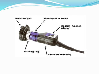
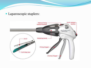
DEPARTMENT OF GENERAL SURGERY
R.N.T. MEDICAL COLLEGE,UDAIPUR
LAPAROSCOPY INSTRUMENTS
-Dr.Sagar Patil
Moderator- Dr.Siddharth Verma Sir
Asso.Professor,
Department of Gen.Surgery
INTRODUCTION
Laparoscopic surgery, also called minimally invasive
surgery (MIS) or keyhole surgery, is a
modern surgical technique in which operations are
performed far from their location through small
incisions (usually 0.5–1.5 cm) elsewhere in the body.
 Three main Components:
 Image production
 Pneumoperitoneum
 Laparoscopic instruments
OPERATING ROOM SET-UP
 The proper hardware and instruments.
 Dedicated interventional laparoscopic operating
rooms.
 A sufficient back up of instrumentation to cover
for equipment failure.
• Facilities for intra-operative imaging .
LAPAROSCOPIC
INSTRUMENTATION
 1. Optical Devices
 2. Equipment for creating / maintaining domain
 3. Instruments for Access
 4. Operative instruments
 5. Energy sources
 6. Tissue approximation/ hemostasis
 7. Miscellaneous
1. Optical Devices
Telescope:
 This endoscope is made of surgical stainless steel
containing an optical lens train comprised of precisely
aligned glass lenses and spacers (Rod lens system)
 Telescopes or laparoscopes come in various sizes
10mm, 5mm, 2-3mm ‘needlescopes’ and with various
visualization capabilities such as zero degree forward
viewing, 30 or 45 degree telescope, zero degree
telescope with 6mm instrument channel(operating
laparoscope)
 Light source:
 White light illumination is provided from a high-
intensity xenon,mercury, or halogen lamp and
delivered via a fiberoptic bundle.
 Light cable:
There are two types of cables
1. Fiberoptic cables are flexible but do not
transmit a precise light spectrum.
2. Fluid cables transmit more light and a
complete spectrum but are more rigid. Fluid
cables require soaking for sterilization and
cannot be gas sterilized.
 Video Camera:
 The basis of laparoscopic cameras is the solidstate
silicon computer chip or CCD (charge-coupled
device).
 The resolution or clarity of the image depends
upon the number of pixels or light receptors on the
chip.
 Standard cameras in laparoscopic use contain
250,000 to 380,000 pixels.
 Television Monitor:
 High-resolution video monitors are required for
suitable reproduction of endoscopic image.
 Three chip cameras require monitors with 700
lines resolution to realize the improved resolution
of extra chip sensors.
 VHS recorder, video printer and sometimes DVD
recorder are standard documentation equipments
housed in the video cart.
2. Equipment for creating /
maintaining domain
 Gas insufflation:
1. CO2 Insufflator: The creation of working space in the
abdominal cavity is generally done using CO2 delivered via
an automatic, high flow,pressure regulated insufflator.
 CO2 is currently the agent of choice due to low risk of gas
embolism,low toxicity to peritoneal tissues, rapid
reabsorption, low cost and inhibits combustion.
 The insufflator delives gas at a flow rate of up to 20 liters /
min.
 level is usually set at 12 to 15 mm Hg.
 Gasless laparoscopy:
This has some theoretical advantages in some high-
risk patients with compromised cardio-respiratory
function.
 It facilitates continuous suction and use of some
conventional open instruments.
3. Instruments for Access
Veress needle:
 The Veress needle is designed to create
pneumoperritoneum prior to insertion of trocar in a closed
fashion.
 It consists of an outer sharp cutting needle and inner blunt
spring-loaded obturator.
Hasson’s cannula:
 used for gaining initial access to the abdominal cavity with
an open cutdown technique.
Optical trocar: allows visualization of the tissues as
the blade cuts through the layers of the abdominal wall.
4. Operative Instruments
 Trocars:
 Available in various diameters and sizes according
to requirements, 10mm and 5 mm being
commonly used.
 They are Bladed and nonbladed types.
 Of the bladed trocars, there are shielded and
nonshielded types.
Graspers: Retraction may be achieved using large
grasping instruments.
Bullet Nose Grasper with either straight or
diamond-cut serrations has a blunt bullet nose tip
design with an atraumatic grasping jaw (one that will
not produce tissue damage). These graspers are ideal
for dissecting or grasping delicate anatomy. It is
important to give attention to the cleaning of the jaw
serrations and hinged areas of the instrument.
Dorsey Intestinal Fenestrated Grasper has
atraumatic horizontal serrations.
 Bullet nose grasper  Dorsey intestinal grasper
Hunter Bowel Grasper has two rows of atraumatic
serrations. The jaw comes in different lengths,depending
on the needs of the surgical procedure.
Crocodile Grasper has long contoured jaws with tissue
herniation channels to ensure superior grasping ability.
Raptor Grasper has long contoured jaws with tissue
herniation channels. It also has two atraumatic teeth at the
tip for grasping difficult anatomy.
Triangular Endo Flex Retractor (angled or straight) is
used for retraction during large organ surgery. Two
common lengths are 60mm and 80mm.
 Hunter bowel grasper  Crocodile graspers
 Raptor grasper  Triangular endoflex
retractor
 Dissectors:
Maryland Dissector has long, curved jaws with fine-
tapered tips. Ideal for precise dissection (resembles
the Crile hemostatic clamp used in open surgeries).
Bipolar Dissector consists of four pieces when
disassembled, and it should be left disassembled
during the sterilization cycle. At the sterile field,
scrub personnel should assemble the dissector
before use and attach a bipolar cord that delivers the
electrosurgical energy to the instrument. It is used
to achieve hemostasis.
 Maryland dissector  Bipolar dissector
 Scissors:
 There are a variety of scissors for dissecting,
mobilizing and cutting tissues, which include
straight and curved types (Endo Shears). Hook
scissors are used to cut sutures, tough fibrous
tissues. Most dissecting scissors have adapters
for diathermy.
 Hook scissors should always be kept in view
while entering and exiting.
 Repeated use of diathermy at the sharp edge
may tend to blunt the scissors.
 Scissors  Hook scissors
 Bowel and lung clamp:
 Tubular structures, bowel and lung can be held
with instruments designed specifically for the
same. (Endo-Babcocks, Endo-Lung, Bowel
Clamp).
 Endo babcock  Endo lung clamp
5. Energy Sources
 Electrosurgery:
 Electrocautery refers to direct current whereas
electrosurgery uses alternating current.
 During electrocautery, current does not enter the patient’s
body. Only the heated wire comes in contact with tissue.
 In electrosurgery,the patient is included in the circuit and
current enters the patient’s body.
 Bipolar:
 In bipolar electrosurgery, both the active electrode and
return electrode functions are performed at the site of
surgery. The two tines of the forceps perform the active and
return electrode functions. Only the tissue grasped is
included in the electrical circuit.
 Monopolar:
 The active electrode is in the wound. The patient
return electrode is attached somewhere else on the
patient. The current must flow through the patient
to the patient return electrode to complete the
circuit.
 Safety Considerations during
Electrosurgical laparoscopic surgery:
 Direct Coupling:
 Capacitive Coupling:
 Insulation Failure:
 Most potential problems can be avoided by following:
· Inspect insulation carefully
· Use lowest possible power setting
· Use a low voltage waveform (cut)
· Use brief intermittent activation vs. prolonged activation
· Do not activate in close proximity or direct contact with
another instrument
· Use bipolar electrosurgery when appropriate
· Select an all-metal cannula system as the safest choice
.Do not use hybrid cannula systems that mix metal with
plastic
· Utilize available technology, such as a tissue response
generator to reduce capacitive coupling or an active
electrode monitoring system, to eliminate concerns about
insulation failure and capacitive coupling.
 Argon-Enhanced Electrosurgery:
 incorporates a stream of argon gas to improve the
surgical effectiveness of the electrosurgical
current.
Argon gas is inert and noncombustible making it a
safe medium through which to pass electrosurgical
current
 Ultrasonic Energy: (The Harmonic scalpel)
 Uses ultrasonic technology, the unique energy form
that allows both cutting and coagulation at the precise
point of impact,resulting in minimal lateral thermal
tissue damage.
 Cuts and coagulates by using lower temperatures than
those used by electrosurgery or lasers.
 Coagulation occurs by means of protein denaturation
when the blade, vibrating at 55,500 Hz, couples with
protein, denaturing it to form a coagulum that seals
small coapted vessels.
 It offers greater precision in tight spaces near vital
structures,fewer instrument changes are needed, less
tissue charring and desiccation occur, and visibility in
the surgical field is improved.
 Harmonic scalpel:
 Power Setting:
The Harmonic Scalpel LCS has five power levels.
Increasing the power level increases cutting speed and
decreases coagulation.
In contrast, less power decreases cutting speed and
increases coagulation.
 Tissue tension:Faster cutting with less coagulation is
achieved by increasing the tissue tension.
 Grip Force/Pressure:Application of a gentle force,or
light pressure, achieves more coagulation with slower
cutting.A firmer grip force achieves less coagulation
with faster cutting.
6. Instruments for Tissue
approximation/ Hemostasis
1. Laparoscopic ligating suture delivery
system:
 A Pre-tied sliding knot with a loop is available with
nylon carrier rod to ligate stump like structures or
tubular structures after cutting. Eg Surgitie.
 The suture is looped around the structure to be ligated
and the knot is slid down to close the loop.
 Useful in appendicectomy.
 Surgitie  Knot pusher
2. Needle drivers:
 Endostitch is a 10mm disposable suturing device.
The needle features a sharp tapering point at each
end with suture attachment at the center of the
needle. The double-ended needle is passed
between the two jaws of the suturing device.
 Its advantages include easy introduction,
atraumatic needle manipulation, good security
and easy accurate needle placement.
 Needle holder  Endo-stitch
3. Clip Applicators:
• Clip appliers are primary modality for ligating blood
vessels and other tubular structures.
Disposable clip appliers contain up to 20 clips, whereas
reusable clip appliers carry one clip at a time.
Clips are made of titanium though now absorbable
clips are also available.
 Clip applicator:
Mechanical Stapling Instruments:
 Laparoscopic staplers are modifications of stapling
devices of open surgery. Staplers are used for
transecting and anastomosing bowel, transecting
mesentery etc.
 A range of staple lengths (2.5-3.8mm)is available
depending on the thickness of the tissue to be
divided.
 Laparoscopic staplers:
7. Miscellaneous
Aspiration / Irrigation probes:
 These are essential for most laparoscopic
procedures in order to maintain a clear operative
field. Irrigation and aspiration channels may be
incorporated into surgical instruments but
working channels are small and subject to
repeated clogging.
 Suction –irrigator:  Nezhat-Dorsey suction
tips
Hand Assisted Laparoscopic surgery:
• The Hand Access Device is intended to provide
extracorporeal extension of pneumoperitoneum and
abdominal access for the surgeon during laparoscopic
surgery.
 It is indicated for use in laparoscopic procedures, where
entry of the surgeon’s hand may facilitate the procedure,
and for extraction of large specimens.
 It has application in colorectal, urological and general
surgical procedures.
 Organ Extraction devices:
These are pre loaded specimen retrival pouches
made of strong material, which is impervious to
cancer cells. The mouth of the pouch is bought out
of the incision site and opened following which
the specimen is extracted.
 Specimen retrieving bag:
Tissue Morcellators:
 These are used to reduce the size of the resected
specimen prior to retrieval.
 It may render pathological examination more difficult.
 Eg.Laparoscopic myomectomy.
 Morcellator:
 Space creators:
 Balloon dissector is a space dissector which functions
when saline or air instilled into the pre-shaped balloon
inserted into the intended region.
 These are used for laparoscopic extraperitoneal
surgery eg hernia repair,endoscopic neck surgery,
subfascial endoscopic ligation of perforators veins,
retroperitoneal surgery, etc.
 Balloon dissector:
THANK YOU

Laparoscopy instruments

  • 1.
    DEPARTMENT OF GENERALSURGERY R.N.T. MEDICAL COLLEGE,UDAIPUR LAPAROSCOPY INSTRUMENTS -Dr.Sagar Patil Moderator- Dr.Siddharth Verma Sir Asso.Professor, Department of Gen.Surgery
  • 2.
    INTRODUCTION Laparoscopic surgery, alsocalled minimally invasive surgery (MIS) or keyhole surgery, is a modern surgical technique in which operations are performed far from their location through small incisions (usually 0.5–1.5 cm) elsewhere in the body.  Three main Components:  Image production  Pneumoperitoneum  Laparoscopic instruments
  • 3.
    OPERATING ROOM SET-UP The proper hardware and instruments.  Dedicated interventional laparoscopic operating rooms.  A sufficient back up of instrumentation to cover for equipment failure. • Facilities for intra-operative imaging .
  • 4.
    LAPAROSCOPIC INSTRUMENTATION  1. OpticalDevices  2. Equipment for creating / maintaining domain  3. Instruments for Access  4. Operative instruments  5. Energy sources  6. Tissue approximation/ hemostasis  7. Miscellaneous
  • 5.
    1. Optical Devices Telescope: This endoscope is made of surgical stainless steel containing an optical lens train comprised of precisely aligned glass lenses and spacers (Rod lens system)  Telescopes or laparoscopes come in various sizes 10mm, 5mm, 2-3mm ‘needlescopes’ and with various visualization capabilities such as zero degree forward viewing, 30 or 45 degree telescope, zero degree telescope with 6mm instrument channel(operating laparoscope)
  • 7.
     Light source: White light illumination is provided from a high- intensity xenon,mercury, or halogen lamp and delivered via a fiberoptic bundle.
  • 9.
     Light cable: Thereare two types of cables 1. Fiberoptic cables are flexible but do not transmit a precise light spectrum. 2. Fluid cables transmit more light and a complete spectrum but are more rigid. Fluid cables require soaking for sterilization and cannot be gas sterilized.
  • 11.
     Video Camera: The basis of laparoscopic cameras is the solidstate silicon computer chip or CCD (charge-coupled device).  The resolution or clarity of the image depends upon the number of pixels or light receptors on the chip.  Standard cameras in laparoscopic use contain 250,000 to 380,000 pixels.
  • 13.
     Television Monitor: High-resolution video monitors are required for suitable reproduction of endoscopic image.  Three chip cameras require monitors with 700 lines resolution to realize the improved resolution of extra chip sensors.  VHS recorder, video printer and sometimes DVD recorder are standard documentation equipments housed in the video cart.
  • 15.
    2. Equipment forcreating / maintaining domain
  • 16.
     Gas insufflation: 1.CO2 Insufflator: The creation of working space in the abdominal cavity is generally done using CO2 delivered via an automatic, high flow,pressure regulated insufflator.  CO2 is currently the agent of choice due to low risk of gas embolism,low toxicity to peritoneal tissues, rapid reabsorption, low cost and inhibits combustion.  The insufflator delives gas at a flow rate of up to 20 liters / min.  level is usually set at 12 to 15 mm Hg.
  • 18.
     Gasless laparoscopy: Thishas some theoretical advantages in some high- risk patients with compromised cardio-respiratory function.  It facilitates continuous suction and use of some conventional open instruments.
  • 19.
    3. Instruments forAccess Veress needle:  The Veress needle is designed to create pneumoperritoneum prior to insertion of trocar in a closed fashion.  It consists of an outer sharp cutting needle and inner blunt spring-loaded obturator. Hasson’s cannula:  used for gaining initial access to the abdominal cavity with an open cutdown technique. Optical trocar: allows visualization of the tissues as the blade cuts through the layers of the abdominal wall.
  • 22.
    4. Operative Instruments Trocars:  Available in various diameters and sizes according to requirements, 10mm and 5 mm being commonly used.  They are Bladed and nonbladed types.  Of the bladed trocars, there are shielded and nonshielded types.
  • 24.
    Graspers: Retraction maybe achieved using large grasping instruments. Bullet Nose Grasper with either straight or diamond-cut serrations has a blunt bullet nose tip design with an atraumatic grasping jaw (one that will not produce tissue damage). These graspers are ideal for dissecting or grasping delicate anatomy. It is important to give attention to the cleaning of the jaw serrations and hinged areas of the instrument. Dorsey Intestinal Fenestrated Grasper has atraumatic horizontal serrations.
  • 25.
     Bullet nosegrasper  Dorsey intestinal grasper
  • 26.
    Hunter Bowel Grasperhas two rows of atraumatic serrations. The jaw comes in different lengths,depending on the needs of the surgical procedure. Crocodile Grasper has long contoured jaws with tissue herniation channels to ensure superior grasping ability. Raptor Grasper has long contoured jaws with tissue herniation channels. It also has two atraumatic teeth at the tip for grasping difficult anatomy. Triangular Endo Flex Retractor (angled or straight) is used for retraction during large organ surgery. Two common lengths are 60mm and 80mm.
  • 27.
     Hunter bowelgrasper  Crocodile graspers
  • 28.
     Raptor grasper Triangular endoflex retractor
  • 29.
     Dissectors: Maryland Dissectorhas long, curved jaws with fine- tapered tips. Ideal for precise dissection (resembles the Crile hemostatic clamp used in open surgeries). Bipolar Dissector consists of four pieces when disassembled, and it should be left disassembled during the sterilization cycle. At the sterile field, scrub personnel should assemble the dissector before use and attach a bipolar cord that delivers the electrosurgical energy to the instrument. It is used to achieve hemostasis.
  • 30.
     Maryland dissector Bipolar dissector
  • 31.
     Scissors:  Thereare a variety of scissors for dissecting, mobilizing and cutting tissues, which include straight and curved types (Endo Shears). Hook scissors are used to cut sutures, tough fibrous tissues. Most dissecting scissors have adapters for diathermy.  Hook scissors should always be kept in view while entering and exiting.  Repeated use of diathermy at the sharp edge may tend to blunt the scissors.
  • 32.
     Scissors Hook scissors
  • 33.
     Bowel andlung clamp:  Tubular structures, bowel and lung can be held with instruments designed specifically for the same. (Endo-Babcocks, Endo-Lung, Bowel Clamp).
  • 34.
     Endo babcock Endo lung clamp
  • 35.
    5. Energy Sources Electrosurgery:  Electrocautery refers to direct current whereas electrosurgery uses alternating current.  During electrocautery, current does not enter the patient’s body. Only the heated wire comes in contact with tissue.  In electrosurgery,the patient is included in the circuit and current enters the patient’s body.
  • 36.
     Bipolar:  Inbipolar electrosurgery, both the active electrode and return electrode functions are performed at the site of surgery. The two tines of the forceps perform the active and return electrode functions. Only the tissue grasped is included in the electrical circuit.
  • 37.
     Monopolar:  Theactive electrode is in the wound. The patient return electrode is attached somewhere else on the patient. The current must flow through the patient to the patient return electrode to complete the circuit.
  • 38.
     Safety Considerationsduring Electrosurgical laparoscopic surgery:  Direct Coupling:  Capacitive Coupling:  Insulation Failure:
  • 39.
     Most potentialproblems can be avoided by following: · Inspect insulation carefully · Use lowest possible power setting · Use a low voltage waveform (cut) · Use brief intermittent activation vs. prolonged activation · Do not activate in close proximity or direct contact with another instrument · Use bipolar electrosurgery when appropriate · Select an all-metal cannula system as the safest choice .Do not use hybrid cannula systems that mix metal with plastic · Utilize available technology, such as a tissue response generator to reduce capacitive coupling or an active electrode monitoring system, to eliminate concerns about insulation failure and capacitive coupling.
  • 40.
     Argon-Enhanced Electrosurgery: incorporates a stream of argon gas to improve the surgical effectiveness of the electrosurgical current. Argon gas is inert and noncombustible making it a safe medium through which to pass electrosurgical current
  • 41.
     Ultrasonic Energy:(The Harmonic scalpel)  Uses ultrasonic technology, the unique energy form that allows both cutting and coagulation at the precise point of impact,resulting in minimal lateral thermal tissue damage.  Cuts and coagulates by using lower temperatures than those used by electrosurgery or lasers.  Coagulation occurs by means of protein denaturation when the blade, vibrating at 55,500 Hz, couples with protein, denaturing it to form a coagulum that seals small coapted vessels.  It offers greater precision in tight spaces near vital structures,fewer instrument changes are needed, less tissue charring and desiccation occur, and visibility in the surgical field is improved.
  • 42.
  • 43.
     Power Setting: TheHarmonic Scalpel LCS has five power levels. Increasing the power level increases cutting speed and decreases coagulation. In contrast, less power decreases cutting speed and increases coagulation.  Tissue tension:Faster cutting with less coagulation is achieved by increasing the tissue tension.  Grip Force/Pressure:Application of a gentle force,or light pressure, achieves more coagulation with slower cutting.A firmer grip force achieves less coagulation with faster cutting.
  • 44.
    6. Instruments forTissue approximation/ Hemostasis 1. Laparoscopic ligating suture delivery system:  A Pre-tied sliding knot with a loop is available with nylon carrier rod to ligate stump like structures or tubular structures after cutting. Eg Surgitie.  The suture is looped around the structure to be ligated and the knot is slid down to close the loop.  Useful in appendicectomy.
  • 45.
     Surgitie Knot pusher
  • 46.
    2. Needle drivers: Endostitch is a 10mm disposable suturing device. The needle features a sharp tapering point at each end with suture attachment at the center of the needle. The double-ended needle is passed between the two jaws of the suturing device.  Its advantages include easy introduction, atraumatic needle manipulation, good security and easy accurate needle placement.
  • 47.
     Needle holder Endo-stitch
  • 48.
    3. Clip Applicators: •Clip appliers are primary modality for ligating blood vessels and other tubular structures. Disposable clip appliers contain up to 20 clips, whereas reusable clip appliers carry one clip at a time. Clips are made of titanium though now absorbable clips are also available.
  • 49.
  • 50.
    Mechanical Stapling Instruments: Laparoscopic staplers are modifications of stapling devices of open surgery. Staplers are used for transecting and anastomosing bowel, transecting mesentery etc.  A range of staple lengths (2.5-3.8mm)is available depending on the thickness of the tissue to be divided.
  • 51.
  • 52.
    7. Miscellaneous Aspiration /Irrigation probes:  These are essential for most laparoscopic procedures in order to maintain a clear operative field. Irrigation and aspiration channels may be incorporated into surgical instruments but working channels are small and subject to repeated clogging.
  • 53.
     Suction –irrigator: Nezhat-Dorsey suction tips
  • 54.
    Hand Assisted Laparoscopicsurgery: • The Hand Access Device is intended to provide extracorporeal extension of pneumoperitoneum and abdominal access for the surgeon during laparoscopic surgery.  It is indicated for use in laparoscopic procedures, where entry of the surgeon’s hand may facilitate the procedure, and for extraction of large specimens.  It has application in colorectal, urological and general surgical procedures.
  • 55.
     Organ Extractiondevices: These are pre loaded specimen retrival pouches made of strong material, which is impervious to cancer cells. The mouth of the pouch is bought out of the incision site and opened following which the specimen is extracted.
  • 56.
  • 57.
    Tissue Morcellators:  Theseare used to reduce the size of the resected specimen prior to retrieval.  It may render pathological examination more difficult.  Eg.Laparoscopic myomectomy.
  • 58.
  • 59.
     Space creators: Balloon dissector is a space dissector which functions when saline or air instilled into the pre-shaped balloon inserted into the intended region.  These are used for laparoscopic extraperitoneal surgery eg hernia repair,endoscopic neck surgery, subfascial endoscopic ligation of perforators veins, retroperitoneal surgery, etc.
  • 60.
  • 61.

Editor's Notes

  • #4 The proper hardware and instruments are essential for performing a safe laparoscopy. Most large medical centers have one or several dedicated interventional laparoscopic operating rooms. There should be a sufficient back up of instrumentation to cover for equipment failure. Using an electric or powered operating room table is a definite asset. In most cases, the surgeon has to frequently change the position of the patient in order to enhance exposure and visualization. If performing laparoscopic bariatric procedures, the weight limit of the operating room table should be checked.