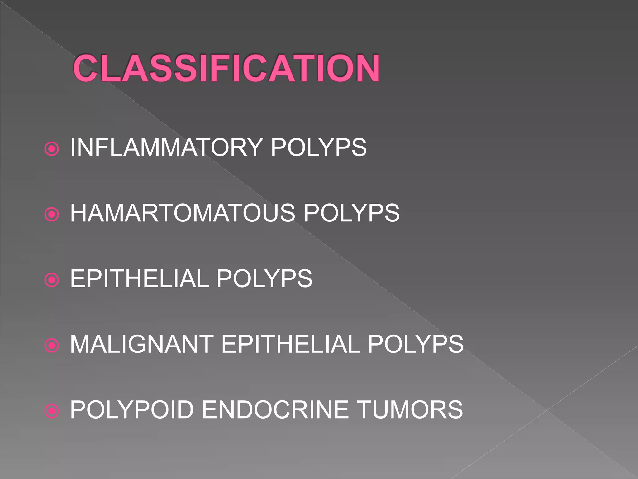
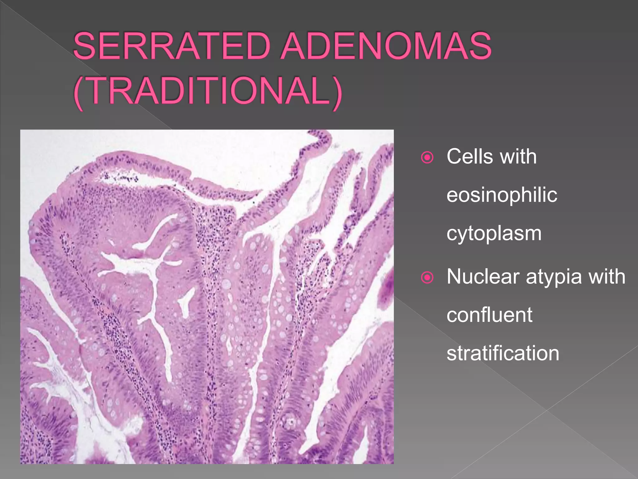
 Colorectal carcinoma(CRC) , one of the most
common cancer worldwide.
 Virtually all CRCs originate from a benign polyp
, which makes this cancer potentially
preventable by appropriate screening
colonoscopy programs in patients at increased
risk.
 The nature of precursor polyp is an essential
classifier of CRC because each tumor is
thought to develop from an unique benign polyp
with its own set of morphologic and molecular
characteristics.
 The two most common polyps identified as
precursors of CRCs-
 Adenomatous polyps and ,
 Serrated polyps.
 INFLAMMATORY POLYPS
 HAMARTOMATOUS POLYPS
 EPITHELIAL POLYPS
 MALIGNANT EPITHELIAL POLYPS
 POLYPOID ENDOCRINE TUMORS
 METASTASIS TO COLON
 LYMPHOID POLYPS
 MESENCHYMAL POLYPS
 MISCELLANEOUS POLYPOID LESIONS
 Junenile polyps and polyposis
 Peutz jeghers syndrome
 Cowden syndrome
 Cronkhite-canada syndrome
Grossly, these polyps grossly appear
rounded, smooth, and unilobular with
an erythematous cap of eroded tissue.
Their cut surface reveals multiple
dilated, mucin-filled crypts, leading to
the term mucus retention polyp. These
polyp share many features with
inflammatory polyp
1. Serrated polyps
2. Conventional Adenomas
3. Adenomatous Polyposis Syndrome
4. Adenomas and adenoma – like DALMS in
Inflammatory Bowel Disease
 Most common type of polyps in colon.
 May progress to malignancy.
 May grow to large sizes, show abnormal
proliferation and maturation (right side), and
may be sessile.
 Decrease in the rate of cell death, leading to,
 Prolonged cell life
 Increase in the no. of epithelial cells
 Serrated phenotypic growth pattern
 Higher susceptibility to DNA methylation
(Unknown reasons)
 Transcriptional silencing of promoters of tumor
suppressor genes (MLH1, BRAF, MGMT)
 Progression to dysplastic serrated polyps and
ultimately adenocarcinomas
 Small, sessile, smooth bumps, flatten on insufflation
of air
1. Microvesicular hyperplastic polyp
2. Goblet cell hyperplastic polyp
3. Mucin poor hyperplastic polyps,
(based on morphologic growth pattern and lack
of proliferative/maturation abnormalities)
 Simple tubular
architecture – no
branching, budding
or crypt dilatation
Luminal serrated
surface (irregular
sawtooth
appearance)
 Columnar cells with
apical vesicular
mucin, eosinophilic
like absorptive cells
and goblet cells
 Large proliferative
zone displaying-
 Mucin depletion
 Nuclear features-
Hyperchromatism ,
Elongation, and
Crowding
 Lesser degree of
luminal serrations
 Elongated crypts rich in
goblet cells, but without
microvesicular mucin
 Least common
 Prominent serrations
along the entire length of
the crypt
 Micropapillary
architecture
 Prominent
serrations
throughout the
crypt
 Mucin cells at the
base of crypt
 Branching at the base
of crypt (flask shaped)
 Irregular distribution of
mature and dystrophic
goblet cells and
mucinous cells
 Greater degree of
nuclear stratification
and atypia
 Sessile serrated polyps with dysplasia
 Serrated adenomas (traditional polyp)
 Conventional adenomas with serrated
architecture
 As a result of dysplastic change within a
sessile serrated polyp.
 Resembles a conventional adenoma,
composed of elongated cigar shaped
hyperchromatic nuclei with stratification and
with architectural serration.
 Sessile serrated
polyp with
prominent
serrations
 Conventional
adenoma-type
with low-grade
dysplasia
 Dysplasia arising
in a sessile
serrated polyp
imparting a
traditional serrated
adenoma
appearance
 Hyperplastic
epithelium
 Dysplastic
epithelium with
stratified nuclei
and hyper-
eosinophilic
cytoplasm
 Sessile serrated polyps with dysplasia
 Serrated adenomas
 Conventional adenomas with serrated
architecture
 Exclusively in the left colon – sigmoid,
rectum
 60-65 yrs
 Pedunculated
 Develop via alternate pathway
 Cells with
eosinophilic
cytoplasm
 Nuclear atypia with
confluent
stratification
 Confluent
nuclear
stratification
 Mucin depletion
 Variable no. of
goblet cells
 Large and
filiform in shape
 Dilated
lymphatics in
lamina propria
 Occasionally serrated adenomas may show
 Adenoma – like foci
 High grade dysplasia characterized by
increased cytological atypia, marked nuclear
stratification and back to back epithelial growth
pattern
 Sessile serrated polyps with dysplasia
 Serrated adenomas
 Conventional adenomas with serrated
architecture
 Rarely adenomas with serrated architecture
 Evidence of a of
carcinogenesis – chromosomal instability
pathway and serrated molecular pathway
(acquisition of KRAS mutation )
“fusion” molecular pathway
 Cigar shaped nuclei
with clumped
chromatin
 Prominent nuclear
stratification
 Mucin depletion
 Due to overlapping features or processing
artifacts.
 Cytologic evidence of dysplasia to place the
lesion into either “nondysplastic” or “dysplastic”
serrated polyp category.
DYSPLASTIC SERRATED
POLYPS
CONVENTIONAL
ADENOMA (CA)
CA WITH SERRATED
ARCHITECTURE
Discrete areas of
hyperplastic change along
with areas of adenoma–type
low grade dysplasia
Dystrophic goblet cells
Hyperchromatic nuclei
Cigar shaped with open
chromatin
Prominent nuclear
stratification
Crypt showing nuclear
features of adenoma along
with serration
1. Serrated polyps
2. Conventional Adenomas
3. Adenomatous Polyposis Syndrome
4. Adenomas and adenoma – like DALMS in
Inflammatory Bowel Disease
 Almost always aymptomatic
 Overt or occult rectal bleeding
 Risk increases with age, at age >50y, 12%
have adenomas, of which 25% are
considered high risk lesion.
 Sessile
 Pedunculated
 Occasionally, multilobulated and/or filiform
 Categorized architecturally as :--
1) Tubular (if >75%)
2) Villous (if >75%)
3) Tubullovillous (25-75%)
 Low grade
 High grade
1) Non complex
crypts
2) Psuedostratified or
partially stratified
nuclei reach only
the lower half of
cytoplasm
3) Minimal nuclear
pleomorphism/loss
of polarity
4) Brisk mitosis but
minimal atypical
mitosis
5) Arranged more in
parallel
configuration
 Marked degree of
nuclear
hyperchromaticity,
pseudostratification,
and loss of polarity
from the base to the
surface of the
mucosa.
 Intraluminal
cribriforming of the
glands is seen
without a definite
breach of the
basement membrane.
More complex intraluminal cribriforming and large gland formation with the
appearance of expanding-type infiltration into the lamina propria without
desmoplasia. In intramucosal carcinoma, the nuclei become more rounded,
rather than cigar shaped, and often contain an open chromatin pattern rather
than a coarse chromatin pattern.
Well circumscribed
lobule of misplaced
glands in the
submucosa of a polyp
stalk
The misplaced crypts
are slightly separated
from one another and
marked congestion and
hemosiderin deposition
are seen around the
edges of the misplaced
epithelium.
Smooth-edged pool of mucin,
lined by low-grade dysplastic
epithelium on the periphery,
The pool of mucin does not
contain floating atypical cells
or glands
Lack of lamina propria
surrounding the invasive
glands; no evidence of
hemorrhage or hemosiderin
deposition is seen.
Irregular and jagged in
contour and infiltrate in a
nonlobular fashion
1. Serrated polyps
2. Conventional Adenomas
3. Adenomatous Polyposis Syndrome
4. Adenomas and adenoma – like DALMS in
Inflammatory Bowel Disease
1) Familial adenomatous polyposis
2) Gardner`s syndrome
3) Turcot`s syndrome
4) Attenuated familial adenomatous polyposis
5) MYH associated polyposis
1. Serrated polyps
2. Conventional Adenomas
3. Adenomatous Polyposis Syndrome
4. Adenomas and adenoma – like DALMS in
Inflammatory Bowel Disease
 clinically important to make a distinction because sporadic
adenomas are generally treated by polypectomy and
continued surveillance, whereas (adenoma-like) polypoid
dysplastic lesions associated with an underlying IBD have
generally been regarded as an indication for colectomy in
medically fit patients
1) Mutations in DNA mismatch repair genes,
leading to a DNA microsatellite instability (MSI)
phenotype (15-20% cases)
2) Mutations in APC and other genes that
activate Wnt pathway, characterized by
chromosomal instability (CIN) phenotype (75-
85%)
3) Global genome hypermethylation, resulting in
switch off of tumor suppressor genes, indicated
as CpG island methylator phenotype (CIMP).
Pre cancerous colonic polyp

Pre cancerous colonic polyp

  • 2.
     Colorectal carcinoma(CRC), one of the most common cancer worldwide.  Virtually all CRCs originate from a benign polyp , which makes this cancer potentially preventable by appropriate screening colonoscopy programs in patients at increased risk.
  • 3.
     The natureof precursor polyp is an essential classifier of CRC because each tumor is thought to develop from an unique benign polyp with its own set of morphologic and molecular characteristics.
  • 4.
     The twomost common polyps identified as precursors of CRCs-  Adenomatous polyps and ,  Serrated polyps.
  • 5.
     INFLAMMATORY POLYPS HAMARTOMATOUS POLYPS  EPITHELIAL POLYPS  MALIGNANT EPITHELIAL POLYPS  POLYPOID ENDOCRINE TUMORS
  • 6.
     METASTASIS TOCOLON  LYMPHOID POLYPS  MESENCHYMAL POLYPS  MISCELLANEOUS POLYPOID LESIONS
  • 7.
     Junenile polypsand polyposis  Peutz jeghers syndrome  Cowden syndrome  Cronkhite-canada syndrome
  • 9.
    Grossly, these polypsgrossly appear rounded, smooth, and unilobular with an erythematous cap of eroded tissue. Their cut surface reveals multiple dilated, mucin-filled crypts, leading to the term mucus retention polyp. These polyp share many features with inflammatory polyp
  • 12.
    1. Serrated polyps 2.Conventional Adenomas 3. Adenomatous Polyposis Syndrome 4. Adenomas and adenoma – like DALMS in Inflammatory Bowel Disease
  • 13.
     Most commontype of polyps in colon.  May progress to malignancy.  May grow to large sizes, show abnormal proliferation and maturation (right side), and may be sessile.
  • 14.
     Decrease inthe rate of cell death, leading to,  Prolonged cell life  Increase in the no. of epithelial cells  Serrated phenotypic growth pattern
  • 15.
     Higher susceptibilityto DNA methylation (Unknown reasons)  Transcriptional silencing of promoters of tumor suppressor genes (MLH1, BRAF, MGMT)  Progression to dysplastic serrated polyps and ultimately adenocarcinomas
  • 18.
     Small, sessile,smooth bumps, flatten on insufflation of air
  • 19.
    1. Microvesicular hyperplasticpolyp 2. Goblet cell hyperplastic polyp 3. Mucin poor hyperplastic polyps, (based on morphologic growth pattern and lack of proliferative/maturation abnormalities)
  • 20.
     Simple tubular architecture– no branching, budding or crypt dilatation
  • 21.
  • 22.
     Columnar cellswith apical vesicular mucin, eosinophilic like absorptive cells and goblet cells
  • 23.
     Large proliferative zonedisplaying-  Mucin depletion  Nuclear features- Hyperchromatism , Elongation, and Crowding
  • 24.
     Lesser degreeof luminal serrations  Elongated crypts rich in goblet cells, but without microvesicular mucin
  • 25.
     Least common Prominent serrations along the entire length of the crypt  Micropapillary architecture
  • 26.
     Prominent serrations throughout the crypt Mucin cells at the base of crypt
  • 27.
     Branching atthe base of crypt (flask shaped)  Irregular distribution of mature and dystrophic goblet cells and mucinous cells
  • 28.
     Greater degreeof nuclear stratification and atypia
  • 30.
     Sessile serratedpolyps with dysplasia  Serrated adenomas (traditional polyp)  Conventional adenomas with serrated architecture
  • 31.
     As aresult of dysplastic change within a sessile serrated polyp.  Resembles a conventional adenoma, composed of elongated cigar shaped hyperchromatic nuclei with stratification and with architectural serration.
  • 32.
     Sessile serrated polypwith prominent serrations  Conventional adenoma-type with low-grade dysplasia
  • 33.
     Dysplasia arising ina sessile serrated polyp imparting a traditional serrated adenoma appearance
  • 34.
     Hyperplastic epithelium  Dysplastic epitheliumwith stratified nuclei and hyper- eosinophilic cytoplasm
  • 35.
     Sessile serratedpolyps with dysplasia  Serrated adenomas  Conventional adenomas with serrated architecture
  • 36.
     Exclusively inthe left colon – sigmoid, rectum  60-65 yrs  Pedunculated  Develop via alternate pathway
  • 37.
     Cells with eosinophilic cytoplasm Nuclear atypia with confluent stratification
  • 38.
     Confluent nuclear stratification  Mucindepletion  Variable no. of goblet cells
  • 39.
     Large and filiformin shape  Dilated lymphatics in lamina propria
  • 40.
     Occasionally serratedadenomas may show  Adenoma – like foci  High grade dysplasia characterized by increased cytological atypia, marked nuclear stratification and back to back epithelial growth pattern
  • 43.
     Sessile serratedpolyps with dysplasia  Serrated adenomas  Conventional adenomas with serrated architecture
  • 44.
     Rarely adenomaswith serrated architecture  Evidence of a of carcinogenesis – chromosomal instability pathway and serrated molecular pathway (acquisition of KRAS mutation ) “fusion” molecular pathway
  • 45.
     Cigar shapednuclei with clumped chromatin  Prominent nuclear stratification  Mucin depletion
  • 46.
     Due tooverlapping features or processing artifacts.  Cytologic evidence of dysplasia to place the lesion into either “nondysplastic” or “dysplastic” serrated polyp category.
  • 47.
    DYSPLASTIC SERRATED POLYPS CONVENTIONAL ADENOMA (CA) CAWITH SERRATED ARCHITECTURE Discrete areas of hyperplastic change along with areas of adenoma–type low grade dysplasia Dystrophic goblet cells Hyperchromatic nuclei Cigar shaped with open chromatin Prominent nuclear stratification Crypt showing nuclear features of adenoma along with serration
  • 48.
    1. Serrated polyps 2.Conventional Adenomas 3. Adenomatous Polyposis Syndrome 4. Adenomas and adenoma – like DALMS in Inflammatory Bowel Disease
  • 49.
     Almost alwaysaymptomatic  Overt or occult rectal bleeding  Risk increases with age, at age >50y, 12% have adenomas, of which 25% are considered high risk lesion.
  • 50.
     Sessile  Pedunculated Occasionally, multilobulated and/or filiform
  • 51.
     Categorized architecturallyas :-- 1) Tubular (if >75%) 2) Villous (if >75%) 3) Tubullovillous (25-75%)
  • 54.
  • 55.
    1) Non complex crypts 2)Psuedostratified or partially stratified nuclei reach only the lower half of cytoplasm 3) Minimal nuclear pleomorphism/loss of polarity 4) Brisk mitosis but minimal atypical mitosis 5) Arranged more in parallel configuration
  • 56.
     Marked degreeof nuclear hyperchromaticity, pseudostratification, and loss of polarity from the base to the surface of the mucosa.  Intraluminal cribriforming of the glands is seen without a definite breach of the basement membrane.
  • 57.
    More complex intraluminalcribriforming and large gland formation with the appearance of expanding-type infiltration into the lamina propria without desmoplasia. In intramucosal carcinoma, the nuclei become more rounded, rather than cigar shaped, and often contain an open chromatin pattern rather than a coarse chromatin pattern.
  • 60.
    Well circumscribed lobule ofmisplaced glands in the submucosa of a polyp stalk
  • 61.
    The misplaced crypts areslightly separated from one another and marked congestion and hemosiderin deposition are seen around the edges of the misplaced epithelium.
  • 62.
    Smooth-edged pool ofmucin, lined by low-grade dysplastic epithelium on the periphery, The pool of mucin does not contain floating atypical cells or glands
  • 63.
    Lack of laminapropria surrounding the invasive glands; no evidence of hemorrhage or hemosiderin deposition is seen. Irregular and jagged in contour and infiltrate in a nonlobular fashion
  • 65.
    1. Serrated polyps 2.Conventional Adenomas 3. Adenomatous Polyposis Syndrome 4. Adenomas and adenoma – like DALMS in Inflammatory Bowel Disease
  • 66.
    1) Familial adenomatouspolyposis 2) Gardner`s syndrome 3) Turcot`s syndrome 4) Attenuated familial adenomatous polyposis 5) MYH associated polyposis
  • 68.
    1. Serrated polyps 2.Conventional Adenomas 3. Adenomatous Polyposis Syndrome 4. Adenomas and adenoma – like DALMS in Inflammatory Bowel Disease
  • 69.
     clinically importantto make a distinction because sporadic adenomas are generally treated by polypectomy and continued surveillance, whereas (adenoma-like) polypoid dysplastic lesions associated with an underlying IBD have generally been regarded as an indication for colectomy in medically fit patients
  • 71.
    1) Mutations inDNA mismatch repair genes, leading to a DNA microsatellite instability (MSI) phenotype (15-20% cases) 2) Mutations in APC and other genes that activate Wnt pathway, characterized by chromosomal instability (CIN) phenotype (75- 85%) 3) Global genome hypermethylation, resulting in switch off of tumor suppressor genes, indicated as CpG island methylator phenotype (CIMP).