SlideShare a Scribd company logo
SURGICAL ANATOMY OF
PANCREAS
Moderator – Dr. B.R.Das, MS
Asstt. Professor
Presenter – Dr. Smaranika Baishya
Introduction
• The word pancreas comes
from the Greek πᾶν (pân,
"all") & κρέας (kréas,
"flesh").
• The pancreas is a
posteriorly situated
retroperitoneal organ that
lies transversely.
• The organ is composed of a
head, neck, body, and tail.
Location
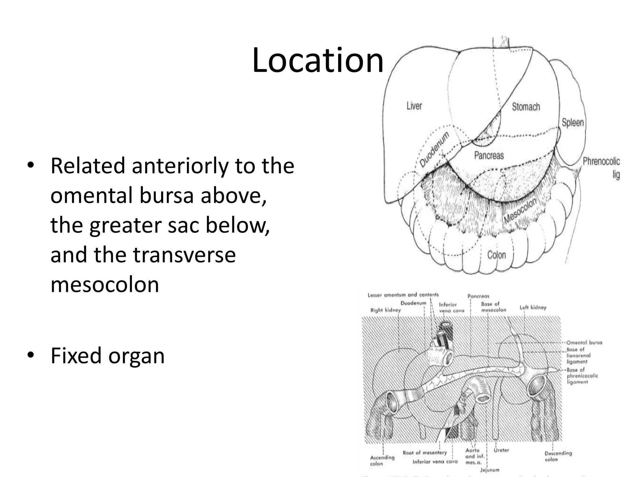
• Related anteriorly to the
omental bursa above,
the greater sac below,
and the transverse
mesocolon
• Fixed organ
Location
• Lies transversely in the
retroperitoneal space,
between the duodenum
on the right and the
spleen on the left
• The head is
encompassed by the
duodenum, whereas
the tail rests in the
splenic hilum.
Location
• A portion of the head
inferiorly is termed the
uncinate process and is
intimately related to the
SMV and SMA.
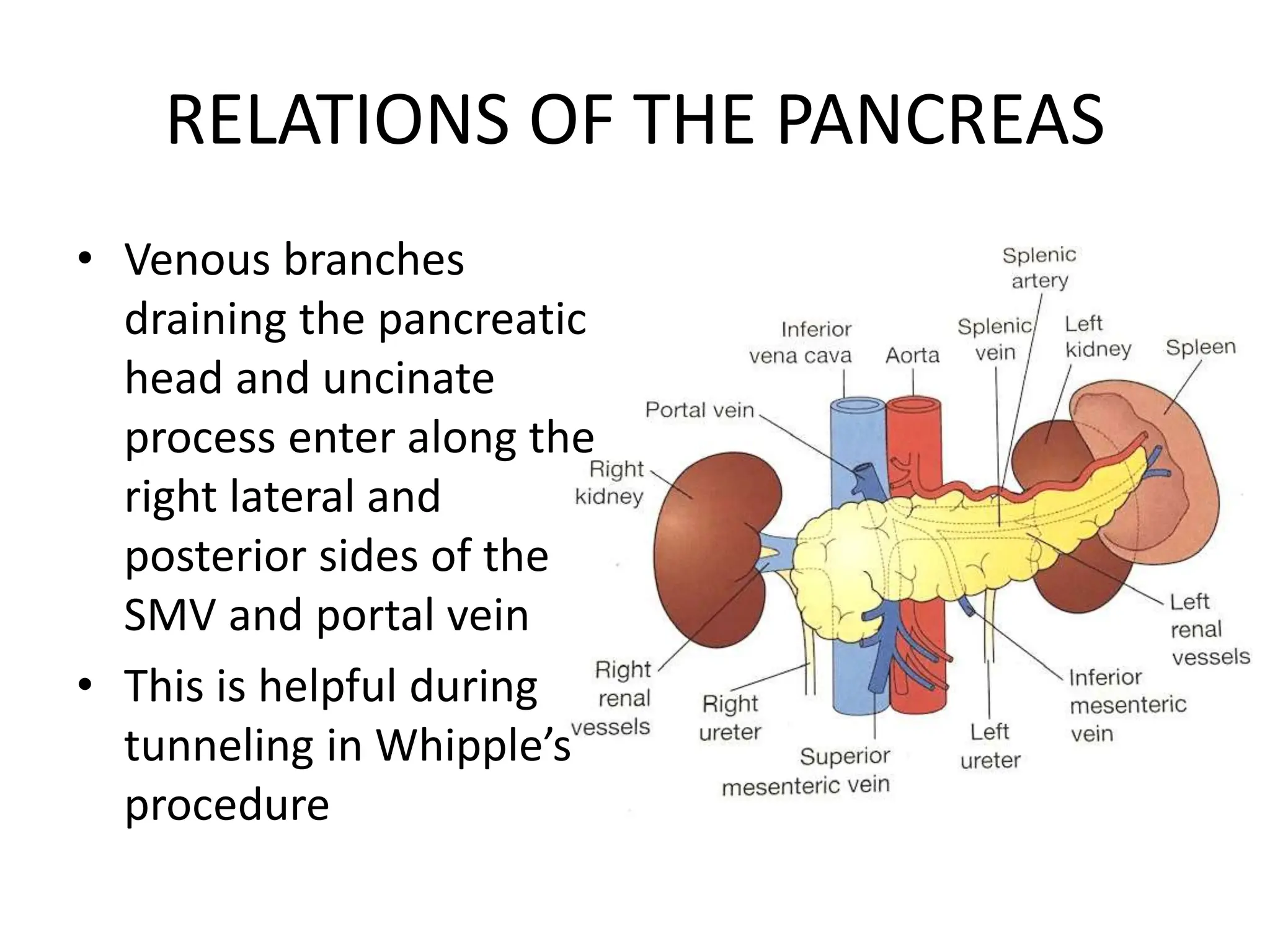
• Posteriorly, the
pancreas is related to
the IVC, aorta, left renal
vein and kidney, and
spleen.
RELATIONS OF THE PANCREAS
• Neck of the pancreas lies
anterior to the SMV and
SMA
• SMV joins with the splenic
vein behind the neck to
form Portal vein
• The neck of the pancreas is
anterior to the vertebral
body of L2,so if there is a
blunt anteroposterior
trauma, then it can
compress the neck of the
pancreas against the spine
RELATIONS OF THE PANCREAS
• Venous branches
draining the pancreatic
head and uncinate
process enter along the
right lateral and
posterior sides of the
SMV and portal vein
• This is helpful during
tunneling in Whipple’s
procedure
RELATIONS OF THE PANCREAS
• The body and tail of the
pancreas lie just
anterior to the splenic
vein and splenic artery
Embryology
• Carnegie stages are a standardized system of 23 stages
used to provide a unified developmental chronology .
• The pancreas is induced within the endoderm at
Carnegie Stage (CS) 12.
• The pancreatic precursors arise within three locations:
1. the ventral endodermal domain,
2. the VMEL[ventral midline of endoderm lip] (both the
ventral endodermal domain and the VMEL are
common progenitors with the liver), and
3. a dorsal domain to give rise to the dorsal and ventral
pancreatic buds.
Steps in the development of pancreas
Development from Ventral and Dorsal
Anlagen
• The pancreas is derived from ventral and dorsal
anlagen, which arise from the foregut
diametrically opposite each other and are distinct
from about day 26 to 32
• The ventral duct is an off-shoot of the bile duct
and maintains this bile duct connection
throughout
• The dorsal anlage will give rise to the head, body,
and tail of the pancreas, whereas the ventral bud
gives rise to the uncinate (Latin – “shaped like a
hook”) process
Development from Ventral and Dorsal
Anlagen
• Actual fusion of pancreatic parenchyma occurs
after a rotation of the ventral duct around the
axis of the foregut at about 50 to 55 days
• This differential heritage can still be evident
histologically by staining for pancreatic
polypeptide (PP)
• PP cells localize to the area derived from the
ventral anlage, while the dorsal pancreas has
larger lobules with PP-poor islets
Pancreatic Ducts
Main pancreatic duct (duct of Wirsung), joins
the CBD to empty into the duodenum at the
ampulla of Vater
Diameter -- 2 to 4 mm
Pressure --15 to 30 mm Hg
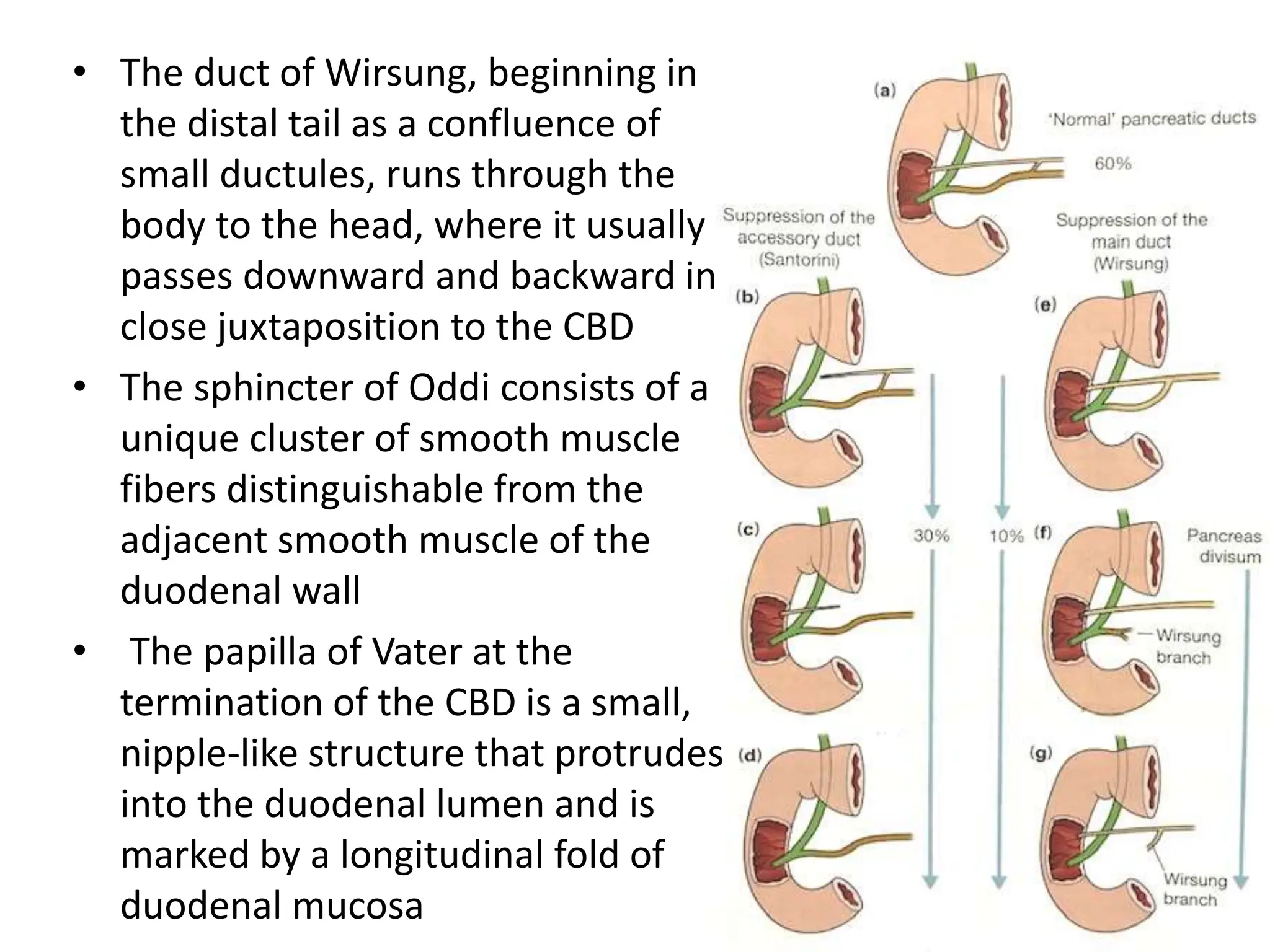
• The duct of Wirsung, beginning in
the distal tail as a confluence of
small ductules, runs through the
body to the head, where it usually
passes downward and backward in
close juxtaposition to the CBD
• The sphincter of Oddi consists of a
unique cluster of smooth muscle
fibers distinguishable from the
adjacent smooth muscle of the
duodenal wall
• The papilla of Vater at the
termination of the CBD is a small,
nipple-like structure that protrudes
into the duodenal lumen and is
marked by a longitudinal fold of
duodenal mucosa
• Several sphincters composed of smooth-
muscle fibres surround the intramural portion
of CBD, the main pancreatic duct, and the
ampulla.
• The complex may be as short as 6 mm or as
long as 30 mm
VARIATIONS IN PANCREATIC
DUCT OPENING
• Type 1 – Pancreatic duct
opens into CBD
• Type 2 – pancreatic and
bile ducts open close to
one another but
seperately without an
ampulla
• Type 3 – Pancreatic and
bile ducts open into the
duodenum seperately
without an ampulla
• The ‘T’ arrangement of
the duodenal mucosal
fold
• Blood supply of Hepato-
pancreatic ampulla
Surgico-anatomic segments of
pancreas
Relation of CBD to the posterior
surface of pancreas
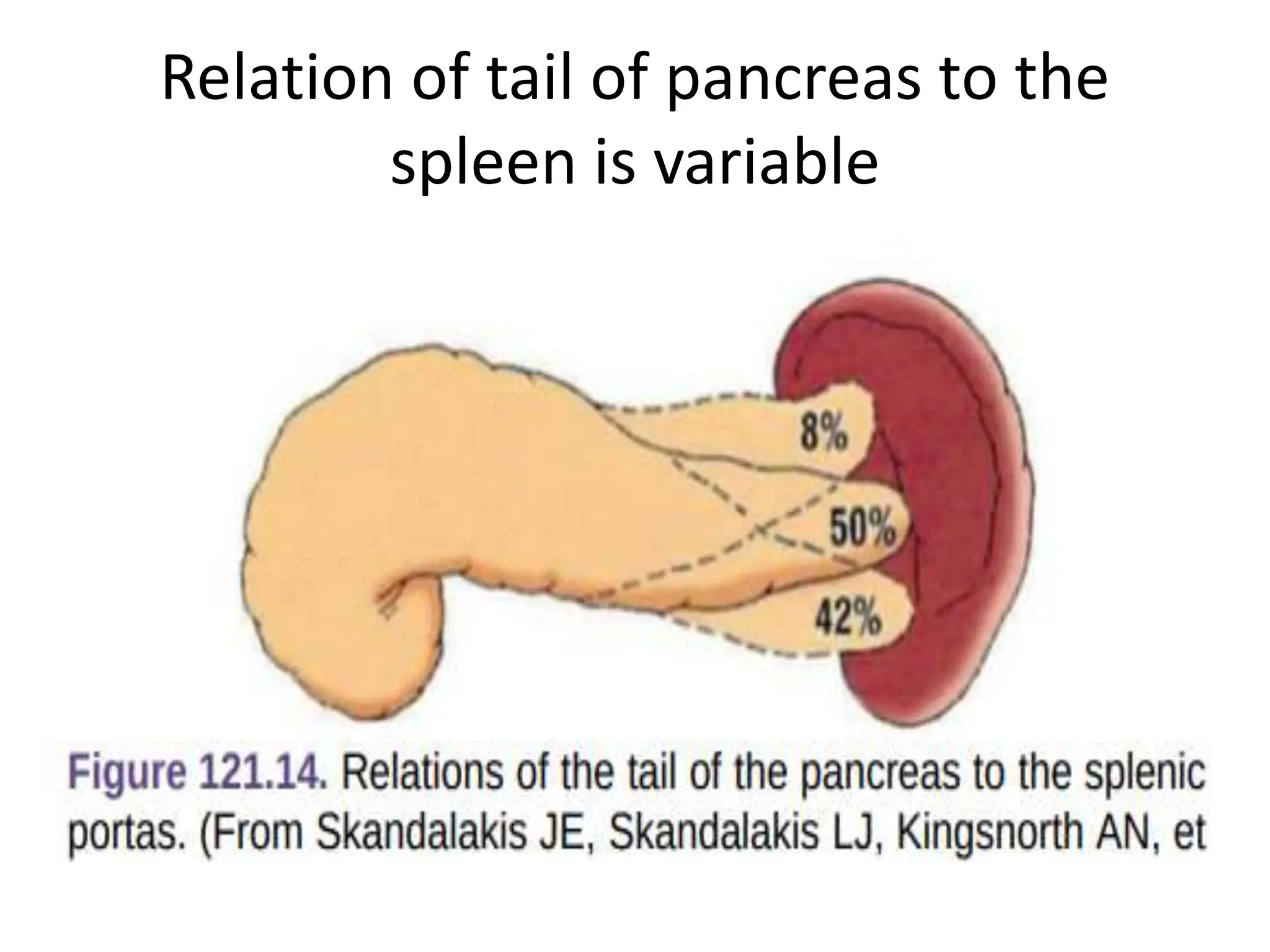
Relation of tail of pancreas to the
spleen is variable
• The duct of Wirsung runs downward and parallel to the
CBD for approximately 2 cm and joins it within the
sphincter segment in 70% to 85% of patients; it enters
the duodenum independently in 10% to 13% of
patients and is replaced by the duct of Santorini in 2%
of patients
• Rarely, the duct of Santorini and the duct of Wirsung
are separate, which is known as pancreas divisum
• The islets of Langerhans, which provide the endocrine
component of the gland, are scattered throughout the
pancreas.
• The pancreatic capsule is loosely attached to
the surface of the pancreas and is contiguous
with the anterior layer of the mesocolon such
that it can be dissected in continuity if
necessary.
• The mesenteric attachments to the pancreas
tend to be contiguous
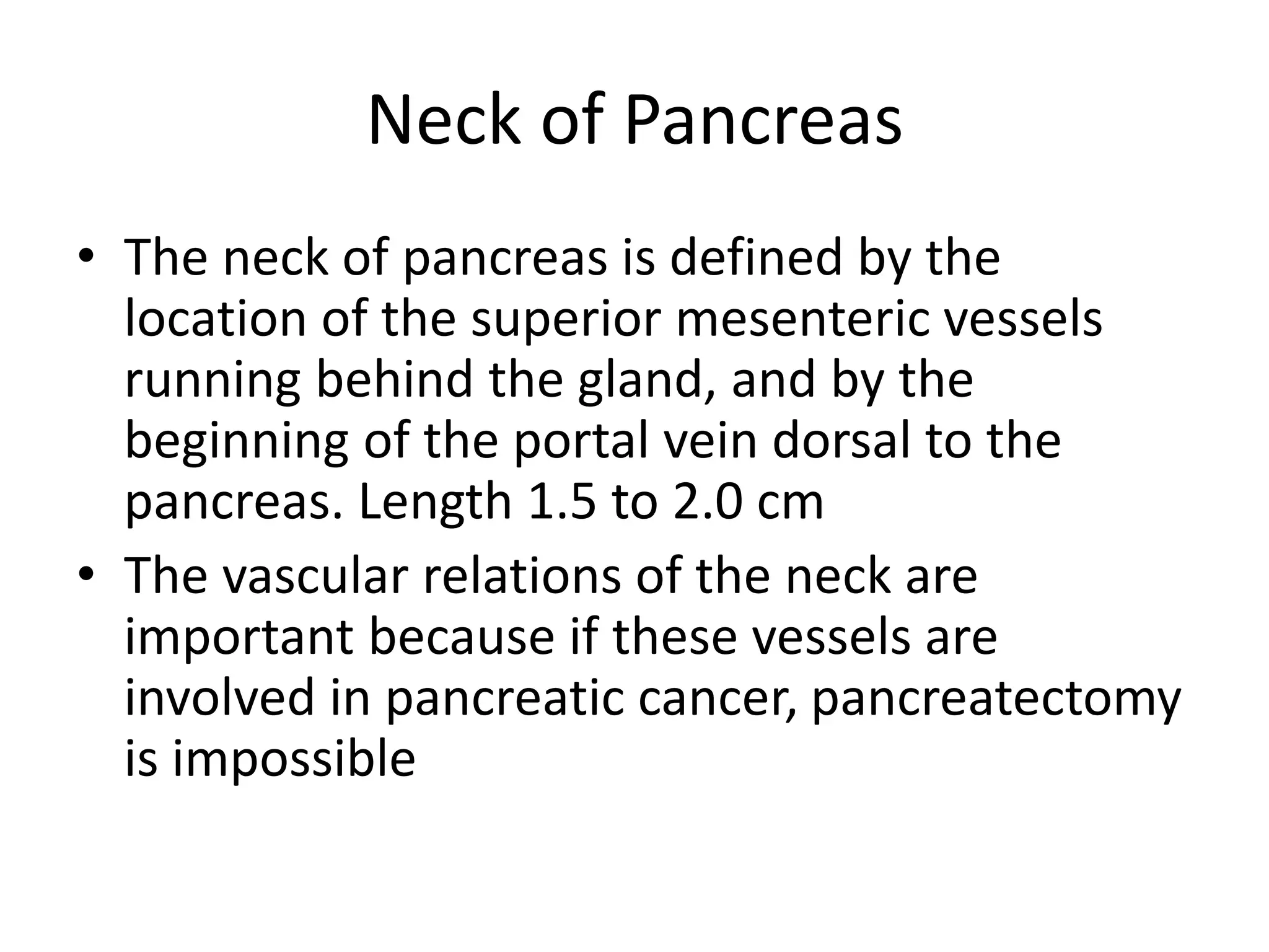
Neck of Pancreas
• The neck of pancreas is defined by the
location of the superior mesenteric vessels
running behind the gland, and by the
beginning of the portal vein dorsal to the
pancreas. Length 1.5 to 2.0 cm
• The vascular relations of the neck are
important because if these vessels are
involved in pancreatic cancer, pancreatectomy
is impossible
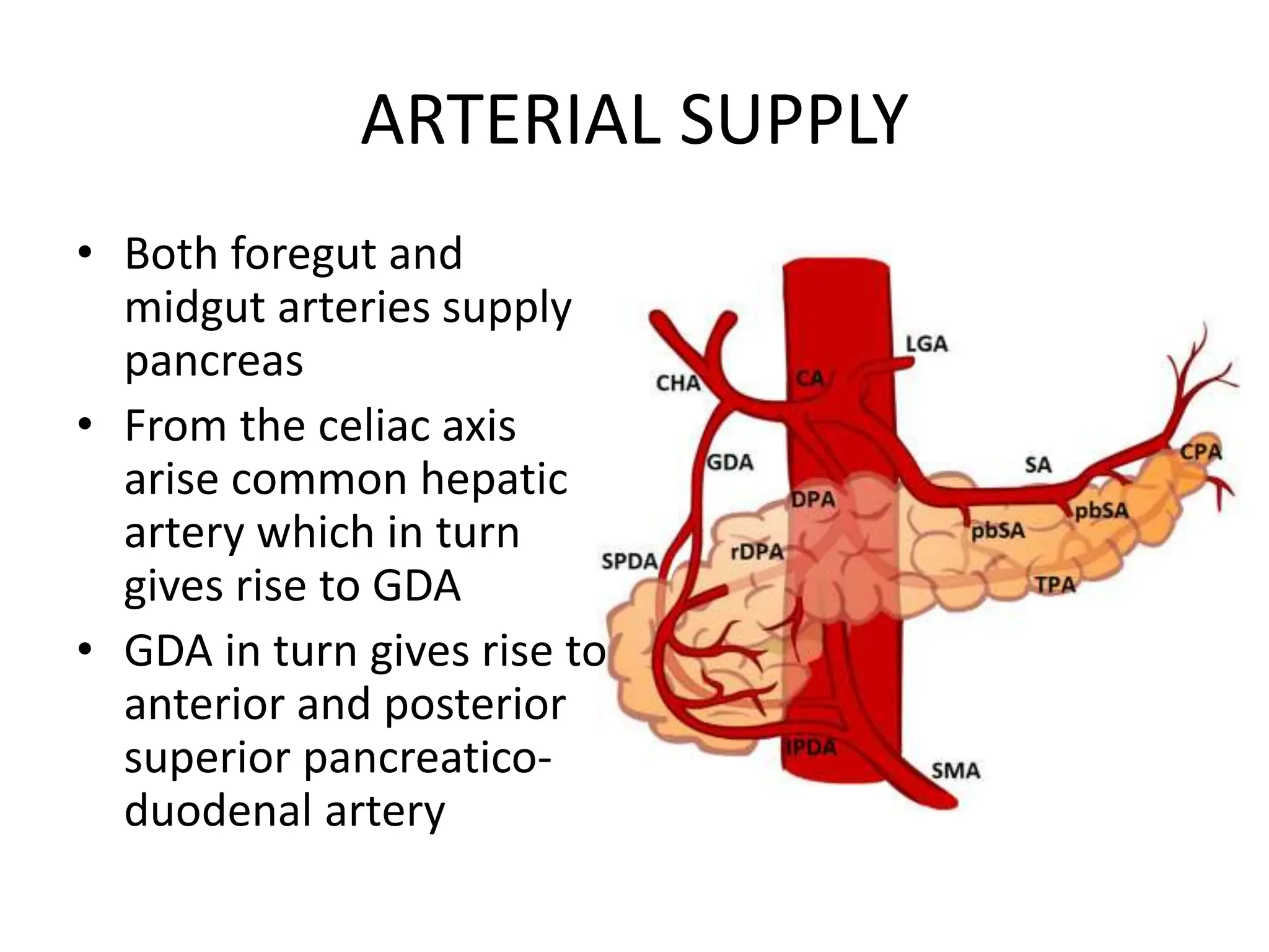
• Both foregut and
midgut arteries supply
pancreas
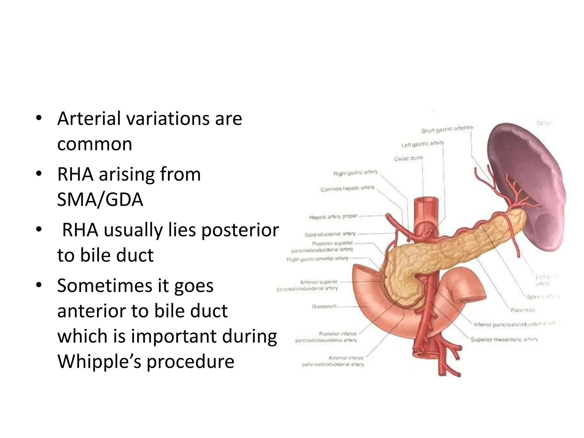
• From the celiac axis
arise common hepatic
artery which in turn
gives rise to GDA
• GDA in turn gives rise to
anterior and posterior
superior pancreatico-
duodenal artery
ARTERIAL SUPPLY
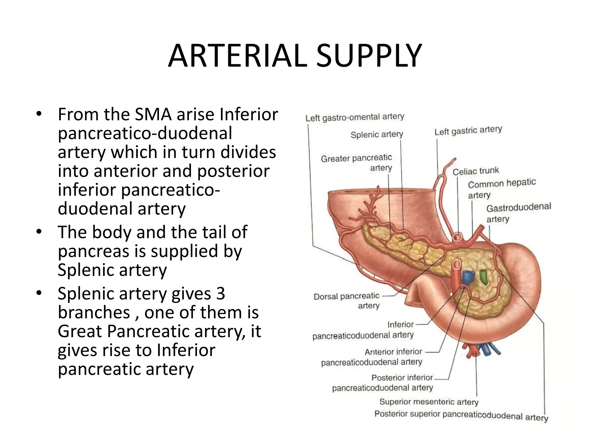
ARTERIAL SUPPLY
• From the SMA arise Inferior
pancreatico-duodenal
artery which in turn divides
into anterior and posterior
inferior pancreatico-
duodenal artery
• The body and the tail of
pancreas is supplied by
Splenic artery
• Splenic artery gives 3
branches , one of them is
Great Pancreatic artery, it
gives rise to Inferior
pancreatic artery
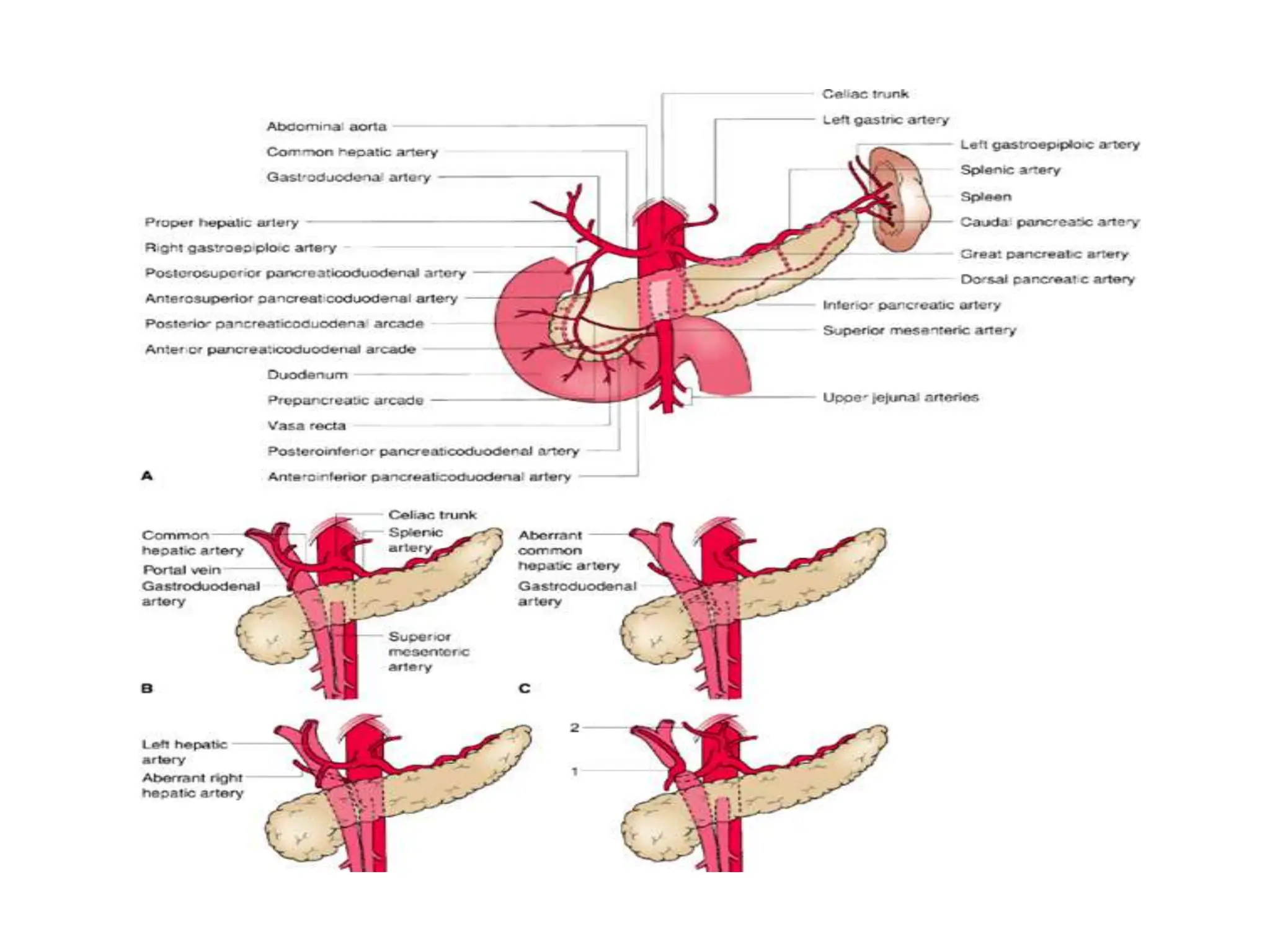
• Arterial variations are
common
• RHA arising from
SMA/GDA
• RHA usually lies posterior
to bile duct
• Sometimes it goes
anterior to bile duct
which is important during
Whipple’s procedure
• In the CT abdomen, if the
RHA is seen anterior to
the portal vein and
posterior to the bile duct
then it is standard course
RHA and if it is seen
posterior to the the portal
vein then it is replaced/
accessory RHA
• If an artery arise from the
left gastric artery and
goes to the falciform,
then it is replaced LHA
Venous Drainage
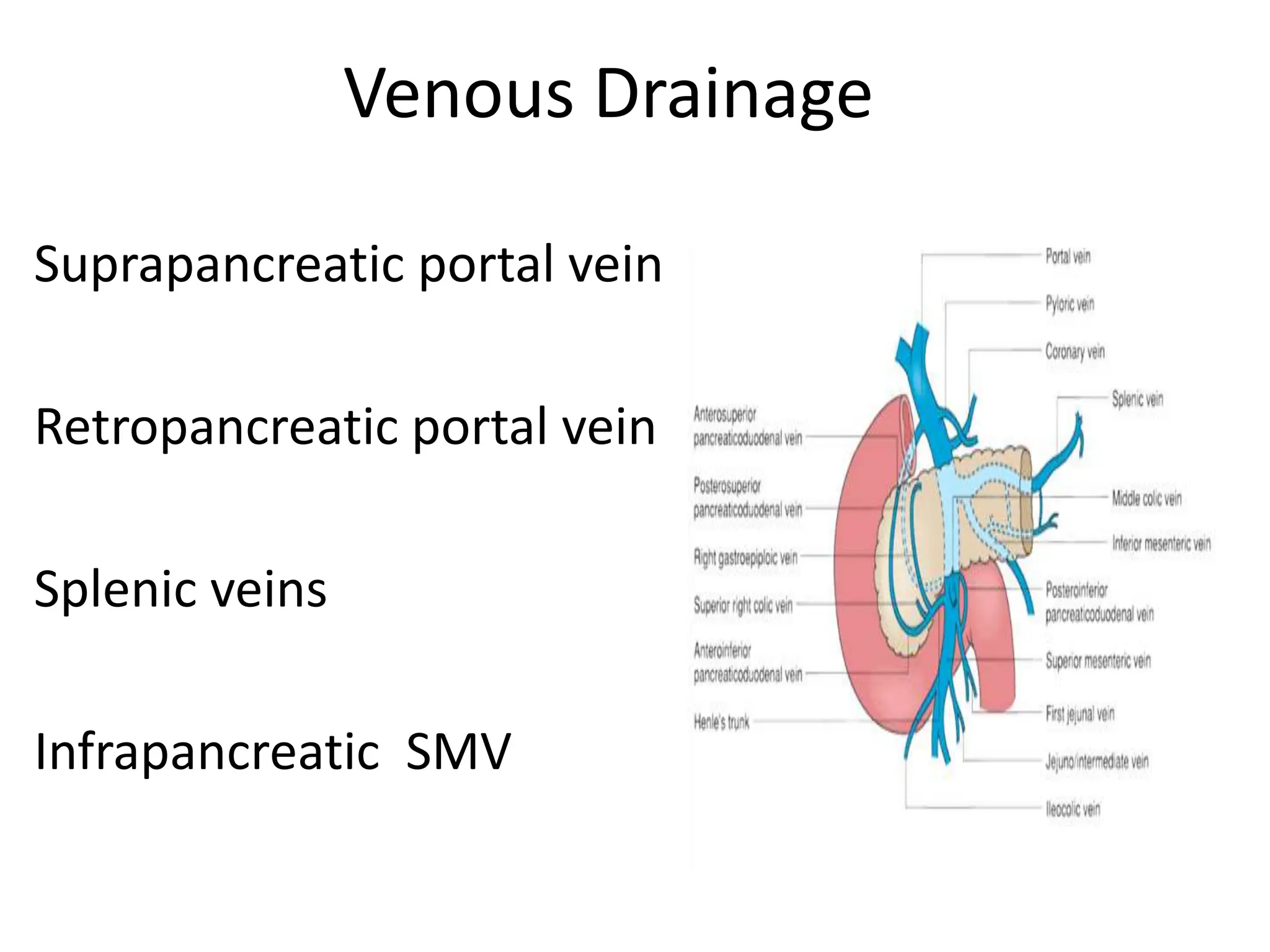
Suprapancreatic portal vein
Retropancreatic portal vein
Splenic veins
Infrapancreatic SMV
Venous Drainage
• Gastrocolic trunk is
important in Whipple’s
procedure
• It is also known as Henle’s
loop or Resident’s vein
• It is formed by Right
gastro epiploic vein,
accessory colic vein or
middle colic vein and
anterior pancreatico-
duodenal vein
Lymphatic Drainage
Drain into five main nodal groups
• Superior nodes drain the upper half of the head of the pancreas
• Anterior lymphatic drain to the prepyloric and infrapyloric nodes
• Inferior group of nodes drain to the superior mesenteric and periaortic
nodes
• Posterior pancreaticoduodenal lymph nodes drain into right periaortic
nodes
• Splenic group of nodes drain into the interceliomesenteric lymph nodes
The absence of a peritoneal barrier on the posterior
surface of the pancreas results in direct communication
of the intrapancreatic lymphatics with retroperitoneal
tissues, and this contributes to the high incidence of
recurrence after presumably curative resections of
pancreatic cancer
Histology
Exocrine Structure
Acinar cells secrete the enzymes responsible for
digestion
Ductular network carry the exocrine secretions into
the duodenum
Constitute 80% to 90% of the pancreatic mass
Endocrine Structure
Islets of Langerhans responsible for the secretion of
hormones that control glucose homeostasis
Contains
alpha ()-- glucagon
beta ()-- insulin
delta ()-- somatostatin
pancreatic polypeptide (PP) or F cells– PP
Accounts for 2% of the pancreatic mass
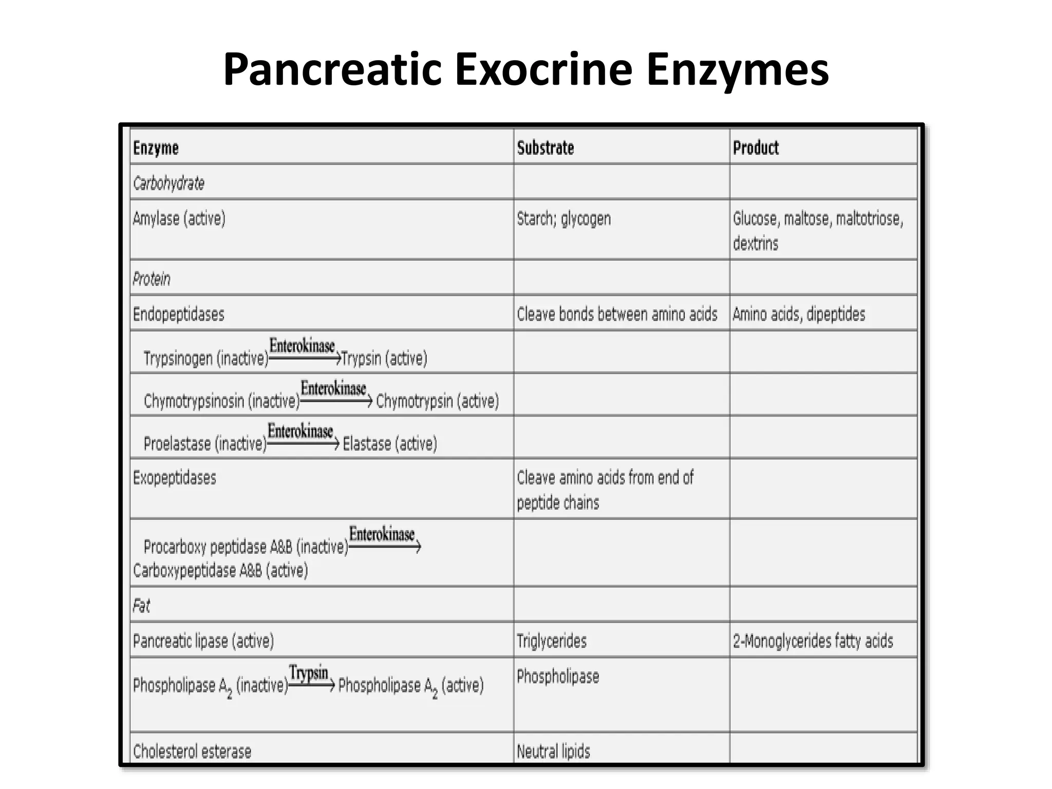
Pancreatic Exocrine Enzymes
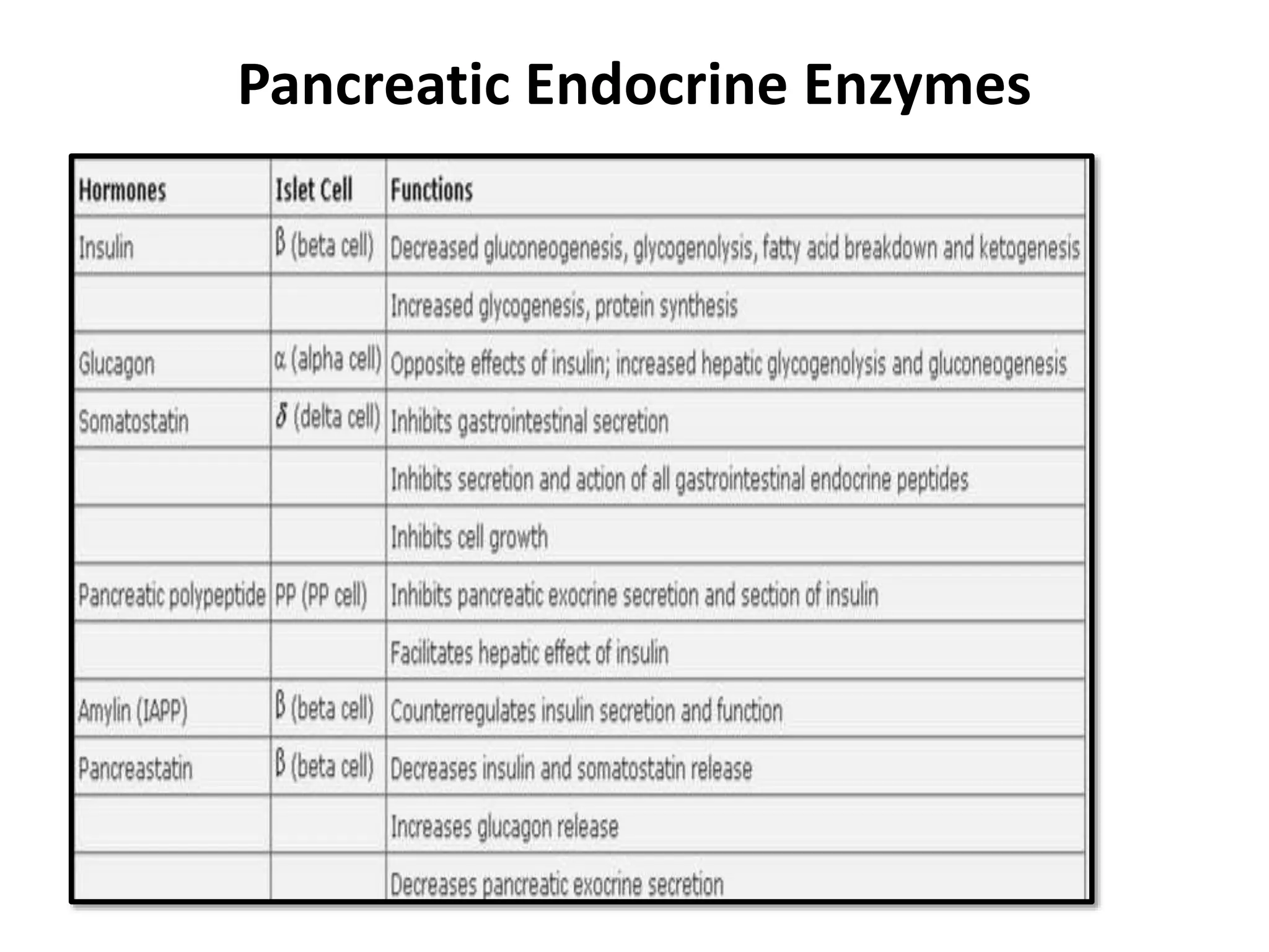
Pancreatic Endocrine Enzymes
PANCREAS DIVISUM
• In this condition, there
is failure of fusion of the
dorsal and ventral ducts
and the dorsal duct
becomes the major
ductal system of the
pancreas
ANNULAR PANCREAS
• Due to maldevelopment of the
primitive pancreatic ducts, a ring
of pancreatic tissue may surround
the descing portion of the
duodenum
• It usually produces no symptoms
but it may occur in association
with duodenal stenosis in which
case duodenal obstruction occurs
• In the presence of duodenal
obstruction, a
duodenojejunostomy is
performed and no attempt is
made to resect the pancreatic
tissue which would result in a
pancreatic fistula
Thank you

More Related Content

Similar to SURGICAL ANATOMY OF PANCREAS INTRO..pptx

Pancreas RADIOLOGY
Pancreas RADIOLOGYPancreas RADIOLOGY
Pancreas RADIOLOGY
AGRAWAL14
 
Embryology liver,pancreas,spleen & respiratory system
Embryology   liver,pancreas,spleen & respiratory systemEmbryology   liver,pancreas,spleen & respiratory system
Embryology liver,pancreas,spleen & respiratory system
MBBS IMS MSU
 
Embryology liver,pancreas,spleen & respiratory system
Embryology   liver,pancreas,spleen & respiratory systemEmbryology   liver,pancreas,spleen & respiratory system
Embryology liver,pancreas,spleen & respiratory system
MBBS IMS MSU
 
BENIGNPROSTATICHYPERPLASIA.pptx
BENIGNPROSTATICHYPERPLASIA.pptxBENIGNPROSTATICHYPERPLASIA.pptx
BENIGNPROSTATICHYPERPLASIA.pptx
Gokul Krishnan
 

Similar to SURGICAL ANATOMY OF PANCREAS INTRO..pptx (20)

Applied Anatomy and Physiology of spleen
Applied Anatomy and Physiology of spleenApplied Anatomy and Physiology of spleen
Applied Anatomy and Physiology of spleen
 
Renal Anatomy - applied aspects
Renal Anatomy - applied aspectsRenal Anatomy - applied aspects
Renal Anatomy - applied aspects
 
PANCREATIC ANOMALY radiology.pptx
PANCREATIC ANOMALY radiology.pptxPANCREATIC ANOMALY radiology.pptx
PANCREATIC ANOMALY radiology.pptx
 
Anatomy and physiology of pancreas
Anatomy and physiology of pancreasAnatomy and physiology of pancreas
Anatomy and physiology of pancreas
 
Endocrine System~4
Endocrine System~4Endocrine System~4
Endocrine System~4
 
Prolapse rectum
Prolapse rectumProlapse rectum
Prolapse rectum
 
Abdomen Presentation anat.ppt
Abdomen Presentation anat.pptAbdomen Presentation anat.ppt
Abdomen Presentation anat.ppt
 
Pancreas RADIOLOGY
Pancreas RADIOLOGYPancreas RADIOLOGY
Pancreas RADIOLOGY
 
Gross anatomy of Urinary System
 Gross anatomy of Urinary System Gross anatomy of Urinary System
Gross anatomy of Urinary System
 
Embryology liver,pancreas,spleen & respiratory system
Embryology   liver,pancreas,spleen & respiratory systemEmbryology   liver,pancreas,spleen & respiratory system
Embryology liver,pancreas,spleen & respiratory system
 
Embryology liver,pancreas,spleen & respiratory system
Embryology   liver,pancreas,spleen & respiratory systemEmbryology   liver,pancreas,spleen & respiratory system
Embryology liver,pancreas,spleen & respiratory system
 
BENIGNPROSTATICHYPERPLASIA.pptx
BENIGNPROSTATICHYPERPLASIA.pptxBENIGNPROSTATICHYPERPLASIA.pptx
BENIGNPROSTATICHYPERPLASIA.pptx
 
Resectional Anatomy of Pancreas
Resectional Anatomy of PancreasResectional Anatomy of Pancreas
Resectional Anatomy of Pancreas
 
Foregut
ForegutForegut
Foregut
 
SURGICAL ANATOMY
SURGICAL ANATOMYSURGICAL ANATOMY
SURGICAL ANATOMY
 
Cme pancreas2
Cme pancreas2Cme pancreas2
Cme pancreas2
 
Foregut
ForegutForegut
Foregut
 
Anatomy physiology pancreas.pptx
Anatomy physiology pancreas.pptxAnatomy physiology pancreas.pptx
Anatomy physiology pancreas.pptx
 
Pancreas.pptx
Pancreas.pptxPancreas.pptx
Pancreas.pptx
 
Radiological anatomy of hepatobiliary system
Radiological anatomy of hepatobiliary systemRadiological anatomy of hepatobiliary system
Radiological anatomy of hepatobiliary system
 

Recently uploaded

Industrial Training Report- AKTU Industrial Training Report
Industrial Training Report- AKTU Industrial Training ReportIndustrial Training Report- AKTU Industrial Training Report
Industrial Training Report- AKTU Industrial Training Report
Avinash Rai
 
Additional Benefits for Employee Website.pdf
Additional Benefits for Employee Website.pdfAdditional Benefits for Employee Website.pdf
Additional Benefits for Employee Website.pdf
joachimlavalley1
 

Recently uploaded (20)

NCERT Solutions Power Sharing Class 10 Notes pdf
NCERT Solutions Power Sharing Class 10 Notes pdfNCERT Solutions Power Sharing Class 10 Notes pdf
NCERT Solutions Power Sharing Class 10 Notes pdf
 
MARUTI SUZUKI- A Successful Joint Venture in India.pptx
MARUTI SUZUKI- A Successful Joint Venture in India.pptxMARUTI SUZUKI- A Successful Joint Venture in India.pptx
MARUTI SUZUKI- A Successful Joint Venture in India.pptx
 
size separation d pharm 1st year pharmaceutics
size separation d pharm 1st year pharmaceuticssize separation d pharm 1st year pharmaceutics
size separation d pharm 1st year pharmaceutics
 
Danh sách HSG Bộ môn cấp trường - Cấp THPT.pdf
Danh sách HSG Bộ môn cấp trường - Cấp THPT.pdfDanh sách HSG Bộ môn cấp trường - Cấp THPT.pdf
Danh sách HSG Bộ môn cấp trường - Cấp THPT.pdf
 
Industrial Training Report- AKTU Industrial Training Report
Industrial Training Report- AKTU Industrial Training ReportIndustrial Training Report- AKTU Industrial Training Report
Industrial Training Report- AKTU Industrial Training Report
 
Matatag-Curriculum and the 21st Century Skills Presentation.pptx
Matatag-Curriculum and the 21st Century Skills Presentation.pptxMatatag-Curriculum and the 21st Century Skills Presentation.pptx
Matatag-Curriculum and the 21st Century Skills Presentation.pptx
 
Research Methods in Psychology | Cambridge AS Level | Cambridge Assessment In...
Research Methods in Psychology | Cambridge AS Level | Cambridge Assessment In...Research Methods in Psychology | Cambridge AS Level | Cambridge Assessment In...
Research Methods in Psychology | Cambridge AS Level | Cambridge Assessment In...
 
How to Manage Notification Preferences in the Odoo 17
How to Manage Notification Preferences in the Odoo 17How to Manage Notification Preferences in the Odoo 17
How to Manage Notification Preferences in the Odoo 17
 
Jose-Rizal-and-Philippine-Nationalism-National-Symbol-2.pptx
Jose-Rizal-and-Philippine-Nationalism-National-Symbol-2.pptxJose-Rizal-and-Philippine-Nationalism-National-Symbol-2.pptx
Jose-Rizal-and-Philippine-Nationalism-National-Symbol-2.pptx
 
aaaaaaaaaaaaaaaaaaaaaaaaaaaaaaaaaaaaaaaaa
aaaaaaaaaaaaaaaaaaaaaaaaaaaaaaaaaaaaaaaaaaaaaaaaaaaaaaaaaaaaaaaaaaaaaaaaaaaaaaaaaa
aaaaaaaaaaaaaaaaaaaaaaaaaaaaaaaaaaaaaaaaa
 
Benefits and Challenges of Using Open Educational Resources
Benefits and Challenges of Using Open Educational ResourcesBenefits and Challenges of Using Open Educational Resources
Benefits and Challenges of Using Open Educational Resources
 
Mattingly "AI & Prompt Design: Limitations and Solutions with LLMs"
Mattingly "AI & Prompt Design: Limitations and Solutions with LLMs"Mattingly "AI & Prompt Design: Limitations and Solutions with LLMs"
Mattingly "AI & Prompt Design: Limitations and Solutions with LLMs"
 
The impact of social media on mental health and well-being has been a topic o...
The impact of social media on mental health and well-being has been a topic o...The impact of social media on mental health and well-being has been a topic o...
The impact of social media on mental health and well-being has been a topic o...
 
The Benefits and Challenges of Open Educational Resources
The Benefits and Challenges of Open Educational ResourcesThe Benefits and Challenges of Open Educational Resources
The Benefits and Challenges of Open Educational Resources
 
Keeping Your Information Safe with Centralized Security Services
Keeping Your Information Safe with Centralized Security ServicesKeeping Your Information Safe with Centralized Security Services
Keeping Your Information Safe with Centralized Security Services
 
Instructions for Submissions thorugh G- Classroom.pptx
Instructions for Submissions thorugh G- Classroom.pptxInstructions for Submissions thorugh G- Classroom.pptx
Instructions for Submissions thorugh G- Classroom.pptx
 
Operations Management - Book1.p - Dr. Abdulfatah A. Salem
Operations Management - Book1.p  - Dr. Abdulfatah A. SalemOperations Management - Book1.p  - Dr. Abdulfatah A. Salem
Operations Management - Book1.p - Dr. Abdulfatah A. Salem
 
2024_Student Session 2_ Set Plan Preparation.pptx
2024_Student Session 2_ Set Plan Preparation.pptx2024_Student Session 2_ Set Plan Preparation.pptx
2024_Student Session 2_ Set Plan Preparation.pptx
 
Additional Benefits for Employee Website.pdf
Additional Benefits for Employee Website.pdfAdditional Benefits for Employee Website.pdf
Additional Benefits for Employee Website.pdf
 
Students, digital devices and success - Andreas Schleicher - 27 May 2024..pptx
Students, digital devices and success - Andreas Schleicher - 27 May 2024..pptxStudents, digital devices and success - Andreas Schleicher - 27 May 2024..pptx
Students, digital devices and success - Andreas Schleicher - 27 May 2024..pptx
 

SURGICAL ANATOMY OF PANCREAS INTRO..pptx

  • 1. SURGICAL ANATOMY OF PANCREAS Moderator – Dr. B.R.Das, MS Asstt. Professor Presenter – Dr. Smaranika Baishya
  • 2. Introduction • The word pancreas comes from the Greek πᾶν (pân, "all") & κρέας (kréas, "flesh"). • The pancreas is a posteriorly situated retroperitoneal organ that lies transversely. • The organ is composed of a head, neck, body, and tail.
  • 3. Location • Related anteriorly to the omental bursa above, the greater sac below, and the transverse mesocolon • Fixed organ
  • 4. Location • Lies transversely in the retroperitoneal space, between the duodenum on the right and the spleen on the left • The head is encompassed by the duodenum, whereas the tail rests in the splenic hilum.
  • 5. Location • A portion of the head inferiorly is termed the uncinate process and is intimately related to the SMV and SMA. • Posteriorly, the pancreas is related to the IVC, aorta, left renal vein and kidney, and spleen.
  • 6. RELATIONS OF THE PANCREAS • Neck of the pancreas lies anterior to the SMV and SMA • SMV joins with the splenic vein behind the neck to form Portal vein • The neck of the pancreas is anterior to the vertebral body of L2,so if there is a blunt anteroposterior trauma, then it can compress the neck of the pancreas against the spine
  • 7. RELATIONS OF THE PANCREAS • Venous branches draining the pancreatic head and uncinate process enter along the right lateral and posterior sides of the SMV and portal vein • This is helpful during tunneling in Whipple’s procedure
  • 8. RELATIONS OF THE PANCREAS • The body and tail of the pancreas lie just anterior to the splenic vein and splenic artery
  • 9. Embryology • Carnegie stages are a standardized system of 23 stages used to provide a unified developmental chronology . • The pancreas is induced within the endoderm at Carnegie Stage (CS) 12. • The pancreatic precursors arise within three locations: 1. the ventral endodermal domain, 2. the VMEL[ventral midline of endoderm lip] (both the ventral endodermal domain and the VMEL are common progenitors with the liver), and 3. a dorsal domain to give rise to the dorsal and ventral pancreatic buds.
  • 10. Steps in the development of pancreas
  • 11. Development from Ventral and Dorsal Anlagen • The pancreas is derived from ventral and dorsal anlagen, which arise from the foregut diametrically opposite each other and are distinct from about day 26 to 32 • The ventral duct is an off-shoot of the bile duct and maintains this bile duct connection throughout • The dorsal anlage will give rise to the head, body, and tail of the pancreas, whereas the ventral bud gives rise to the uncinate (Latin – “shaped like a hook”) process
  • 12. Development from Ventral and Dorsal Anlagen • Actual fusion of pancreatic parenchyma occurs after a rotation of the ventral duct around the axis of the foregut at about 50 to 55 days • This differential heritage can still be evident histologically by staining for pancreatic polypeptide (PP) • PP cells localize to the area derived from the ventral anlage, while the dorsal pancreas has larger lobules with PP-poor islets
  • 13.
  • 14. Pancreatic Ducts Main pancreatic duct (duct of Wirsung), joins the CBD to empty into the duodenum at the ampulla of Vater Diameter -- 2 to 4 mm Pressure --15 to 30 mm Hg
  • 15. • The duct of Wirsung, beginning in the distal tail as a confluence of small ductules, runs through the body to the head, where it usually passes downward and backward in close juxtaposition to the CBD • The sphincter of Oddi consists of a unique cluster of smooth muscle fibers distinguishable from the adjacent smooth muscle of the duodenal wall • The papilla of Vater at the termination of the CBD is a small, nipple-like structure that protrudes into the duodenal lumen and is marked by a longitudinal fold of duodenal mucosa
  • 16. • Several sphincters composed of smooth- muscle fibres surround the intramural portion of CBD, the main pancreatic duct, and the ampulla. • The complex may be as short as 6 mm or as long as 30 mm
  • 17. VARIATIONS IN PANCREATIC DUCT OPENING • Type 1 – Pancreatic duct opens into CBD • Type 2 – pancreatic and bile ducts open close to one another but seperately without an ampulla • Type 3 – Pancreatic and bile ducts open into the duodenum seperately without an ampulla
  • 18. • The ‘T’ arrangement of the duodenal mucosal fold • Blood supply of Hepato- pancreatic ampulla
  • 20. Relation of CBD to the posterior surface of pancreas
  • 21. Relation of tail of pancreas to the spleen is variable
  • 22. • The duct of Wirsung runs downward and parallel to the CBD for approximately 2 cm and joins it within the sphincter segment in 70% to 85% of patients; it enters the duodenum independently in 10% to 13% of patients and is replaced by the duct of Santorini in 2% of patients • Rarely, the duct of Santorini and the duct of Wirsung are separate, which is known as pancreas divisum • The islets of Langerhans, which provide the endocrine component of the gland, are scattered throughout the pancreas.
  • 23. • The pancreatic capsule is loosely attached to the surface of the pancreas and is contiguous with the anterior layer of the mesocolon such that it can be dissected in continuity if necessary. • The mesenteric attachments to the pancreas tend to be contiguous
  • 24. Neck of Pancreas • The neck of pancreas is defined by the location of the superior mesenteric vessels running behind the gland, and by the beginning of the portal vein dorsal to the pancreas. Length 1.5 to 2.0 cm • The vascular relations of the neck are important because if these vessels are involved in pancreatic cancer, pancreatectomy is impossible
  • 25. • Both foregut and midgut arteries supply pancreas • From the celiac axis arise common hepatic artery which in turn gives rise to GDA • GDA in turn gives rise to anterior and posterior superior pancreatico- duodenal artery ARTERIAL SUPPLY
  • 26. ARTERIAL SUPPLY • From the SMA arise Inferior pancreatico-duodenal artery which in turn divides into anterior and posterior inferior pancreatico- duodenal artery • The body and the tail of pancreas is supplied by Splenic artery • Splenic artery gives 3 branches , one of them is Great Pancreatic artery, it gives rise to Inferior pancreatic artery
  • 27.
  • 28. • Arterial variations are common • RHA arising from SMA/GDA • RHA usually lies posterior to bile duct • Sometimes it goes anterior to bile duct which is important during Whipple’s procedure
  • 29. • In the CT abdomen, if the RHA is seen anterior to the portal vein and posterior to the bile duct then it is standard course RHA and if it is seen posterior to the the portal vein then it is replaced/ accessory RHA • If an artery arise from the left gastric artery and goes to the falciform, then it is replaced LHA
  • 30. Venous Drainage Suprapancreatic portal vein Retropancreatic portal vein Splenic veins Infrapancreatic SMV
  • 31. Venous Drainage • Gastrocolic trunk is important in Whipple’s procedure • It is also known as Henle’s loop or Resident’s vein • It is formed by Right gastro epiploic vein, accessory colic vein or middle colic vein and anterior pancreatico- duodenal vein
  • 32. Lymphatic Drainage Drain into five main nodal groups • Superior nodes drain the upper half of the head of the pancreas • Anterior lymphatic drain to the prepyloric and infrapyloric nodes • Inferior group of nodes drain to the superior mesenteric and periaortic nodes • Posterior pancreaticoduodenal lymph nodes drain into right periaortic nodes • Splenic group of nodes drain into the interceliomesenteric lymph nodes
  • 33. The absence of a peritoneal barrier on the posterior surface of the pancreas results in direct communication of the intrapancreatic lymphatics with retroperitoneal tissues, and this contributes to the high incidence of recurrence after presumably curative resections of pancreatic cancer
  • 34.
  • 35. Histology Exocrine Structure Acinar cells secrete the enzymes responsible for digestion Ductular network carry the exocrine secretions into the duodenum Constitute 80% to 90% of the pancreatic mass
  • 36. Endocrine Structure Islets of Langerhans responsible for the secretion of hormones that control glucose homeostasis Contains alpha ()-- glucagon beta ()-- insulin delta ()-- somatostatin pancreatic polypeptide (PP) or F cells– PP Accounts for 2% of the pancreatic mass
  • 39. PANCREAS DIVISUM • In this condition, there is failure of fusion of the dorsal and ventral ducts and the dorsal duct becomes the major ductal system of the pancreas
  • 40. ANNULAR PANCREAS • Due to maldevelopment of the primitive pancreatic ducts, a ring of pancreatic tissue may surround the descing portion of the duodenum • It usually produces no symptoms but it may occur in association with duodenal stenosis in which case duodenal obstruction occurs • In the presence of duodenal obstruction, a duodenojejunostomy is performed and no attempt is made to resect the pancreatic tissue which would result in a pancreatic fistula