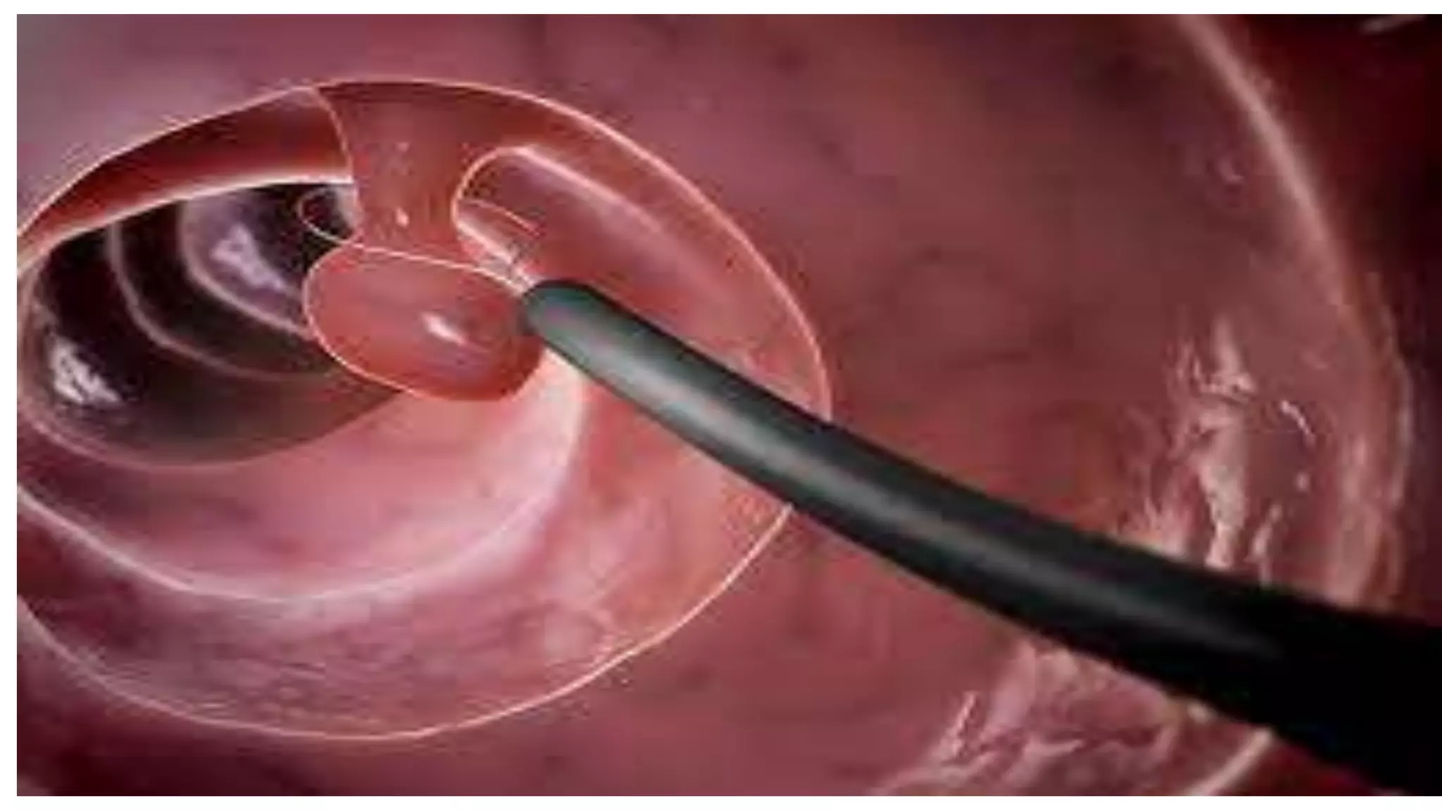
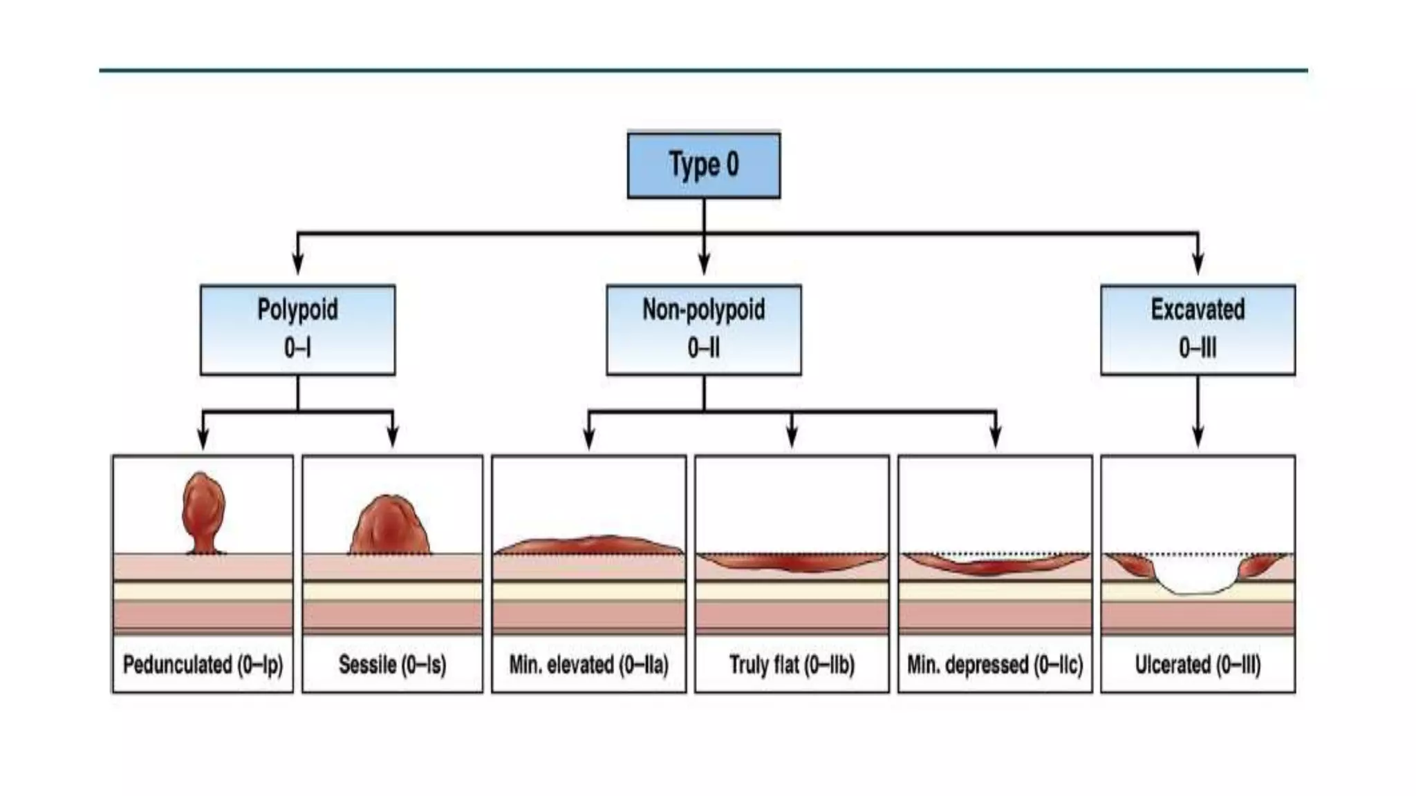
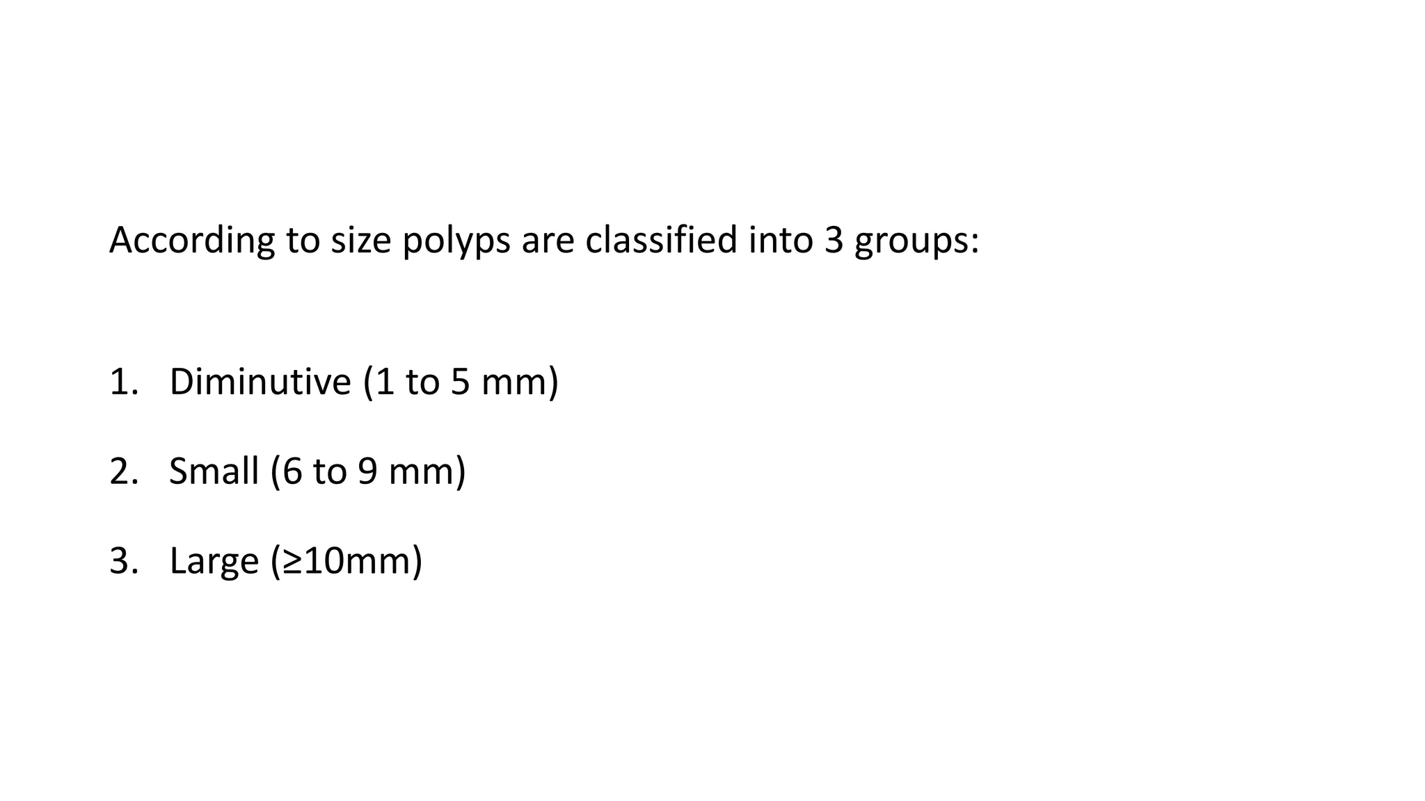
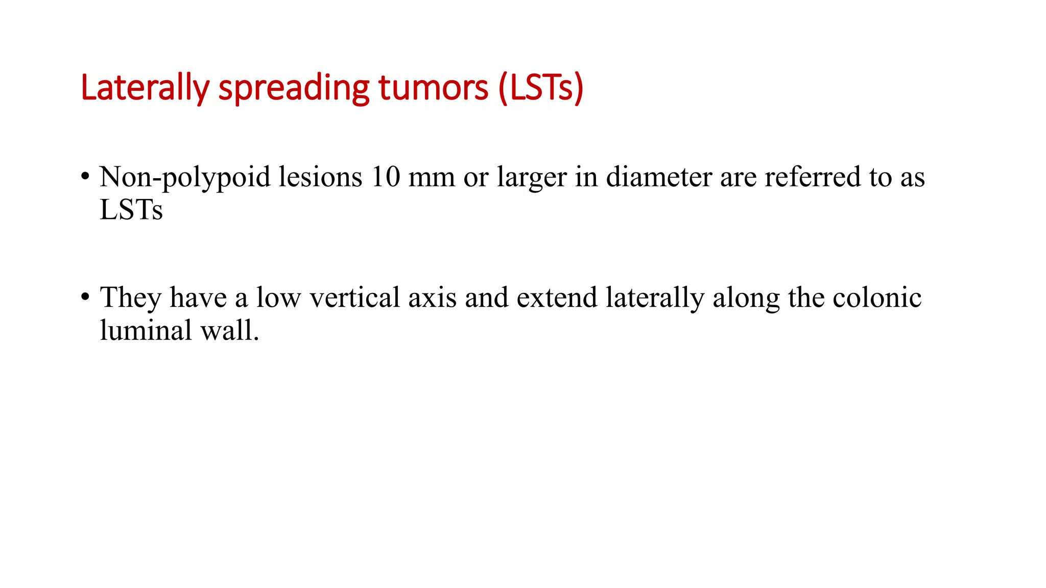
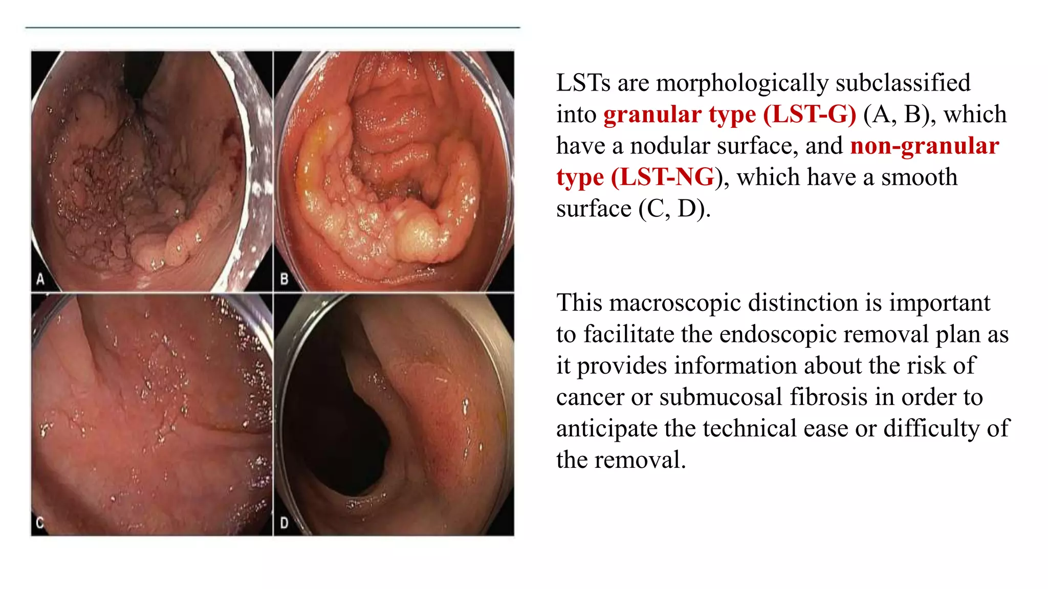
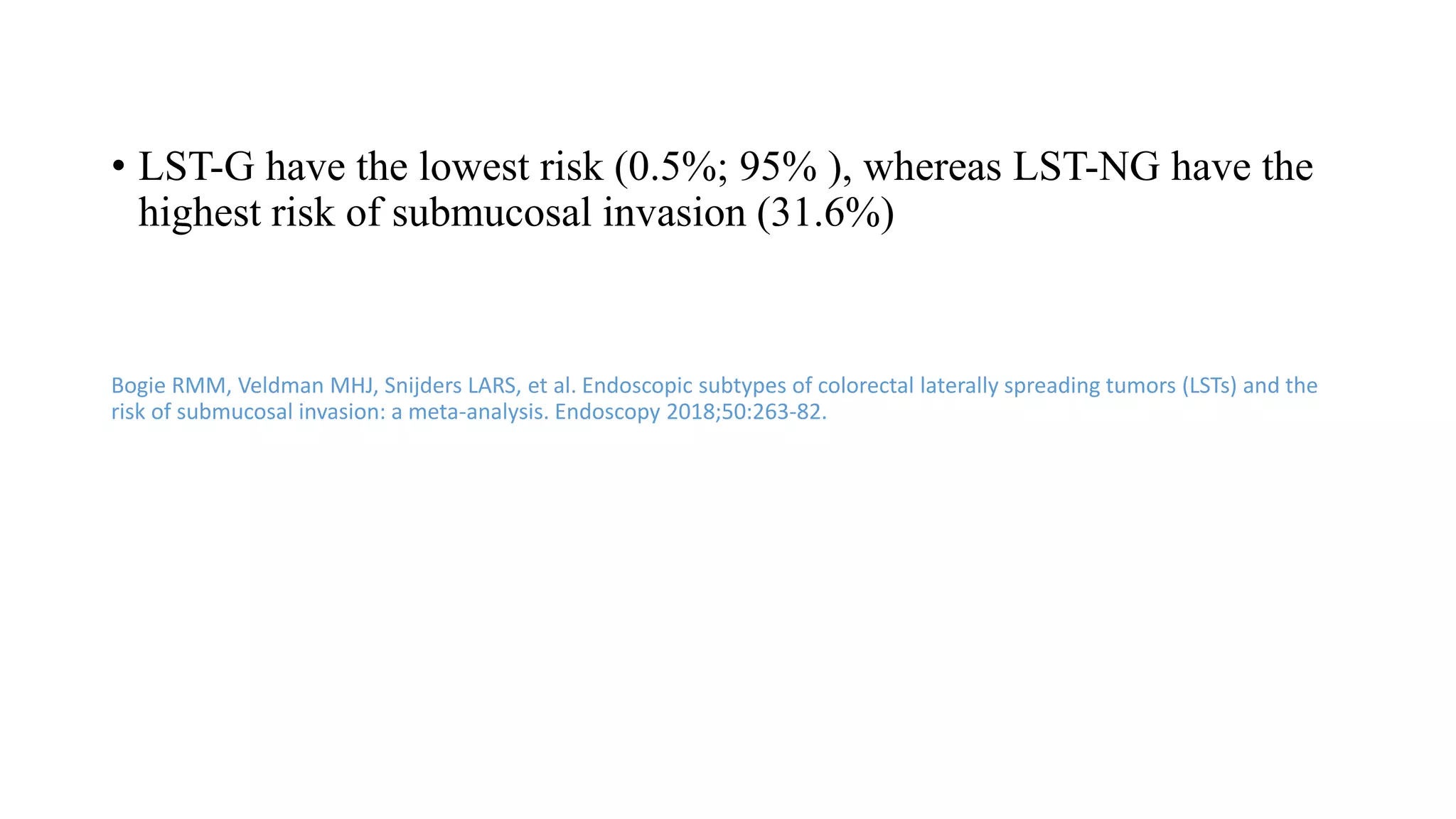
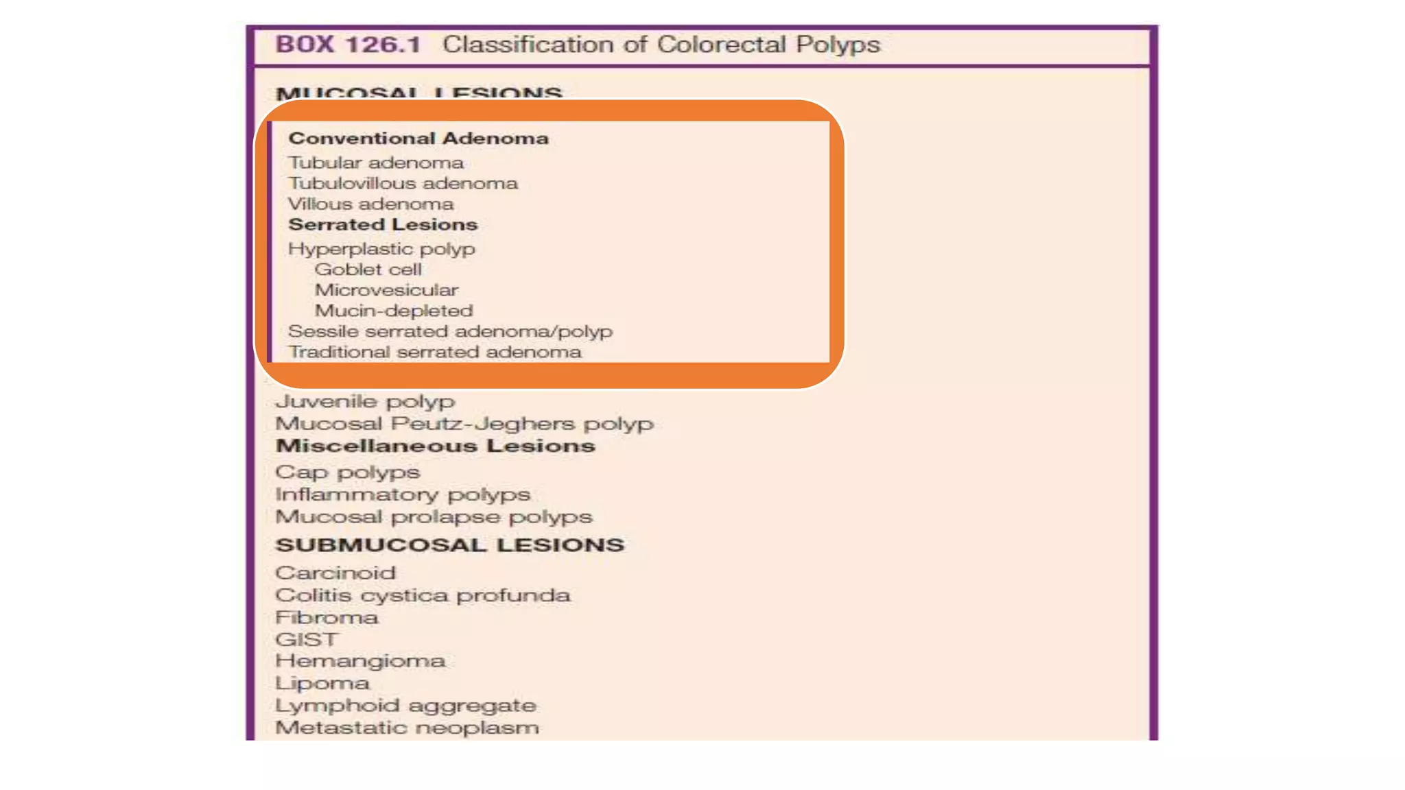
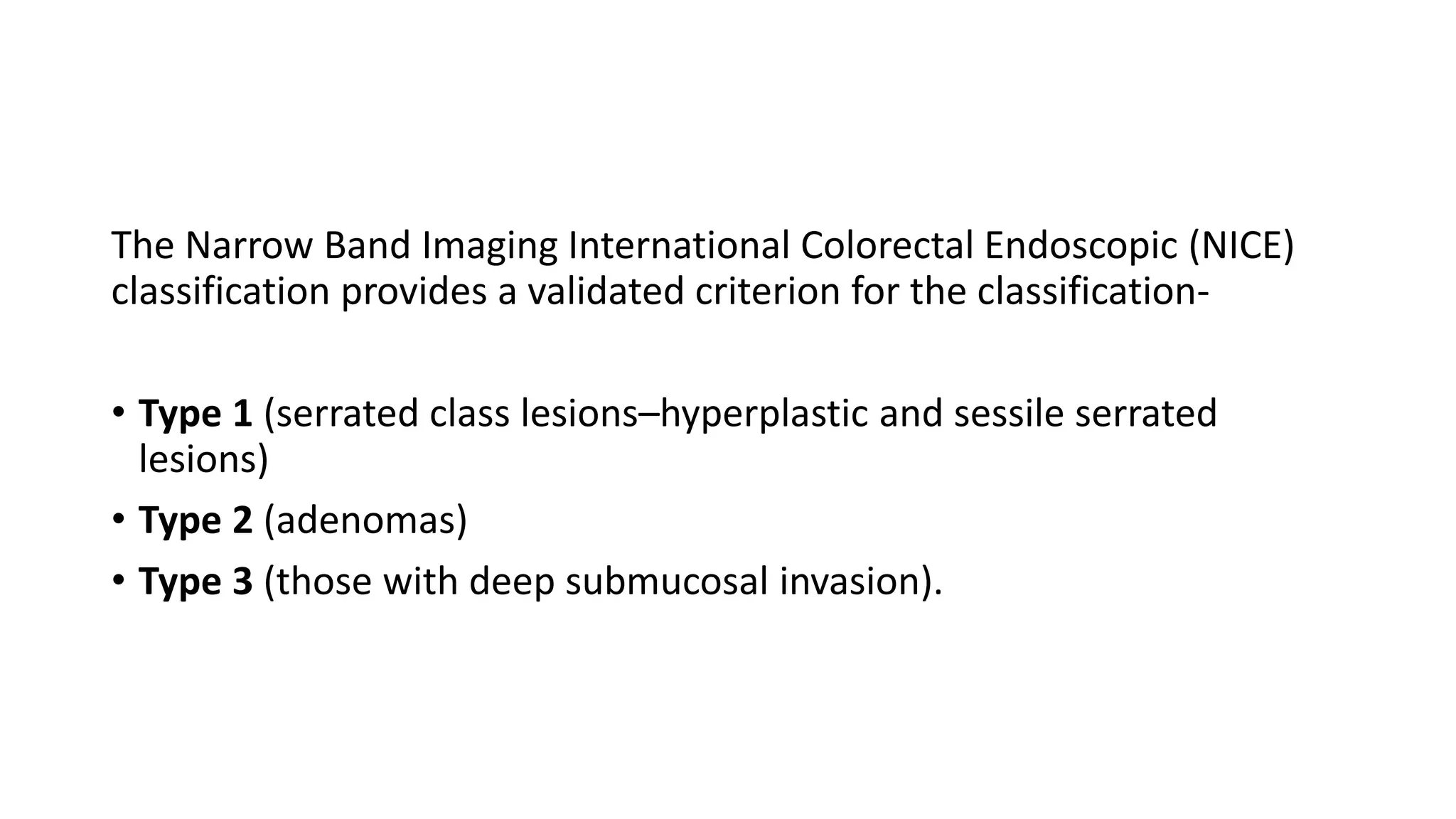
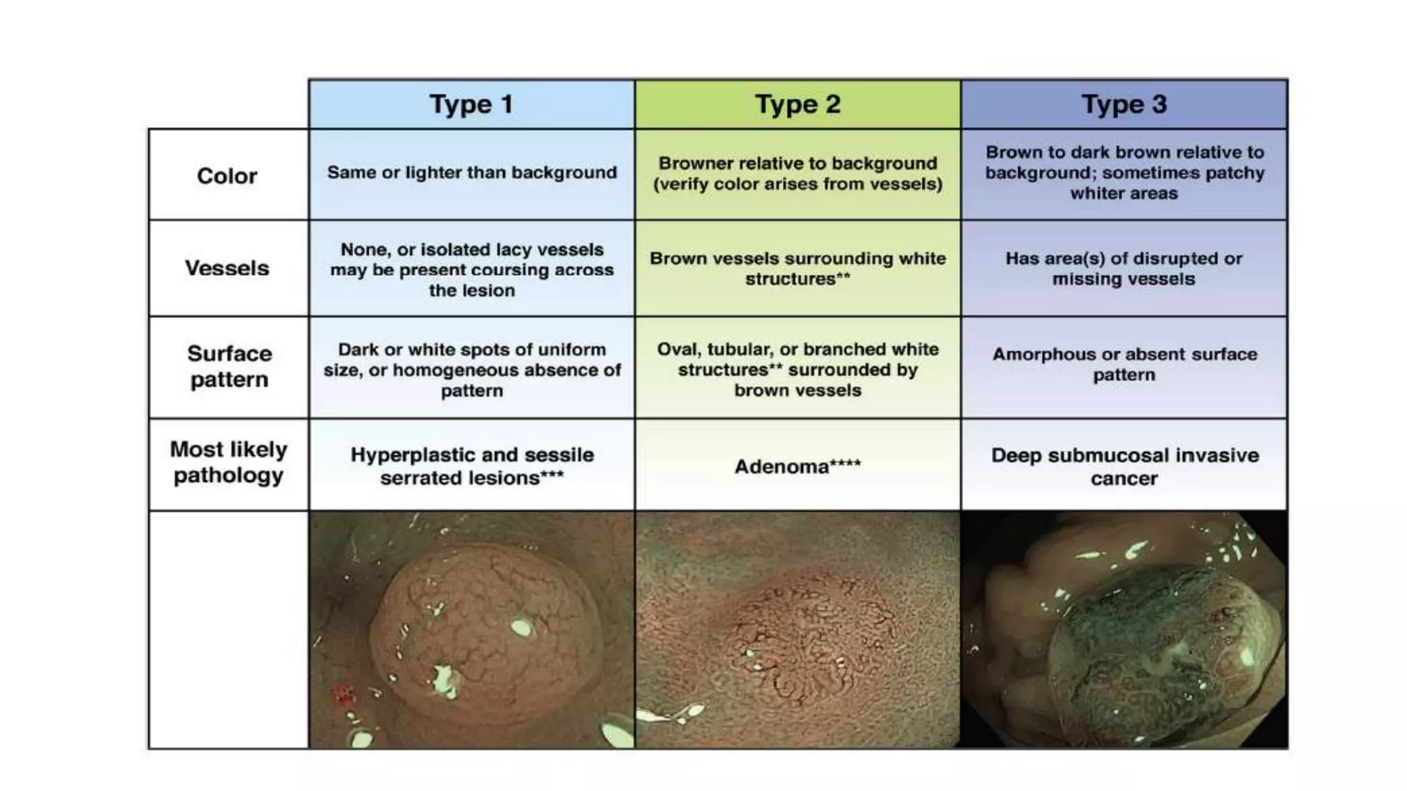
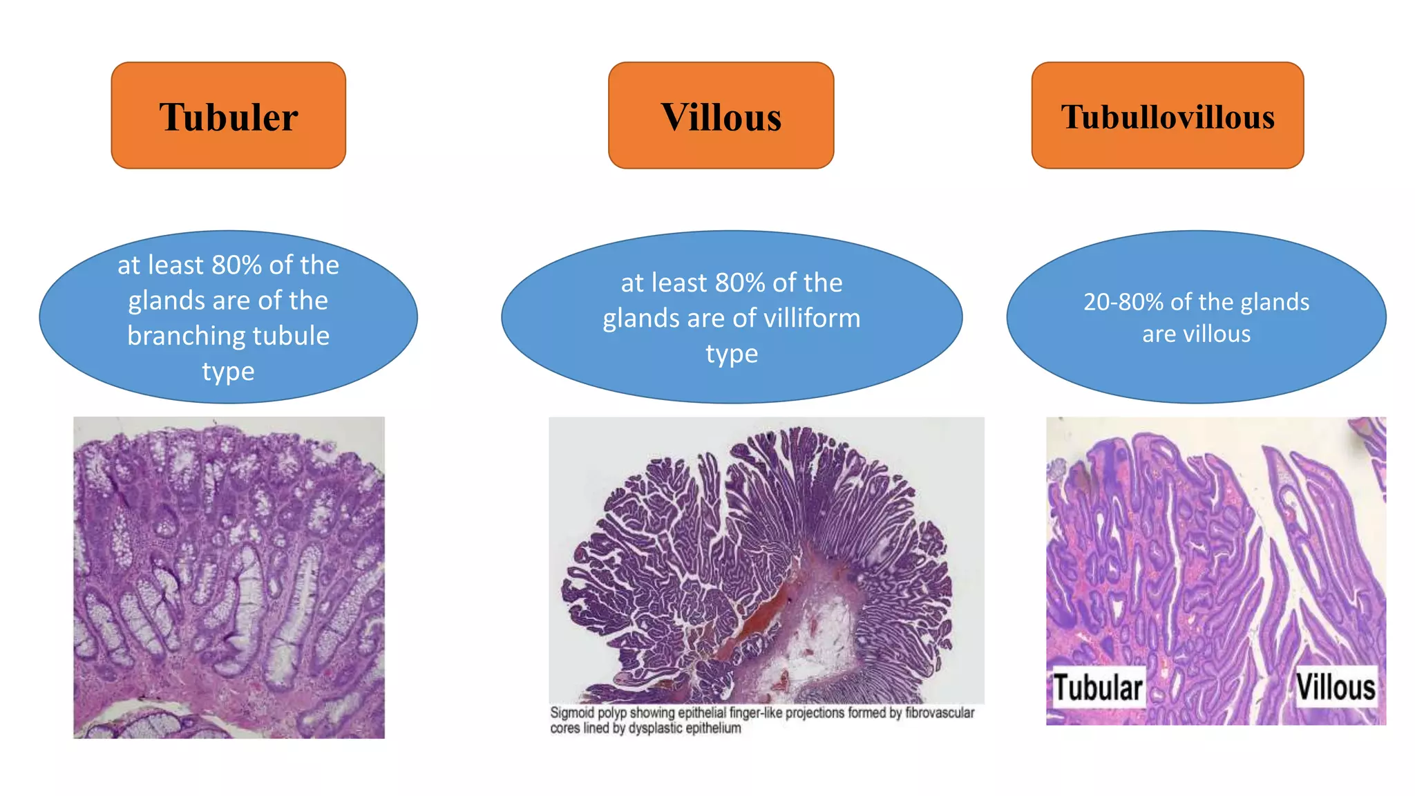
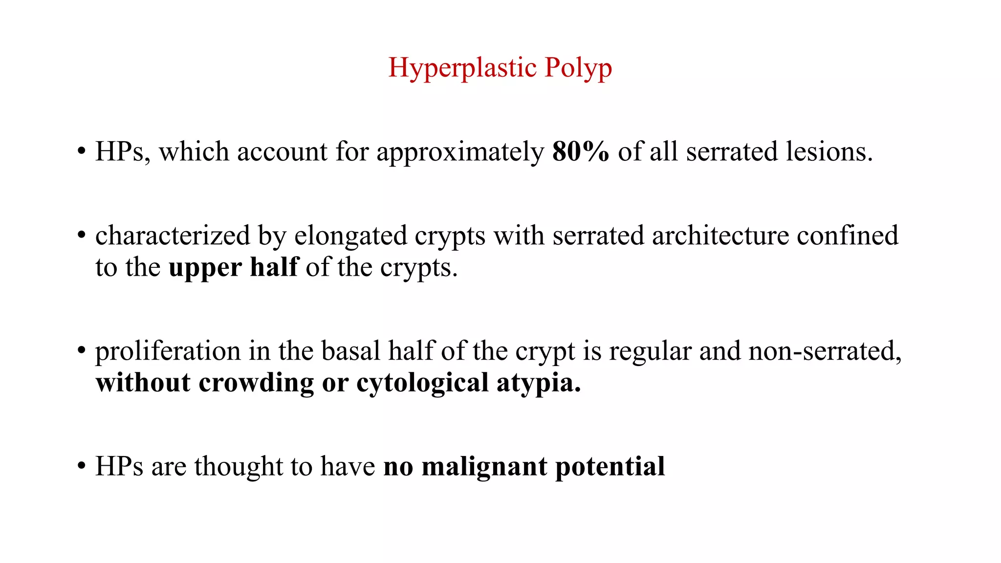
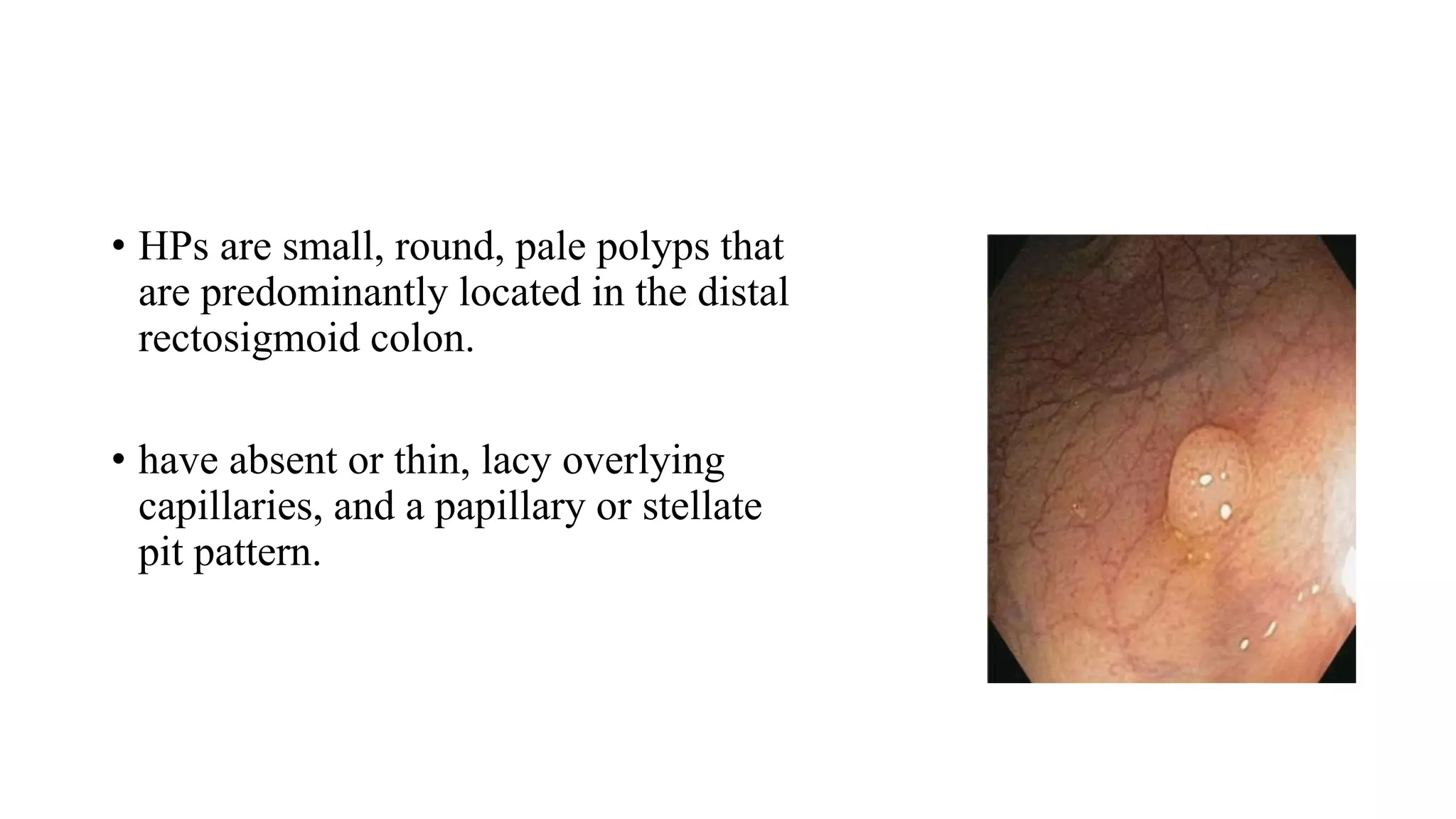
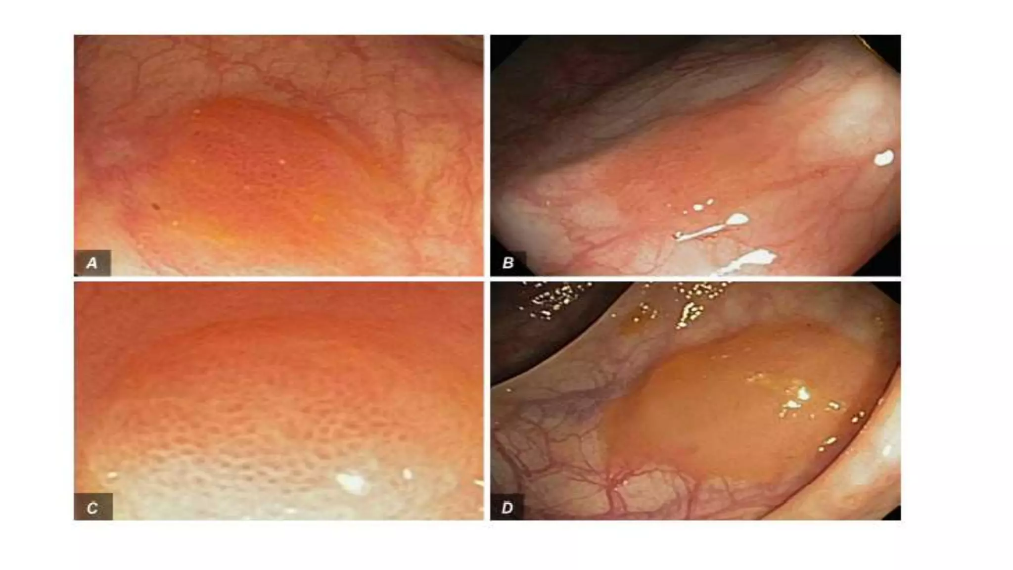
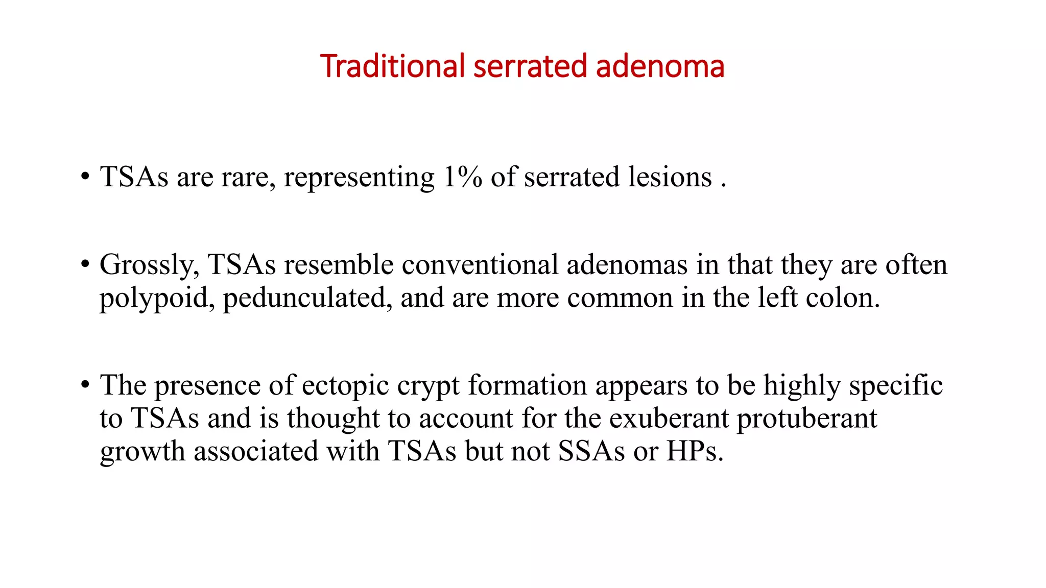
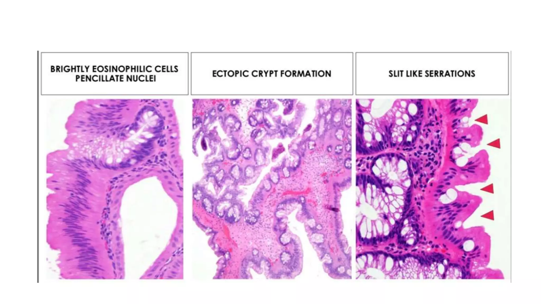
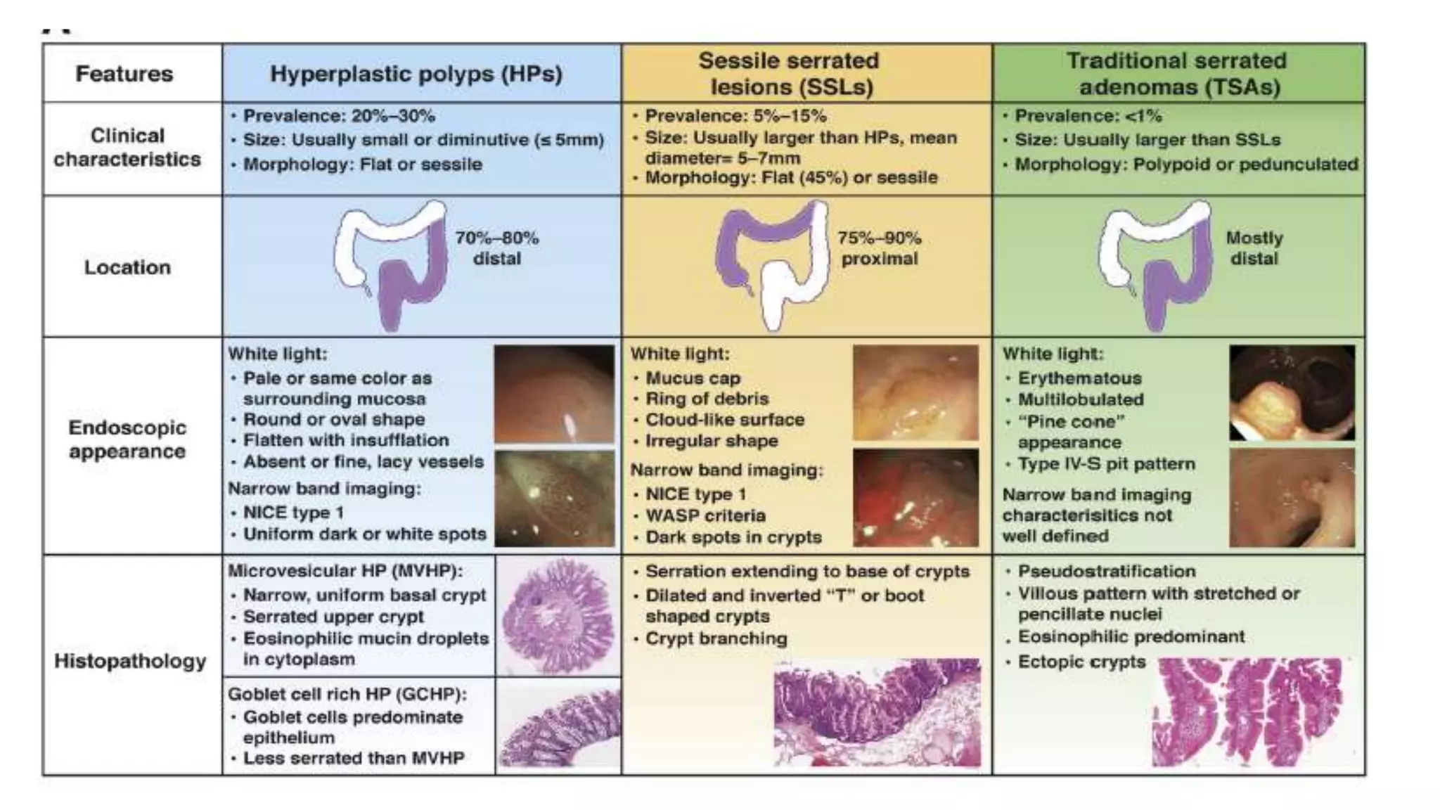
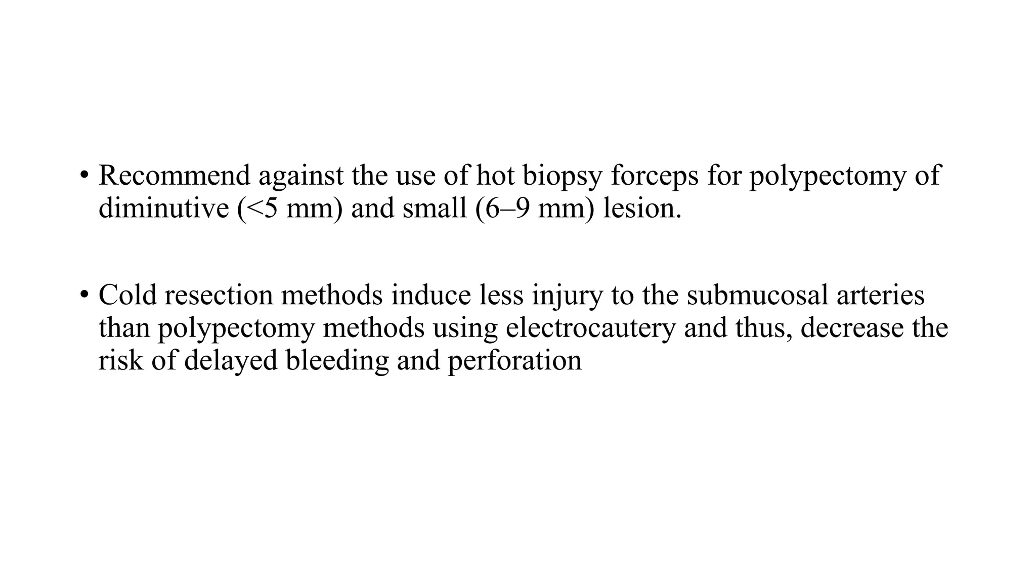
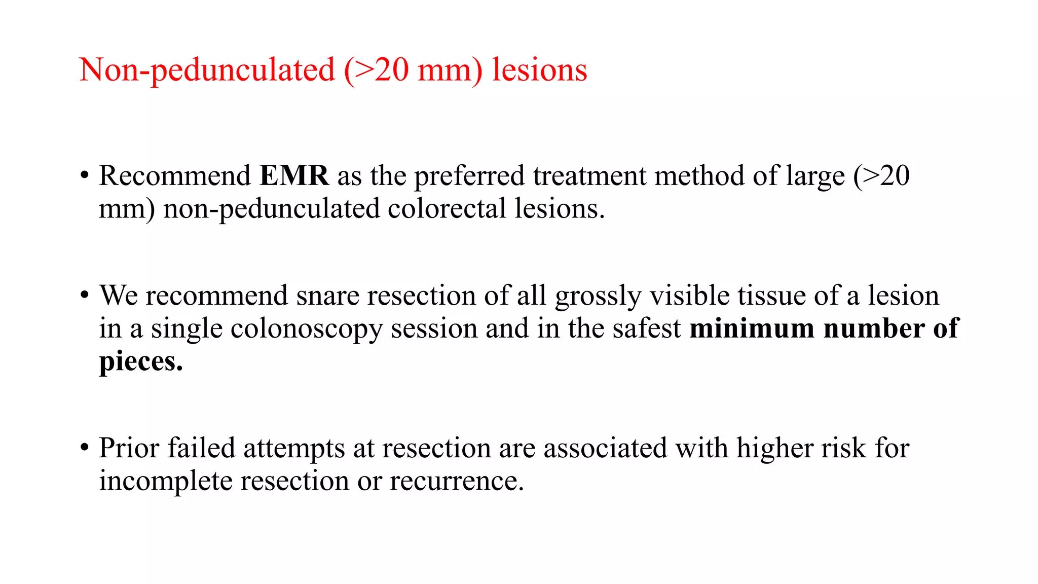
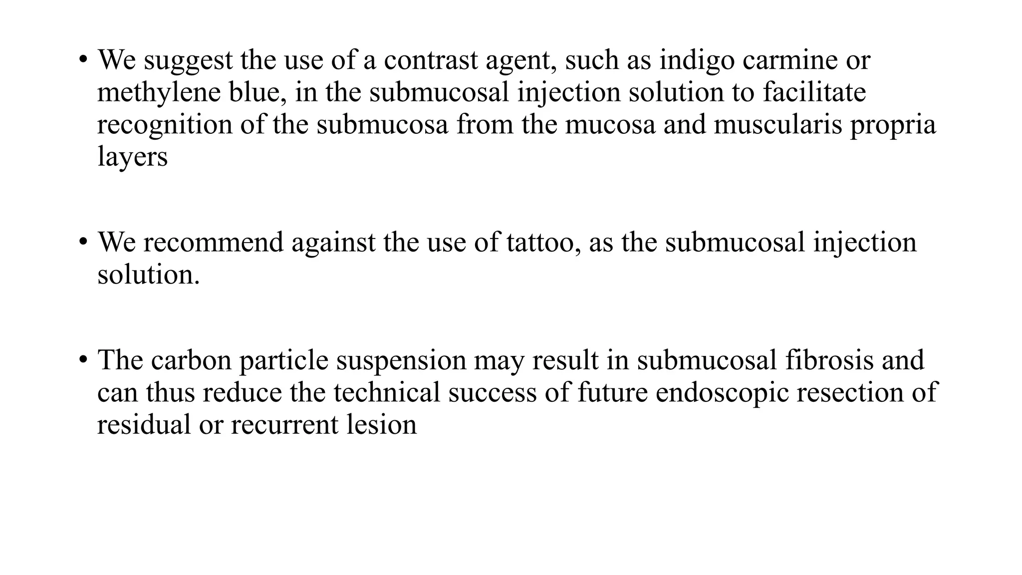
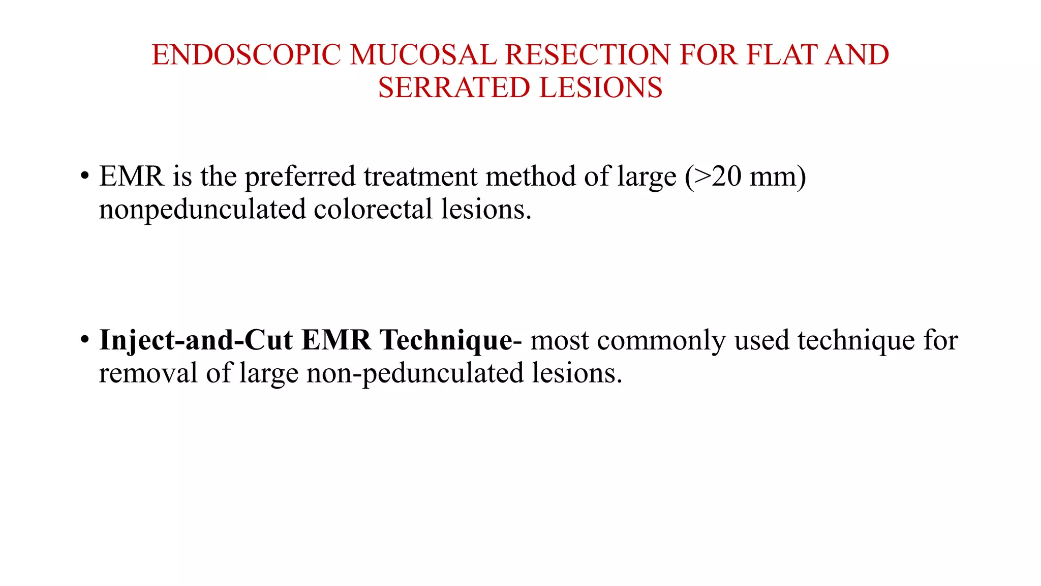
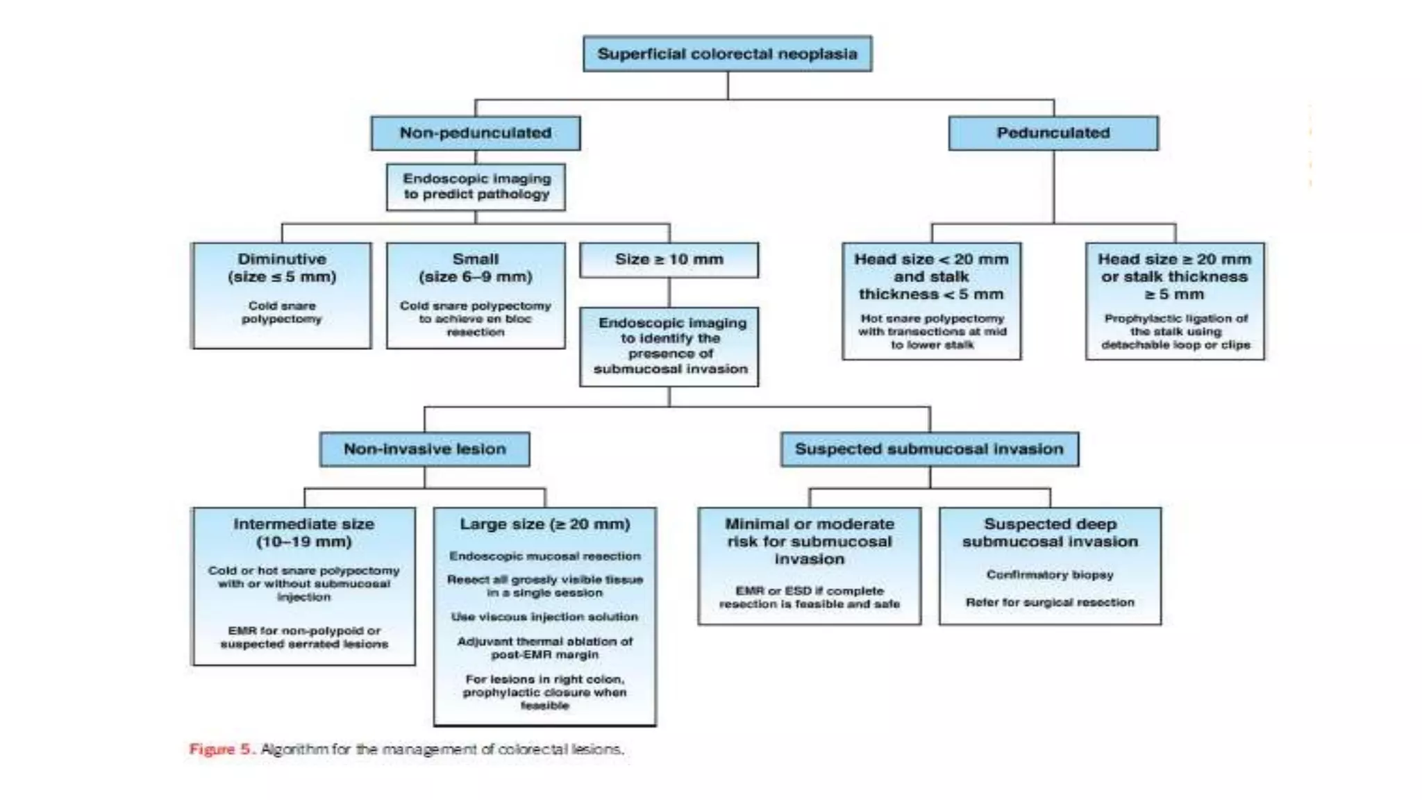
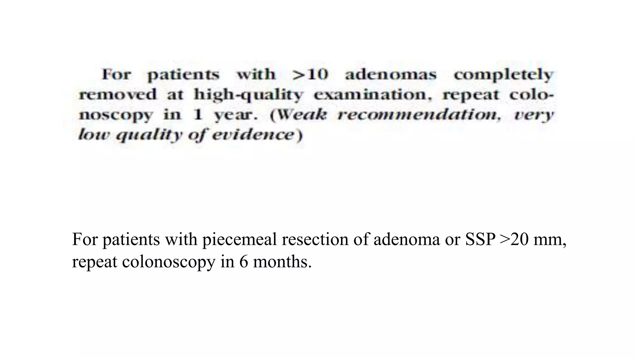
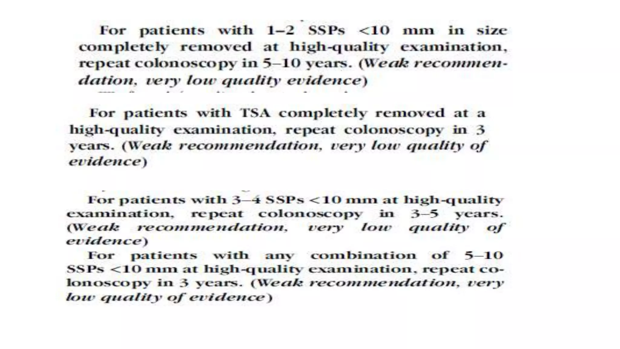
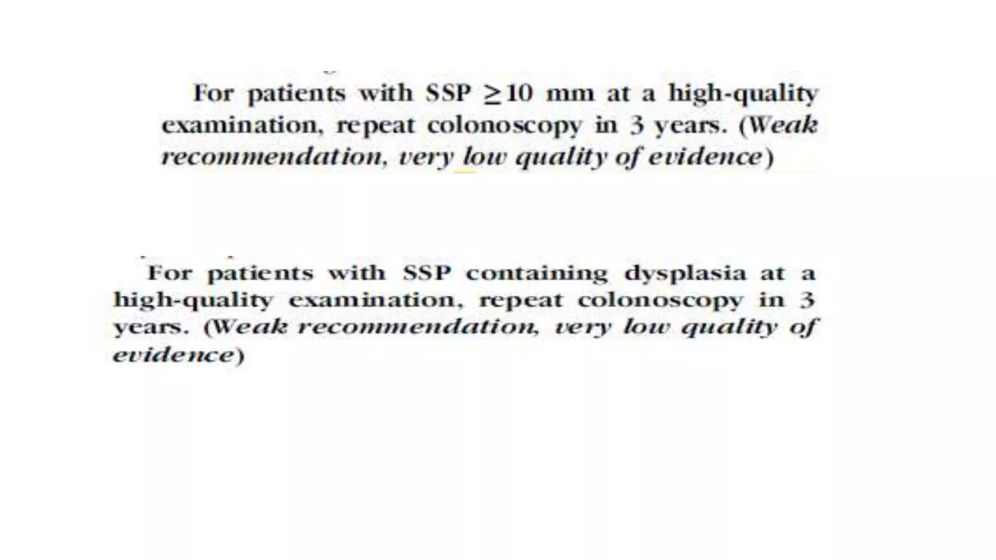
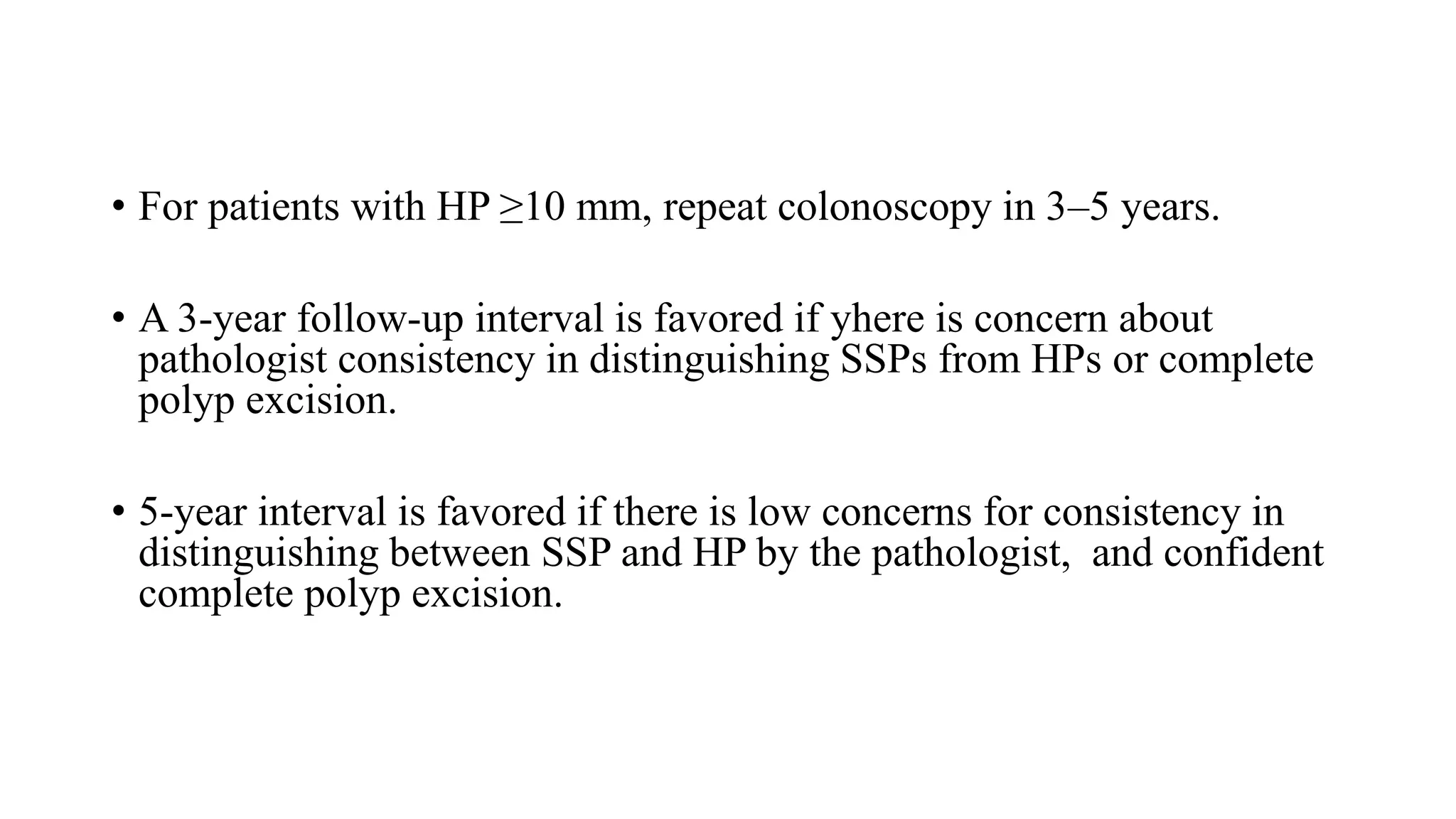
SlideShare a Scribd company logo
Approach to Colorectal Polyp
Dr Awadhesh Narayan
MD. DrNB (Gastro)
• A polyp is a discrete mass of tissue that protrudes into the lumen of the
bowel.
• Polyp’ is a term derived from the Greek word polypous, which means
‘morbid lump.’
• Generally, this term describes any mass protruding into the lumen of a
hollow vessel, anywhere in the gastro-intestinal, genito-urinary or
respiratory tracts
Methods for Detection
• Colorectal polyps usually are clinically silent.
• They most often are detected either incidentally during investigation
for non-specific symptoms.
• As part of the evaluation for iron deficiency anemia
• or in asymptomatic people being screened for colorectal neoplasia
Fecal Occult Blood Testing
• In general, polyps smaller than 1 cm do not bleed.
• polyp detected after a positive test result may be coincidental and that the FOBT
result is not directly attributable to bleeding from the polyp
• Upon colonoscopic evaluation, less than half of these people will have a colorectal
neoplasm, and among the lesions found, adenomas outnumber carcinomas by 3:1.
• false positives can occur if the patient recently had ingested vegetable peroxidases (in
turnips, radishes, melons, broccoli,carrots, cauliflower, cucumbers, grapefruit,
mushrooms) or red meat (containing myoglobin), and false negatives can occur in the
presence of high doses of antioxidants such as vitamin C.
Morikawa T, Kato J, Yamaji Y. Sensitivity of immunochemical fecal occult blood test to small colorectal adenomas. Am J Gastroenterol
2007;102(10):2259–64.
Fecal Immunochemical Testing
• To avoid some of the drawbacks of FOBTs, fecal immunochemical
testing (FIT) uses antibody-based detection of human hemoglobin in
the stool.
• the overall sensitivity for detecting any adenoma ranged from 11.4%
to 58.0%, and 25.4% to 71.5% for advanced adenomas.
• detects more cancers than adenomas
Hundt S, Haug U, Brenner H. Comparative evaluation of immunochemical fecal occult blood tests for colorectal
adenoma detection.Ann Intern Med 2009;150(3):162–9.
Barium Enema
• The detection of adenomas by barium enema (BE) depends on their
size.
• In the National Polyp Study, the detection ratesof adenomas smaller
than 6 mm, 6 to 10 mm, and larger than 10 mm were 32%, 53%, and
48%, respectively.
Winawer SJ, Stewart ET, Zauber AG, et al. A comparison of colonoscopy and double-contrast barium enema for surveillance afterpolypectomy.
N Engl J Med 2000;342:1766–72.
Colonoscopy
• Considered the gold standard for detecting polyps
• Colonoscopy also can miss neoplasms, especially those located at
flexures or behind folds.
• In general, missed adenomas tend to be small.
• Studies had demonstrated adenoma miss rates of 0% to 6% for
adenomas larger than 1 cm, 12% to 13% for adenomas 6 to 9 mm, and
15% to 27% for adenomas smaller than 6 mm.
• NBI did not show an improvement in ADR over high-definition white-
light endoscopy
Rex DK. Maximizing detection of adenomas and cancers during colonoscopy. Am J Gastroenterol 2006;101:2866–77.
CT Colonography
• Also known as virtual colonoscopy, CTC involves scanning the colon
with a helical or spiral CT scanner to produce both 2- and 3-
dimensional images of the colon and rectum.
• In the first large study to involve a pure asymptomatic screening
population, CTC had a sensitivity of 86% for polyps 5 to 9 mm and
92% for polyps larger than 10 mm
Pickhardt PJ, Choi JR, Hwang I, et al. Computed tomographic virtual colonoscopy to screen for colorectal neoplasia in asymptomatic adults. N
Engl J Med 2003;349:2191–200.
Stool DNA Testing
• DNA-based CRC screening approaches are based on the premise that
tumor cells are shed into the lumen and abnormal DNA from these
neoplastic lesions will be detectable in the stool.
• fecal DNA testing detected only 42% of advanced adenomatous
polyps.
Macroscopic classification
Paris classification-
2 macroscopic types:
• (1) type 0- the superficial lesions
• (2) types 1–5, the advanced cancers
According to size polyps are classified into 3 groups:
1. Diminutive (1 to 5 mm)
2. Small (6 to 9 mm)
3. Large (≥10mm)
Laterally spreading tumors (LSTs)
• Non-polypoid lesions 10 mm or larger in diameter are referred to as
LSTs
• They have a low vertical axis and extend laterally along the colonic
luminal wall.
LSTs are morphologically subclassified
into granular type (LST-G) (A, B), which
have a nodular surface, and non-granular
type (LST-NG), which have a smooth
surface (C, D).
This macroscopic distinction is important
to facilitate the endoscopic removal plan as
it provides information about the risk of
cancer or submucosal fibrosis in order to
anticipate the technical ease or difficulty of
the removal.
• LST-G have the lowest risk (0.5%; 95% ), whereas LST-NG have the
highest risk of submucosal invasion (31.6%)
Bogie RMM, Veldman MHJ, Snijders LARS, et al. Endoscopic subtypes of colorectal laterally spreading tumors (LSTs) and the
risk of submucosal invasion: a meta-analysis. Endoscopy 2018;50:263-82.
Optical diagnosis
• Endoscopic prediction of the histologic class of a polyp may influence
the resection approach to ensure complete removal.
• optical diagnosis of colorectal lesions is feasible in routine clinical
practice and comparable to the current reference standard,
histopathology.
The Narrow Band Imaging International Colorectal Endoscopic (NICE)
classification provides a validated criterion for the classification-
• Type 1 (serrated class lesions–hyperplastic and sessile serrated
lesions)
• Type 2 (adenomas)
• Type 3 (those with deep submucosal invasion).
• This classification have achieved 93% concordance of optical
diagnosis and pathology, and a >90% negative predictive value for
rectosigmoid lesions.
ASGE Technology Committee; Abu Dayyeh BK, Thosani N, et al. ASGE Technology Committee systematic review and meta-analysis
Gastrointest Endosc 2015;81:502; e1–e16
Workgroup Serrated Polyps and
Polyposis-WASP) criteria added 4
sessile serrated lesion features (ie,
clouded surface, indistinctive
borders, irregular shape, and
dark spots inside crypts) to the
NICE classification
• valley sign is highly specific (>90%)
for conventional adenoma in
diminutive (<5 mm) lesions,
suggesting it to be a valid predictor of
adenomatous histology in diminutive
colorectal lesions
Its application has been shown to be useful in assessing the most
clinically relevant approaches:
• leave hyperplastic diminutive lesions of the rectum and sigmoid colon
• remove all adenomas anywhere in the colon and any serrated lesions
proximal to sigmoid colon and >5 mm.
• biopsy and refer to surgery lesions with deep submucosal invasion.
CONVENTIONALADENOMAS
Histologic Features
• Characterized by abnormal cellular proliferation and renewal, resulting
in hypercellularity of colonic crypts, with cells that appear
hyperchromatic and depleted of mucin with elongated nuclei arranged
in a picket-fence pattern.
• In a study involving 13,992 participants who underwent screening
colonoscopy, 5891 nontumorous polyps were removed; of these, 3469
(59.0%) were adenomatous.
Lieberman D, Moravec M, Holub J, Michaels L, Eisen G. Polyp size and advanced histology in patients undergoing colonoscopy screening:
Gastroenterology 2008; 135: 1100-5.
Tubuler Villous Tubullovillous
at least 80% of the
glands are of the
branching tubule
type
20-80% of the glands
are villous
at least 80% of the
glands are of villiform
type
• Of all adenomatous polyps, TAs account for 80-86%, tubulovillous for
8-16%, and villous adenomas for 3-16%.
• By definition, all conventional adenomas are dysplastic.
• TAs usually are small and exhibit mild dysplasia, whereas villous
architecture is more often encountered in large adenomas and tends to
be associated with an increased frequency of HGD
Konishi F, Morson BCJ. Pathology of colorectal adenomas: a colonoscopic survey. J Clin Pathol 1982;35:830–41
Adenomas are considered advanced-
• if they are 10 mm or more in diameter
• adenomas that are <1 cm in diameter are considered to be advanced if
they contain at least 25% villous features, high-grade dysplasia, or
carcinoma.
Colorectal AdenomasN Engl J Med 2016
• If a focus of neoplastic cells grows beyond the basement membrane
into the lamina propria, the lesion is termed intramucosal carcinoma.
• Both HGD and intramucosal carcinoma are noninvasive lesions
without metastatic potential.
• Only when a focus of neoplastic cells has spread through the
muscularis mucosae is the lesion considered invasive carcinoma.
• An adenoma that contains a focus of invasive carcinoma commonly is
referred to as a malignant polyp
Malignant Potential of Adenomatous Polyp
• The 3 features that correlate with malignant potential for an
adenomatous polyp are size, histologic type, and degree of dysplasia.
• malignant potential is correlated directly with larger adenoma size,
more villous histology and higher degrees of dysplasia.
• These 3 criteria usually are interdependent.
Molecular Pathogenesis
Conventional adenomas and a subset of sessile serrated adenomas
develop through to 3 major pathways:
1. chromosomal instability (CIN)
2. microsatellite instability(MSI).
3. aberrant hypermethylation of CpG island methylator phenotype
(CIMP)
• 85% of sporadic colon cancers arise from conventional adenomas
through the classic adenoma-carcinoma sequence,
SERRASTED ADENOMAS
Hyperplastic polyp
Sessile serrated
adenoma
Traditional serrated
adenoma
Histologically these are distinguished
from conventional adenomas
primarily
by a saw-tooth or stellate appearance
of their colonic crypts
Hyperplastic Polyp
• HPs, which account for approximately 80% of all serrated lesions.
• characterized by elongated crypts with serrated architecture confined
to the upper half of the crypts.
• proliferation in the basal half of the crypt is regular and non-serrated,
without crowding or cytological atypia.
• HPs are thought to have no malignant potential
• HPs are small, round, pale polyps that
are predominantly located in the distal
rectosigmoid colon.
• have absent or thin, lacy overlying
capillaries, and a papillary or stellate
pit pattern.
Sessile Serrated Adenoma
• second most common type of serrated lesion (15% to 20%).
• along with TSAs, are considered precursor lesion to CRC arising via
the serrated neoplasia pathway.
• The pathologic hallmarks of SSAs are hyperserration and dilatation
extending to the lower third or the base of the crypt.
• SSAs are also found more commonly in the proximal colon and are
frequently multiple.
• SSA are typically larger than HPs,but are also pale and have either a
flat or sessile morphology.
• Distinguishing features of SSA include a cloud-like surface, an
overlying mucus cap, a rim of debris or stool around the lesion, and
obscuration of the underlying mucosal vasculature
Traditional serrated adenoma
• TSAs are rare, representing 1% of serrated lesions .
• Grossly, TSAs resemble conventional adenomas in that they are often
polypoid, pedunculated, and are more common in the left colon.
• The presence of ectopic crypt formation appears to be highly specific
to TSAs and is thought to account for the exuberant protuberant
growth associated with TSAs but not SSAs or HPs.
GASTROINTESTINAL ENDOSCOPY Volume 91, No. 3 : 2020
Diminutive (<5 mm) and small (6–9 mm) lesions
• Cold snare polypectomy to remove diminutive (<5 mm) and small
(6–9 mm) lesions.
• Recommend against the use of cold forceps polypectomy to remove
diminutive lesions due to high rates of incomplete resection.
• For diminutive lesions <2 mm, if cold snare polypectomy is
technically difficult, jumbo or large-capacity forceps polypectomymay
be considered
• Recommend against the use of hot biopsy forceps for polypectomy of
diminutive (<5 mm) and small (6–9 mm) lesion.
• Cold resection methods induce less injury to the submucosal arteries
than polypectomy methods using electrocautery and thus, decrease the
risk of delayed bleeding and perforation
Non-pedunculated (10–19 mm) lesions
• Suggest cold or hot snare polypectomy (with or without submucosal
injection) to remove 10–19 mm nonpedunculated lesions.
• EMR should be considered for non-polypoid and serrated lesions in
the 10- to 19-mm size.
Non-pedunculated (>20 mm) lesions
• Recommend EMR as the preferred treatment method of large (>20
mm) non-pedunculated colorectal lesions.
• We recommend snare resection of all grossly visible tissue of a lesion
in a single colonoscopy session and in the safest minimum number of
pieces.
• Prior failed attempts at resection are associated with higher risk for
incomplete resection or recurrence.
• We suggest the use of a contrast agent, such as indigo carmine or
methylene blue, in the submucosal injection solution to facilitate
recognition of the submucosa from the mucosa and muscularis propria
layers
• We recommend against the use of tattoo, as the submucosal injection
solution.
• The carbon particle suspension may result in submucosal fibrosis and
can thus reduce the technical success of future endoscopic resection of
residual or recurrent lesion
• We suggest the use of a viscous injection solution (eg, hydroxyethyl
starch, Eleview, ORISE Gel) for lesions>20mm to remove the lesion.
• We recommend against the use of ablative techniques (eg APC, snare
tip soft coagulation) on endoscopically visible residual tissue of a
lesion, as they have been associated with an increased risk of
recurrence.
• We suggest the use of adjuvant thermal ablation of the post- EMR
margin, where no endoscopically visible adenoma remains despite
meticulous inspection.
ENDOSCOPIC MUCOSAL RESECTION FOR FLAT AND
SERRATED LESIONS
• EMR is the preferred treatment method of large (>20 mm)
nonpedunculated colorectal lesions.
• Inject-and-Cut EMR Technique- most commonly used technique for
removal of large non-pedunculated lesions.
Underwater Endoscopic Mucosal Resection
• It obviates the step of submucosal injection before snare resection.
• When the lumen is distended with water the mucosa and submucosa
floats as folds into the non-distended colon, while the muscularis
propria remains circular.
• The segment of lumen with the lesion is completely immersed under
water, the borders of the polyp are marked using APC or snare tip
coagulation, and the hot snare resection is completed
Cold Snare Endoscopic Mucosal Resection
• Cold snare with injection is a recently described method to remove large lesions
without electrocautery tominimize the risk of delayed bleeding and perforation
• prospective study- removed 163 serrated lesions >10 mm (median size, 15 mm;
range, 10–40 mm) using an injection of succinylated gelatin and diluted methylene
blue before piecemeal snare resection without diathermy. Short-term surveillance
colonoscopy in 82% of the lesions (n= 134) at 6 months showed a single
recurrence (0.6%).
Tutticci NJ, Hewett DG. Cold EMR of large sessile serrated polyps at colonoscopy. Gastrointest Endosc 2018;87:837–842
Endoscopic Submucosal Dissection
• The indications for colorectal ESD are-
• Large-sized (>20 mm in diameter) lesions that are indicated for
endoscopic rather than surgical resection, and in which en bloc
resection using inject-and-cut EMR is difficult.
• These include, lesion suspected to have submucosal invasion (ie, large
depressed lesion or pseudodepressed LST-NG lesion), mucosal lesions
with fibrosis, local residual early carcinoma after endoscopic
resection, and non-polypoid colorectal dysplasia in patients with
inflammatory bowel disease.
Pedunculated lesions
• We recommend hot snare polypectomy to remove pedunculated
lesions >10 mm.
• We recommend prophylactic mechanical ligation of the stalk with a
detachable loop or clips on pedunculated lesions with head >20 mm
or with stalk thickness >5 mm to reduce immediate and delayed post-
polypectomy bleeding
• studies have shown that individuals with adenoma, despite adenoma
removal, may have increased risk for CRC compared to the general
population
• Surveillance colonoscopy after baseline removal of adenoma with
high-risk features (eg, size ≥10 mm) reduce risk for incident CRC.
• Impact of surveillance colonoscopy after removal of adenoma with
low-risk features (such as 1–2 adenomas <10 mm) on risk for incident
CRC is uncertain
For patients with piecemeal resection of adenoma or SSP >20 mm,
repeat colonoscopy in 6 months.
• For patients with HP ≥10 mm, repeat colonoscopy in 3–5 years.
• A 3-year follow-up interval is favored if yhere is concern about
pathologist consistency in distinguishing SSPs from HPs or complete
polyp excision.
• 5-year interval is favored if there is low concerns for consistency in
distinguishing between SSP and HP by the pathologist, and confident
complete polyp excision.
Colorectal Polyp.pptx

More Related Content

What's hot

CARCINOMA PENIS
CARCINOMA PENISCARCINOMA PENIS
CARCINOMA PENIS
Vikas Kumar
 
periampullary carcinoma
periampullary carcinomaperiampullary carcinoma
periampullary carcinoma
Gauri Kulkarni
 
Carcinoma penis
Carcinoma penisCarcinoma penis
Carcinoma penis
Arkaprovo Roy
 
Management of pancreatic fistulas
Management of pancreatic fistulasManagement of pancreatic fistulas
Management of pancreatic fistulas
Abhilash Cheriyan
 
Prostatic carcinoma
Prostatic carcinoma Prostatic carcinoma
Prostatic carcinoma
Meena Reddy
 
Pancreatic Surgery
Pancreatic SurgeryPancreatic Surgery
Pancreatic Surgery
Dr Harsh Shah
 
Lynch syndrome
Lynch syndromeLynch syndrome
Lynch syndrome
Ashish Pokharkar
 
Ivor lewis esophagectomy
Ivor lewis esophagectomyIvor lewis esophagectomy
Ivor lewis esophagectomy
rajat1906
 
Gastric Cancer Surgery
Gastric Cancer SurgeryGastric Cancer Surgery
Gastric Cancer Surgery
Joshua Ellenhorn
 
Short bowel syndrome
Short bowel syndromeShort bowel syndrome
Short bowel syndrome
Jibran Mohsin
 
Post gastrectomy syndrome
Post gastrectomy syndrome   Post gastrectomy syndrome
Post gastrectomy syndrome
Youttam Laudari
 
gastric resection, reconstruction and post gastrectomy syndromes
gastric resection, reconstruction and post gastrectomy syndromesgastric resection, reconstruction and post gastrectomy syndromes
gastric resection, reconstruction and post gastrectomy syndromes
sanyal1981
 
Carcinoma penis
Carcinoma penisCarcinoma penis
Carcinoma penis
Shubham Lavania
 
Pancreatic pseudocyst
Pancreatic pseudocystPancreatic pseudocyst
Pancreatic pseudocystdraakif
 
Parastomal hernia ppt
Parastomal hernia pptParastomal hernia ppt
Parastomal hernia ppt
piyushpatwa
 
Neoplasm of pancreas
Neoplasm of pancreasNeoplasm of pancreas
Neoplasm of pancreas
Priyatham Kasaraneni
 
RETROPERITONEAL FIBROSIS
RETROPERITONEAL FIBROSISRETROPERITONEAL FIBROSIS
RETROPERITONEAL FIBROSIS
GAURAV NAHAR
 
testicular tumors
testicular tumorstesticular tumors
testicular tumors
DrAyush Garg
 

What's hot (20)

CARCINOMA PENIS
CARCINOMA PENISCARCINOMA PENIS
CARCINOMA PENIS
 
periampullary carcinoma
periampullary carcinomaperiampullary carcinoma
periampullary carcinoma
 
Carcinoma penis
Carcinoma penisCarcinoma penis
Carcinoma penis
 
Management of pancreatic fistulas
Management of pancreatic fistulasManagement of pancreatic fistulas
Management of pancreatic fistulas
 
Prostatic carcinoma
Prostatic carcinoma Prostatic carcinoma
Prostatic carcinoma
 
Pancreatic Surgery
Pancreatic SurgeryPancreatic Surgery
Pancreatic Surgery
 
Lynch syndrome
Lynch syndromeLynch syndrome
Lynch syndrome
 
Ivor lewis esophagectomy
Ivor lewis esophagectomyIvor lewis esophagectomy
Ivor lewis esophagectomy
 
Gastric Cancer Surgery
Gastric Cancer SurgeryGastric Cancer Surgery
Gastric Cancer Surgery
 
Short bowel syndrome
Short bowel syndromeShort bowel syndrome
Short bowel syndrome
 
Post gastrectomy syndrome
Post gastrectomy syndrome   Post gastrectomy syndrome
Post gastrectomy syndrome
 
gastric resection, reconstruction and post gastrectomy syndromes
gastric resection, reconstruction and post gastrectomy syndromesgastric resection, reconstruction and post gastrectomy syndromes
gastric resection, reconstruction and post gastrectomy syndromes
 
Carcinoma penis
Carcinoma penisCarcinoma penis
Carcinoma penis
 
Psoas abscess
Psoas abscessPsoas abscess
Psoas abscess
 
Choledochal cyst
Choledochal cystCholedochal cyst
Choledochal cyst
 
Pancreatic pseudocyst
Pancreatic pseudocystPancreatic pseudocyst
Pancreatic pseudocyst
 
Parastomal hernia ppt
Parastomal hernia pptParastomal hernia ppt
Parastomal hernia ppt
 
Neoplasm of pancreas
Neoplasm of pancreasNeoplasm of pancreas
Neoplasm of pancreas
 
RETROPERITONEAL FIBROSIS
RETROPERITONEAL FIBROSISRETROPERITONEAL FIBROSIS
RETROPERITONEAL FIBROSIS
 
testicular tumors
testicular tumorstesticular tumors
testicular tumors
 

Similar to Colorectal Polyp.pptx

Rectal cancer management , Nccn guideline 2024 .pptx
Rectal cancer management , Nccn guideline 2024 .pptxRectal cancer management , Nccn guideline 2024 .pptx
Rectal cancer management , Nccn guideline 2024 .pptx
Dr Maria Tamanna
 
Anal canal cancer
Anal canal cancerAnal canal cancer
Anal canal cancer
Dr. Aaditya Prakash
 
Bladder cancer
Bladder cancerBladder cancer
Bladder cancer
Ministry of Health
 
Colon cancer in 2018
Colon cancer in 2018Colon cancer in 2018
Colon cancer in 2018
Ali Musavi
 
ANAL CARCINOMAAAAAAAAAAAAAAAAAAAAAAAAAAAAAAAA
ANAL CARCINOMAAAAAAAAAAAAAAAAAAAAAAAAAAAAAAAAANAL CARCINOMAAAAAAAAAAAAAAAAAAAAAAAAAAAAAAAA
ANAL CARCINOMAAAAAAAAAAAAAAAAAAAAAAAAAAAAAAAA
UswatunAortatikaKhas
 
Gallbladder tumors
Gallbladder tumorsGallbladder tumors
Gallbladder tumors
Tawfiq Nawafleh
 
Gastric carcinoma
Gastric carcinoma Gastric carcinoma
Gastric carcinoma
Dr.Saadvik Raghuram
 
Tumors of small bowel.pptx
Tumors of small bowel.pptxTumors of small bowel.pptx
Tumors of small bowel.pptx
AkshaySarraf1
 
GALLBLADDER CANCER.pptx
GALLBLADDER CANCER.pptxGALLBLADDER CANCER.pptx
GALLBLADDER CANCER.pptx
SujanPandey11
 
Breast diseases
Breast diseasesBreast diseases
Breast diseases
Tariq Ibrahim
 
Ca endometrium
Ca endometriumCa endometrium
Ca endometrium
Satyajeet Rath
 
Endometrium part 1 2018
Endometrium part 1 2018Endometrium part 1 2018
Endometrium part 1 2018
Dr- Mustafa Ahmed Alazam
 
Tumor board Ca stomach
Tumor board Ca stomachTumor board Ca stomach
Tumor board Ca stomach
Dr. Varughese George
 
Ampullary carcinoma
Ampullary carcinomaAmpullary carcinoma
Ampullary carcinoma
Dr Tauqeer A Siddiqui MD FACP
 
Carcinoma esophagus 2020
Carcinoma esophagus 2020Carcinoma esophagus 2020
Carcinoma esophagus 2020
Dr.Anjali Reddy Baddiggam
 
Rathod Gastric Cancer Presentation final.pptx
Rathod Gastric Cancer Presentation final.pptxRathod Gastric Cancer Presentation final.pptx
Rathod Gastric Cancer Presentation final.pptx
Aadarsh Kavoram
 
Principle of oncology
Principle of oncologyPrinciple of oncology
Principle of oncology
RajeevPandit10
 
benign and Malignant Salivary Gland Tumors.pptx
benign and Malignant Salivary Gland Tumors.pptxbenign and Malignant Salivary Gland Tumors.pptx
benign and Malignant Salivary Gland Tumors.pptx
ulster University
 
Adnexal Masses
Adnexal MassesAdnexal Masses
Adnexal Masses
NARENDRA C MALHOTRA
 
Adnexal Masses in Reproductive Age
Adnexal Masses in Reproductive AgeAdnexal Masses in Reproductive Age
Adnexal Masses in Reproductive Age
NARENDRA C MALHOTRA
 

Similar to Colorectal Polyp.pptx (20)

Rectal cancer management , Nccn guideline 2024 .pptx
Rectal cancer management , Nccn guideline 2024 .pptxRectal cancer management , Nccn guideline 2024 .pptx
Rectal cancer management , Nccn guideline 2024 .pptx
 
Anal canal cancer
Anal canal cancerAnal canal cancer
Anal canal cancer
 
Bladder cancer
Bladder cancerBladder cancer
Bladder cancer
 
Colon cancer in 2018
Colon cancer in 2018Colon cancer in 2018
Colon cancer in 2018
 
ANAL CARCINOMAAAAAAAAAAAAAAAAAAAAAAAAAAAAAAAA
ANAL CARCINOMAAAAAAAAAAAAAAAAAAAAAAAAAAAAAAAAANAL CARCINOMAAAAAAAAAAAAAAAAAAAAAAAAAAAAAAAA
ANAL CARCINOMAAAAAAAAAAAAAAAAAAAAAAAAAAAAAAAA
 
Gallbladder tumors
Gallbladder tumorsGallbladder tumors
Gallbladder tumors
 
Gastric carcinoma
Gastric carcinoma Gastric carcinoma
Gastric carcinoma
 
Tumors of small bowel.pptx
Tumors of small bowel.pptxTumors of small bowel.pptx
Tumors of small bowel.pptx
 
GALLBLADDER CANCER.pptx
GALLBLADDER CANCER.pptxGALLBLADDER CANCER.pptx
GALLBLADDER CANCER.pptx
 
Breast diseases
Breast diseasesBreast diseases
Breast diseases
 
Ca endometrium
Ca endometriumCa endometrium
Ca endometrium
 
Endometrium part 1 2018
Endometrium part 1 2018Endometrium part 1 2018
Endometrium part 1 2018
 
Tumor board Ca stomach
Tumor board Ca stomachTumor board Ca stomach
Tumor board Ca stomach
 
Ampullary carcinoma
Ampullary carcinomaAmpullary carcinoma
Ampullary carcinoma
 
Carcinoma esophagus 2020
Carcinoma esophagus 2020Carcinoma esophagus 2020
Carcinoma esophagus 2020
 
Rathod Gastric Cancer Presentation final.pptx
Rathod Gastric Cancer Presentation final.pptxRathod Gastric Cancer Presentation final.pptx
Rathod Gastric Cancer Presentation final.pptx
 
Principle of oncology
Principle of oncologyPrinciple of oncology
Principle of oncology
 
benign and Malignant Salivary Gland Tumors.pptx
benign and Malignant Salivary Gland Tumors.pptxbenign and Malignant Salivary Gland Tumors.pptx
benign and Malignant Salivary Gland Tumors.pptx
 
Adnexal Masses
Adnexal MassesAdnexal Masses
Adnexal Masses
 
Adnexal Masses in Reproductive Age
Adnexal Masses in Reproductive AgeAdnexal Masses in Reproductive Age
Adnexal Masses in Reproductive Age
 

Recently uploaded

basicmodesofventilation2022-220313203758.pdf
basicmodesofventilation2022-220313203758.pdfbasicmodesofventilation2022-220313203758.pdf
basicmodesofventilation2022-220313203758.pdf
aljamhori teaching hospital
 
How STIs Influence the Development of Pelvic Inflammatory Disease.pptx
How STIs Influence the Development of Pelvic Inflammatory Disease.pptxHow STIs Influence the Development of Pelvic Inflammatory Disease.pptx
How STIs Influence the Development of Pelvic Inflammatory Disease.pptx
FFragrant
 
Flu Vaccine Alert in Bangalore Karnataka
Flu Vaccine Alert in Bangalore KarnatakaFlu Vaccine Alert in Bangalore Karnataka
Flu Vaccine Alert in Bangalore Karnataka
addon Scans
 
Pictures of Superficial & Deep Fascia.ppt.pdf
Pictures of Superficial & Deep Fascia.ppt.pdfPictures of Superficial & Deep Fascia.ppt.pdf
Pictures of Superficial & Deep Fascia.ppt.pdf
Dr. Rabia Inam Gandapore
 
Tom Selleck Health: A Comprehensive Look at the Iconic Actor’s Wellness Journey
Tom Selleck Health: A Comprehensive Look at the Iconic Actor’s Wellness JourneyTom Selleck Health: A Comprehensive Look at the Iconic Actor’s Wellness Journey
Tom Selleck Health: A Comprehensive Look at the Iconic Actor’s Wellness Journey
greendigital
 
Pharynx and Clinical Correlations BY Dr.Rabia Inam Gandapore.pptx
Pharynx and Clinical Correlations BY Dr.Rabia Inam Gandapore.pptxPharynx and Clinical Correlations BY Dr.Rabia Inam Gandapore.pptx
Pharynx and Clinical Correlations BY Dr.Rabia Inam Gandapore.pptx
Dr. Rabia Inam Gandapore
 
Novas diretrizes da OMS para os cuidados perinatais de mais qualidade
Novas diretrizes da OMS para os cuidados perinatais de mais qualidadeNovas diretrizes da OMS para os cuidados perinatais de mais qualidade
Novas diretrizes da OMS para os cuidados perinatais de mais qualidade
Prof. Marcus Renato de Carvalho
 
Hemodialysis: Chapter 3, Dialysis Water Unit - Dr.Gawad
Hemodialysis: Chapter 3, Dialysis Water Unit - Dr.GawadHemodialysis: Chapter 3, Dialysis Water Unit - Dr.Gawad
Hemodialysis: Chapter 3, Dialysis Water Unit - Dr.Gawad
NephroTube - Dr.Gawad
 
MANAGEMENT OF ATRIOVENTRICULAR CONDUCTION BLOCK.pdf
MANAGEMENT OF ATRIOVENTRICULAR CONDUCTION BLOCK.pdfMANAGEMENT OF ATRIOVENTRICULAR CONDUCTION BLOCK.pdf
MANAGEMENT OF ATRIOVENTRICULAR CONDUCTION BLOCK.pdf
Jim Jacob Roy
 
ARTIFICIAL INTELLIGENCE IN HEALTHCARE.pdf
ARTIFICIAL INTELLIGENCE IN  HEALTHCARE.pdfARTIFICIAL INTELLIGENCE IN  HEALTHCARE.pdf
ARTIFICIAL INTELLIGENCE IN HEALTHCARE.pdf
Anujkumaranit
 
Light House Retreats: Plant Medicine Retreat Europe
Light House Retreats: Plant Medicine Retreat EuropeLight House Retreats: Plant Medicine Retreat Europe
Light House Retreats: Plant Medicine Retreat Europe
Lighthouse Retreat
 
BRACHYTHERAPY OVERVIEW AND APPLICATORS
BRACHYTHERAPY OVERVIEW  AND  APPLICATORSBRACHYTHERAPY OVERVIEW  AND  APPLICATORS
BRACHYTHERAPY OVERVIEW AND APPLICATORS
Krishan Murari
 
HOT NEW PRODUCT! BIG SALES FAST SHIPPING NOW FROM CHINA!! EU KU DB BK substit...
HOT NEW PRODUCT! BIG SALES FAST SHIPPING NOW FROM CHINA!! EU KU DB BK substit...HOT NEW PRODUCT! BIG SALES FAST SHIPPING NOW FROM CHINA!! EU KU DB BK substit...
HOT NEW PRODUCT! BIG SALES FAST SHIPPING NOW FROM CHINA!! EU KU DB BK substit...
GL Anaacs
 
Knee anatomy and clinical tests 2024.pdf
Knee anatomy and clinical tests 2024.pdfKnee anatomy and clinical tests 2024.pdf
Knee anatomy and clinical tests 2024.pdf
vimalpl1234
 
For Better Surat #ℂall #Girl Service ❤85270-49040❤ Surat #ℂall #Girls
For Better Surat #ℂall #Girl Service ❤85270-49040❤ Surat #ℂall #GirlsFor Better Surat #ℂall #Girl Service ❤85270-49040❤ Surat #ℂall #Girls
For Better Surat #ℂall #Girl Service ❤85270-49040❤ Surat #ℂall #Girls
Savita Shen $i11
 
263778731218 Abortion Clinic /Pills In Harare ,
263778731218 Abortion Clinic /Pills In Harare ,263778731218 Abortion Clinic /Pills In Harare ,
263778731218 Abortion Clinic /Pills In Harare ,
sisternakatoto
 
heat stroke and heat exhaustion in children
heat stroke and heat exhaustion in childrenheat stroke and heat exhaustion in children
heat stroke and heat exhaustion in children
SumeraAhmad5
 
Surat @ℂall @Girls ꧁❤8527049040❤꧂@ℂall @Girls Service Vip Top Model Safe
Surat @ℂall @Girls ꧁❤8527049040❤꧂@ℂall @Girls Service Vip Top Model SafeSurat @ℂall @Girls ꧁❤8527049040❤꧂@ℂall @Girls Service Vip Top Model Safe
Surat @ℂall @Girls ꧁❤8527049040❤꧂@ℂall @Girls Service Vip Top Model Safe
Savita Shen $i11
 
Evaluation of antidepressant activity of clitoris ternatea in animals
Evaluation of antidepressant activity of clitoris ternatea in animalsEvaluation of antidepressant activity of clitoris ternatea in animals
Evaluation of antidepressant activity of clitoris ternatea in animals
Shweta
 
Triangles of Neck and Clinical Correlation by Dr. RIG.pptx
Triangles of Neck and Clinical Correlation by Dr. RIG.pptxTriangles of Neck and Clinical Correlation by Dr. RIG.pptx
Triangles of Neck and Clinical Correlation by Dr. RIG.pptx
Dr. Rabia Inam Gandapore
 

Recently uploaded (20)

basicmodesofventilation2022-220313203758.pdf
basicmodesofventilation2022-220313203758.pdfbasicmodesofventilation2022-220313203758.pdf
basicmodesofventilation2022-220313203758.pdf
 
How STIs Influence the Development of Pelvic Inflammatory Disease.pptx
How STIs Influence the Development of Pelvic Inflammatory Disease.pptxHow STIs Influence the Development of Pelvic Inflammatory Disease.pptx
How STIs Influence the Development of Pelvic Inflammatory Disease.pptx
 
Flu Vaccine Alert in Bangalore Karnataka
Flu Vaccine Alert in Bangalore KarnatakaFlu Vaccine Alert in Bangalore Karnataka
Flu Vaccine Alert in Bangalore Karnataka
 
Pictures of Superficial & Deep Fascia.ppt.pdf
Pictures of Superficial & Deep Fascia.ppt.pdfPictures of Superficial & Deep Fascia.ppt.pdf
Pictures of Superficial & Deep Fascia.ppt.pdf
 
Tom Selleck Health: A Comprehensive Look at the Iconic Actor’s Wellness Journey
Tom Selleck Health: A Comprehensive Look at the Iconic Actor’s Wellness JourneyTom Selleck Health: A Comprehensive Look at the Iconic Actor’s Wellness Journey
Tom Selleck Health: A Comprehensive Look at the Iconic Actor’s Wellness Journey
 
Pharynx and Clinical Correlations BY Dr.Rabia Inam Gandapore.pptx
Pharynx and Clinical Correlations BY Dr.Rabia Inam Gandapore.pptxPharynx and Clinical Correlations BY Dr.Rabia Inam Gandapore.pptx
Pharynx and Clinical Correlations BY Dr.Rabia Inam Gandapore.pptx
 
Novas diretrizes da OMS para os cuidados perinatais de mais qualidade
Novas diretrizes da OMS para os cuidados perinatais de mais qualidadeNovas diretrizes da OMS para os cuidados perinatais de mais qualidade
Novas diretrizes da OMS para os cuidados perinatais de mais qualidade
 
Hemodialysis: Chapter 3, Dialysis Water Unit - Dr.Gawad
Hemodialysis: Chapter 3, Dialysis Water Unit - Dr.GawadHemodialysis: Chapter 3, Dialysis Water Unit - Dr.Gawad
Hemodialysis: Chapter 3, Dialysis Water Unit - Dr.Gawad
 
MANAGEMENT OF ATRIOVENTRICULAR CONDUCTION BLOCK.pdf
MANAGEMENT OF ATRIOVENTRICULAR CONDUCTION BLOCK.pdfMANAGEMENT OF ATRIOVENTRICULAR CONDUCTION BLOCK.pdf
MANAGEMENT OF ATRIOVENTRICULAR CONDUCTION BLOCK.pdf
 
ARTIFICIAL INTELLIGENCE IN HEALTHCARE.pdf
ARTIFICIAL INTELLIGENCE IN  HEALTHCARE.pdfARTIFICIAL INTELLIGENCE IN  HEALTHCARE.pdf
ARTIFICIAL INTELLIGENCE IN HEALTHCARE.pdf
 
Light House Retreats: Plant Medicine Retreat Europe
Light House Retreats: Plant Medicine Retreat EuropeLight House Retreats: Plant Medicine Retreat Europe
Light House Retreats: Plant Medicine Retreat Europe
 
BRACHYTHERAPY OVERVIEW AND APPLICATORS
BRACHYTHERAPY OVERVIEW  AND  APPLICATORSBRACHYTHERAPY OVERVIEW  AND  APPLICATORS
BRACHYTHERAPY OVERVIEW AND APPLICATORS
 
HOT NEW PRODUCT! BIG SALES FAST SHIPPING NOW FROM CHINA!! EU KU DB BK substit...
HOT NEW PRODUCT! BIG SALES FAST SHIPPING NOW FROM CHINA!! EU KU DB BK substit...HOT NEW PRODUCT! BIG SALES FAST SHIPPING NOW FROM CHINA!! EU KU DB BK substit...
HOT NEW PRODUCT! BIG SALES FAST SHIPPING NOW FROM CHINA!! EU KU DB BK substit...
 
Knee anatomy and clinical tests 2024.pdf
Knee anatomy and clinical tests 2024.pdfKnee anatomy and clinical tests 2024.pdf
Knee anatomy and clinical tests 2024.pdf
 
For Better Surat #ℂall #Girl Service ❤85270-49040❤ Surat #ℂall #Girls
For Better Surat #ℂall #Girl Service ❤85270-49040❤ Surat #ℂall #GirlsFor Better Surat #ℂall #Girl Service ❤85270-49040❤ Surat #ℂall #Girls
For Better Surat #ℂall #Girl Service ❤85270-49040❤ Surat #ℂall #Girls
 
263778731218 Abortion Clinic /Pills In Harare ,
263778731218 Abortion Clinic /Pills In Harare ,263778731218 Abortion Clinic /Pills In Harare ,
263778731218 Abortion Clinic /Pills In Harare ,
 
heat stroke and heat exhaustion in children
heat stroke and heat exhaustion in childrenheat stroke and heat exhaustion in children
heat stroke and heat exhaustion in children
 
Surat @ℂall @Girls ꧁❤8527049040❤꧂@ℂall @Girls Service Vip Top Model Safe
Surat @ℂall @Girls ꧁❤8527049040❤꧂@ℂall @Girls Service Vip Top Model SafeSurat @ℂall @Girls ꧁❤8527049040❤꧂@ℂall @Girls Service Vip Top Model Safe
Surat @ℂall @Girls ꧁❤8527049040❤꧂@ℂall @Girls Service Vip Top Model Safe
 
Evaluation of antidepressant activity of clitoris ternatea in animals
Evaluation of antidepressant activity of clitoris ternatea in animalsEvaluation of antidepressant activity of clitoris ternatea in animals
Evaluation of antidepressant activity of clitoris ternatea in animals
 
Triangles of Neck and Clinical Correlation by Dr. RIG.pptx
Triangles of Neck and Clinical Correlation by Dr. RIG.pptxTriangles of Neck and Clinical Correlation by Dr. RIG.pptx
Triangles of Neck and Clinical Correlation by Dr. RIG.pptx
 

Colorectal Polyp.pptx

  • 1. Approach to Colorectal Polyp Dr Awadhesh Narayan MD. DrNB (Gastro)
  • 2.
  • 3. • A polyp is a discrete mass of tissue that protrudes into the lumen of the bowel. • Polyp’ is a term derived from the Greek word polypous, which means ‘morbid lump.’ • Generally, this term describes any mass protruding into the lumen of a hollow vessel, anywhere in the gastro-intestinal, genito-urinary or respiratory tracts
  • 4. Methods for Detection • Colorectal polyps usually are clinically silent. • They most often are detected either incidentally during investigation for non-specific symptoms. • As part of the evaluation for iron deficiency anemia • or in asymptomatic people being screened for colorectal neoplasia
  • 5. Fecal Occult Blood Testing • In general, polyps smaller than 1 cm do not bleed. • polyp detected after a positive test result may be coincidental and that the FOBT result is not directly attributable to bleeding from the polyp • Upon colonoscopic evaluation, less than half of these people will have a colorectal neoplasm, and among the lesions found, adenomas outnumber carcinomas by 3:1. • false positives can occur if the patient recently had ingested vegetable peroxidases (in turnips, radishes, melons, broccoli,carrots, cauliflower, cucumbers, grapefruit, mushrooms) or red meat (containing myoglobin), and false negatives can occur in the presence of high doses of antioxidants such as vitamin C. Morikawa T, Kato J, Yamaji Y. Sensitivity of immunochemical fecal occult blood test to small colorectal adenomas. Am J Gastroenterol 2007;102(10):2259–64.
  • 6. Fecal Immunochemical Testing • To avoid some of the drawbacks of FOBTs, fecal immunochemical testing (FIT) uses antibody-based detection of human hemoglobin in the stool. • the overall sensitivity for detecting any adenoma ranged from 11.4% to 58.0%, and 25.4% to 71.5% for advanced adenomas. • detects more cancers than adenomas Hundt S, Haug U, Brenner H. Comparative evaluation of immunochemical fecal occult blood tests for colorectal adenoma detection.Ann Intern Med 2009;150(3):162–9.
  • 7. Barium Enema • The detection of adenomas by barium enema (BE) depends on their size. • In the National Polyp Study, the detection ratesof adenomas smaller than 6 mm, 6 to 10 mm, and larger than 10 mm were 32%, 53%, and 48%, respectively. Winawer SJ, Stewart ET, Zauber AG, et al. A comparison of colonoscopy and double-contrast barium enema for surveillance afterpolypectomy. N Engl J Med 2000;342:1766–72.
  • 8. Colonoscopy • Considered the gold standard for detecting polyps • Colonoscopy also can miss neoplasms, especially those located at flexures or behind folds. • In general, missed adenomas tend to be small. • Studies had demonstrated adenoma miss rates of 0% to 6% for adenomas larger than 1 cm, 12% to 13% for adenomas 6 to 9 mm, and 15% to 27% for adenomas smaller than 6 mm. • NBI did not show an improvement in ADR over high-definition white- light endoscopy Rex DK. Maximizing detection of adenomas and cancers during colonoscopy. Am J Gastroenterol 2006;101:2866–77.
  • 9. CT Colonography • Also known as virtual colonoscopy, CTC involves scanning the colon with a helical or spiral CT scanner to produce both 2- and 3- dimensional images of the colon and rectum. • In the first large study to involve a pure asymptomatic screening population, CTC had a sensitivity of 86% for polyps 5 to 9 mm and 92% for polyps larger than 10 mm Pickhardt PJ, Choi JR, Hwang I, et al. Computed tomographic virtual colonoscopy to screen for colorectal neoplasia in asymptomatic adults. N Engl J Med 2003;349:2191–200.
  • 10. Stool DNA Testing • DNA-based CRC screening approaches are based on the premise that tumor cells are shed into the lumen and abnormal DNA from these neoplastic lesions will be detectable in the stool. • fecal DNA testing detected only 42% of advanced adenomatous polyps.
  • 11. Macroscopic classification Paris classification- 2 macroscopic types: • (1) type 0- the superficial lesions • (2) types 1–5, the advanced cancers
  • 12.
  • 13. According to size polyps are classified into 3 groups: 1. Diminutive (1 to 5 mm) 2. Small (6 to 9 mm) 3. Large (≥10mm)
  • 14. Laterally spreading tumors (LSTs) • Non-polypoid lesions 10 mm or larger in diameter are referred to as LSTs • They have a low vertical axis and extend laterally along the colonic luminal wall.
  • 15. LSTs are morphologically subclassified into granular type (LST-G) (A, B), which have a nodular surface, and non-granular type (LST-NG), which have a smooth surface (C, D). This macroscopic distinction is important to facilitate the endoscopic removal plan as it provides information about the risk of cancer or submucosal fibrosis in order to anticipate the technical ease or difficulty of the removal.
  • 16. • LST-G have the lowest risk (0.5%; 95% ), whereas LST-NG have the highest risk of submucosal invasion (31.6%) Bogie RMM, Veldman MHJ, Snijders LARS, et al. Endoscopic subtypes of colorectal laterally spreading tumors (LSTs) and the risk of submucosal invasion: a meta-analysis. Endoscopy 2018;50:263-82.
  • 17.
  • 18. Optical diagnosis • Endoscopic prediction of the histologic class of a polyp may influence the resection approach to ensure complete removal. • optical diagnosis of colorectal lesions is feasible in routine clinical practice and comparable to the current reference standard, histopathology.
  • 19. The Narrow Band Imaging International Colorectal Endoscopic (NICE) classification provides a validated criterion for the classification- • Type 1 (serrated class lesions–hyperplastic and sessile serrated lesions) • Type 2 (adenomas) • Type 3 (those with deep submucosal invasion).
  • 20.
  • 21. • This classification have achieved 93% concordance of optical diagnosis and pathology, and a >90% negative predictive value for rectosigmoid lesions. ASGE Technology Committee; Abu Dayyeh BK, Thosani N, et al. ASGE Technology Committee systematic review and meta-analysis Gastrointest Endosc 2015;81:502; e1–e16
  • 22. Workgroup Serrated Polyps and Polyposis-WASP) criteria added 4 sessile serrated lesion features (ie, clouded surface, indistinctive borders, irregular shape, and dark spots inside crypts) to the NICE classification
  • 23. • valley sign is highly specific (>90%) for conventional adenoma in diminutive (<5 mm) lesions, suggesting it to be a valid predictor of adenomatous histology in diminutive colorectal lesions
  • 24. Its application has been shown to be useful in assessing the most clinically relevant approaches: • leave hyperplastic diminutive lesions of the rectum and sigmoid colon • remove all adenomas anywhere in the colon and any serrated lesions proximal to sigmoid colon and >5 mm. • biopsy and refer to surgery lesions with deep submucosal invasion.
  • 25. CONVENTIONALADENOMAS Histologic Features • Characterized by abnormal cellular proliferation and renewal, resulting in hypercellularity of colonic crypts, with cells that appear hyperchromatic and depleted of mucin with elongated nuclei arranged in a picket-fence pattern. • In a study involving 13,992 participants who underwent screening colonoscopy, 5891 nontumorous polyps were removed; of these, 3469 (59.0%) were adenomatous. Lieberman D, Moravec M, Holub J, Michaels L, Eisen G. Polyp size and advanced histology in patients undergoing colonoscopy screening: Gastroenterology 2008; 135: 1100-5.
  • 26. Tubuler Villous Tubullovillous at least 80% of the glands are of the branching tubule type 20-80% of the glands are villous at least 80% of the glands are of villiform type
  • 27. • Of all adenomatous polyps, TAs account for 80-86%, tubulovillous for 8-16%, and villous adenomas for 3-16%. • By definition, all conventional adenomas are dysplastic. • TAs usually are small and exhibit mild dysplasia, whereas villous architecture is more often encountered in large adenomas and tends to be associated with an increased frequency of HGD Konishi F, Morson BCJ. Pathology of colorectal adenomas: a colonoscopic survey. J Clin Pathol 1982;35:830–41
  • 28. Adenomas are considered advanced- • if they are 10 mm or more in diameter • adenomas that are <1 cm in diameter are considered to be advanced if they contain at least 25% villous features, high-grade dysplasia, or carcinoma. Colorectal AdenomasN Engl J Med 2016
  • 29. • If a focus of neoplastic cells grows beyond the basement membrane into the lamina propria, the lesion is termed intramucosal carcinoma. • Both HGD and intramucosal carcinoma are noninvasive lesions without metastatic potential. • Only when a focus of neoplastic cells has spread through the muscularis mucosae is the lesion considered invasive carcinoma. • An adenoma that contains a focus of invasive carcinoma commonly is referred to as a malignant polyp
  • 30.
  • 31. Malignant Potential of Adenomatous Polyp • The 3 features that correlate with malignant potential for an adenomatous polyp are size, histologic type, and degree of dysplasia. • malignant potential is correlated directly with larger adenoma size, more villous histology and higher degrees of dysplasia. • These 3 criteria usually are interdependent.
  • 32. Molecular Pathogenesis Conventional adenomas and a subset of sessile serrated adenomas develop through to 3 major pathways: 1. chromosomal instability (CIN) 2. microsatellite instability(MSI). 3. aberrant hypermethylation of CpG island methylator phenotype (CIMP) • 85% of sporadic colon cancers arise from conventional adenomas through the classic adenoma-carcinoma sequence,
  • 33.
  • 34. SERRASTED ADENOMAS Hyperplastic polyp Sessile serrated adenoma Traditional serrated adenoma Histologically these are distinguished from conventional adenomas primarily by a saw-tooth or stellate appearance of their colonic crypts
  • 35. Hyperplastic Polyp • HPs, which account for approximately 80% of all serrated lesions. • characterized by elongated crypts with serrated architecture confined to the upper half of the crypts. • proliferation in the basal half of the crypt is regular and non-serrated, without crowding or cytological atypia. • HPs are thought to have no malignant potential
  • 36. • HPs are small, round, pale polyps that are predominantly located in the distal rectosigmoid colon. • have absent or thin, lacy overlying capillaries, and a papillary or stellate pit pattern.
  • 37. Sessile Serrated Adenoma • second most common type of serrated lesion (15% to 20%). • along with TSAs, are considered precursor lesion to CRC arising via the serrated neoplasia pathway. • The pathologic hallmarks of SSAs are hyperserration and dilatation extending to the lower third or the base of the crypt. • SSAs are also found more commonly in the proximal colon and are frequently multiple.
  • 38. • SSA are typically larger than HPs,but are also pale and have either a flat or sessile morphology. • Distinguishing features of SSA include a cloud-like surface, an overlying mucus cap, a rim of debris or stool around the lesion, and obscuration of the underlying mucosal vasculature
  • 39.
  • 40. Traditional serrated adenoma • TSAs are rare, representing 1% of serrated lesions . • Grossly, TSAs resemble conventional adenomas in that they are often polypoid, pedunculated, and are more common in the left colon. • The presence of ectopic crypt formation appears to be highly specific to TSAs and is thought to account for the exuberant protuberant growth associated with TSAs but not SSAs or HPs.
  • 41.
  • 42.
  • 44. Diminutive (<5 mm) and small (6–9 mm) lesions • Cold snare polypectomy to remove diminutive (<5 mm) and small (6–9 mm) lesions. • Recommend against the use of cold forceps polypectomy to remove diminutive lesions due to high rates of incomplete resection. • For diminutive lesions <2 mm, if cold snare polypectomy is technically difficult, jumbo or large-capacity forceps polypectomymay be considered
  • 45. • Recommend against the use of hot biopsy forceps for polypectomy of diminutive (<5 mm) and small (6–9 mm) lesion. • Cold resection methods induce less injury to the submucosal arteries than polypectomy methods using electrocautery and thus, decrease the risk of delayed bleeding and perforation
  • 46. Non-pedunculated (10–19 mm) lesions • Suggest cold or hot snare polypectomy (with or without submucosal injection) to remove 10–19 mm nonpedunculated lesions. • EMR should be considered for non-polypoid and serrated lesions in the 10- to 19-mm size.
  • 47. Non-pedunculated (>20 mm) lesions • Recommend EMR as the preferred treatment method of large (>20 mm) non-pedunculated colorectal lesions. • We recommend snare resection of all grossly visible tissue of a lesion in a single colonoscopy session and in the safest minimum number of pieces. • Prior failed attempts at resection are associated with higher risk for incomplete resection or recurrence.
  • 48. • We suggest the use of a contrast agent, such as indigo carmine or methylene blue, in the submucosal injection solution to facilitate recognition of the submucosa from the mucosa and muscularis propria layers • We recommend against the use of tattoo, as the submucosal injection solution. • The carbon particle suspension may result in submucosal fibrosis and can thus reduce the technical success of future endoscopic resection of residual or recurrent lesion
  • 49. • We suggest the use of a viscous injection solution (eg, hydroxyethyl starch, Eleview, ORISE Gel) for lesions>20mm to remove the lesion.
  • 50. • We recommend against the use of ablative techniques (eg APC, snare tip soft coagulation) on endoscopically visible residual tissue of a lesion, as they have been associated with an increased risk of recurrence. • We suggest the use of adjuvant thermal ablation of the post- EMR margin, where no endoscopically visible adenoma remains despite meticulous inspection.
  • 51. ENDOSCOPIC MUCOSAL RESECTION FOR FLAT AND SERRATED LESIONS • EMR is the preferred treatment method of large (>20 mm) nonpedunculated colorectal lesions. • Inject-and-Cut EMR Technique- most commonly used technique for removal of large non-pedunculated lesions.
  • 52. Underwater Endoscopic Mucosal Resection • It obviates the step of submucosal injection before snare resection. • When the lumen is distended with water the mucosa and submucosa floats as folds into the non-distended colon, while the muscularis propria remains circular. • The segment of lumen with the lesion is completely immersed under water, the borders of the polyp are marked using APC or snare tip coagulation, and the hot snare resection is completed
  • 53. Cold Snare Endoscopic Mucosal Resection • Cold snare with injection is a recently described method to remove large lesions without electrocautery tominimize the risk of delayed bleeding and perforation • prospective study- removed 163 serrated lesions >10 mm (median size, 15 mm; range, 10–40 mm) using an injection of succinylated gelatin and diluted methylene blue before piecemeal snare resection without diathermy. Short-term surveillance colonoscopy in 82% of the lesions (n= 134) at 6 months showed a single recurrence (0.6%). Tutticci NJ, Hewett DG. Cold EMR of large sessile serrated polyps at colonoscopy. Gastrointest Endosc 2018;87:837–842
  • 54.
  • 55. Endoscopic Submucosal Dissection • The indications for colorectal ESD are- • Large-sized (>20 mm in diameter) lesions that are indicated for endoscopic rather than surgical resection, and in which en bloc resection using inject-and-cut EMR is difficult. • These include, lesion suspected to have submucosal invasion (ie, large depressed lesion or pseudodepressed LST-NG lesion), mucosal lesions with fibrosis, local residual early carcinoma after endoscopic resection, and non-polypoid colorectal dysplasia in patients with inflammatory bowel disease.
  • 56. Pedunculated lesions • We recommend hot snare polypectomy to remove pedunculated lesions >10 mm. • We recommend prophylactic mechanical ligation of the stalk with a detachable loop or clips on pedunculated lesions with head >20 mm or with stalk thickness >5 mm to reduce immediate and delayed post- polypectomy bleeding
  • 57.
  • 58.
  • 59. • studies have shown that individuals with adenoma, despite adenoma removal, may have increased risk for CRC compared to the general population • Surveillance colonoscopy after baseline removal of adenoma with high-risk features (eg, size ≥10 mm) reduce risk for incident CRC. • Impact of surveillance colonoscopy after removal of adenoma with low-risk features (such as 1–2 adenomas <10 mm) on risk for incident CRC is uncertain
  • 60.
  • 61.
  • 62.
  • 63. For patients with piecemeal resection of adenoma or SSP >20 mm, repeat colonoscopy in 6 months.
  • 64.
  • 65.
  • 66.
  • 67.
  • 68. • For patients with HP ≥10 mm, repeat colonoscopy in 3–5 years. • A 3-year follow-up interval is favored if yhere is concern about pathologist consistency in distinguishing SSPs from HPs or complete polyp excision. • 5-year interval is favored if there is low concerns for consistency in distinguishing between SSP and HP by the pathologist, and confident complete polyp excision.