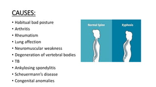
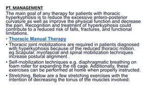
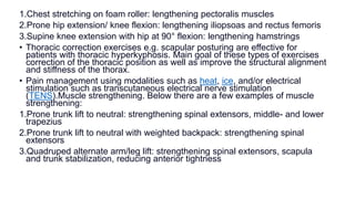
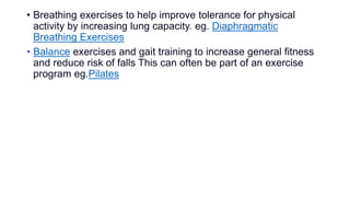
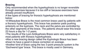
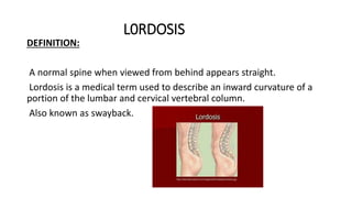
PRESENTATION ON
KYPHOSIS AND LORDOSIS
SUBMITTED BY- KANCHAN JASWAL(MPT ORTHO)
SUBMITTED TO- DR. ANIRBAN PATRA
DEFINITION:
Kyphosis is a spinal disorder in which an excessive outward curve of the
spine results in an abnormal rounding of the upper back.
The condition is sometimes known as “roundback” or in the case of severe
curve- as “hunchback”.
Kyphosis can occur at any age, but is common during adolescence.
Normal kyphosis angle vary between 20 and 40 degree in young public 48 to
50 degree in women and about 44 degree in men in older population.
TYPES of Kyphosis
There are several types of Kyphosis. The factors that contribute to kyphosis
vary depending on the type someone has.
1. Postural Kyphosis: This is the most common form of kyphosis. It often
starts in adolescence, when the muscles surrounding the spine develop
differently due to a postural imbalance, such as slouching. Slouching
increases the forward curvature, stretching the extensor muscles and
posterior ligaments of the spine, which weaken over time.
2. Scheuermann’s kyphosis: This also tends to develop during adolescence.
However, it can become more severe than postural kyphosis.
3. Age-related kyphosis: This type of kyphosis causes a curve in the
spine that progressively gets worse in older age, often as a result of
conditions that effect the bones in the spine. A common example is
osteoporosis, which causes the bones to lose density and become
weaker.
4. Congenital kyphosis: This occurs when the spine does not develop
properly before birth, causing kyphosis at birth. It can rapidly worsen
with age.
CAUSES:
• Habitual bad posture
• Arthritis
• Rheumatism
• Lung affection
• Neuromuscular weakness
• Degeneration of vertebral bodies
• TB
• Ankylosing spondylitis
• Scheuermann’s disease
• Congenital anomalies
Symptoms of kyphosis
The primary sign of kyphosis is a visible forward curve in the upper part of the spine. It
causes the upper back to appear curved over, with the shoulders rounded forward.
In mild cases, the excess spinal curve is not very noticeable. In other instances, a person
may look as if they are bending forwards.
Kyphosis often occurs without any other symptoms. However, other symptoms can include:
-back pain
-stiffness in the upper back
-a rounded back
-tight hamstrings
INVESTIGATION
-Radiography/ X-ray: The most common radiographs are upright
posteroanterior and lateral images of the entire spine.
-Magnetic resonance imaging: MRI can be a useful adjunct in planning
treatment for patients with kyphosis.
If a neurological abnormality is present, MRI may aid in localizing
impingement on neural structures.
TREATMENT
MEDICAL MANAGEMENT:
Nonsurgical management may involve:
- Observation
- Physical therapy
- Bracing
- Nonsteroidal anti-inflammatory drugs(NSAIDS): can help reduce any
pain caused by kyphosis.
PT. MANAGEMENT:
The main goal of any therapy for patients with thoracic
hyperkyphosis is to reduce the excessive antero-posterior
curvature as well as improve the physical function and decrease
the pain. Recognition and treatment of hyperkyphosis could
contribute to a reduced risk of falls, fractures, and functional
limitations.
• Thoracic Manual Therapy
• Thoracic joint mobilizations are required in patients diagnosed
with hyperkyphosis because of the reduced thoracic motion.
eg.Scapular, myofascial and spinal mobilization techniques
increase postural alignment .
• Self-mobilization techniques e.g. diaphragmatic breathing on
foam roller for expanding the rib cage. Additionally, these
exercises can be performed at home when properly instructed.
• Stretching. Below are a few stretching exercises with the
intention of decreasing the tonus of the muscles involved:
1.Chest stretching on foam roller: lengthening pectoralis muscles
2.Prone hip extension/ knee flexion: lengthening iliopsoas and rectus femoris
3.Supine knee extension with hip at 90° flexion: lengthening hamstrings
• Thoracic correction exercises e.g. scapular posturing are effective for
patients with thoracic hyperkyphosis. Main goal of these types of exercises
correction of the thoracic position as well as improve the structural alignment
and stiffness of the thorax.
• Pain management using modalities such as heat, ice, and/or electrical
stimulation such as transcutaneous electrical nerve stimulation
(TENS).Muscle strengthening. Below there are a few examples of muscle
strengthening:
1.Prone trunk lift to neutral: strengthening spinal extensors, middle- and lower
trapezius
2.Prone trunk lift to neutral with weighted backpack: strengthening spinal
extensors
3.Quadruped alternate arm/leg lift: strengthening spinal extensors, scapula
and trunk stabilization, reducing anterior tightness
• Breathing exercises to help improve tolerance for physical
activity by increasing lung capacity. eg. Diaphragmatic
Breathing Exercises
• Balance exercises and gait training to increase general fitness
and reduce risk of falls This can often be part of an exercise
program eg.Pilates
Bracing
Only recommended when the hyperkyphosis is no longer reversible
through exercises because it is too stiff or because exercises have
already proved insufficient.
A few types of bracing for thoracic hyperkyphosis are mentioned
below:
•A Milwaukee Brace is the most common brace used by patients with
a thoracic hyperkyphosis. This brace has posterior pads pushing
anteriorly on the kyphosis. The neck and the pelvis are controlled by
the other segments of the brace. The patients should wear this brace
23 hours a day for 1-2 years.
•The results of the Lyon Antikyphosis Brace were very satisfactory in
most patients with thoracic hyperkyphosis.
•Also a new bracing design called the Kyphologic Brace has been
shown to have a good in-brace correction on average.
•Another kind of brace using the two 3-point pressure system is the
‘Gschwend type’ brace. This brace is mostly used in Germany.
• Taping
• May also reduce kyphosis. Tape from the anterior aspect of the
acromioclavicular joint, over the muscle bulk of the upper
trapezius, and diagonally over the spinous process of T6.
• Spinal Orthosis
• A SpinoMed is a spinal orthosis which can be used for a
thoracic hyperkyphosis. There is some evidence found about
the use of this device. The patient should wear this for 2 hours a
day during 6 months. This will result in a decrease in kyphosis
angle, an improvement of standing height, an increase in spinal
extensor strength and a decreased postural sway.
L0RDOSIS
DEFINITION:
A normal spine when viewed from behind appears straight.
Lordosis is a medical term used to describe an inward curvature of a
portion of the lumbar and cervical vertebral column.
Also known as swayback.
TYPES
There are 5 primary types :
1. Postural Lordosis
This often comes from over- weight and lack of muscle conditioning in
the stomach and back muscles.
2. Congenital/ Traumatic Lordosis
A trauma/ injury to the connecting link of the spine can cause them to
break(fracture) causing pain in the spine.
3. Post- surgical Laminectomy Hyperlordosis
Laminectomy is a surgical procedure where part of the vertebra are
removed to give access to the spinal cord or nerve roots.
When this is done over several levels in spine, it can cause the spine to
be unstable and increase the normal curve of hyperlordotic position.
4. Neuromuscular Lordosis
This group include a large variety of conditions/ disorders that can lead
to many different types of spinal curvature problems.
5. Lordosis Secondary to Hip Flexion Contracture
This contracture can come from a variety of reasons including
infection, injury, or muscle imbalance issues from a several different
disorders.
ETIOLOGY
- Cause of lordosis has been linked to achondroplasia and
spondylolisthesis.
- Lordosis is also associated with poor posture, a congenital problem
with the vertebrae, neuromuscular problem, back surgery or a hip
problem.
- In children rickets may also leads to lordosis.
-However, lordosis can affect people of any age.
- Poor posture
- Obesity
- Osteoporosis (weakening of the bones with age)
- Spondylolisthesis (a condition in which one vertebra slip forward or
backward relative to the next vertebra)
- Achondroplasia (a form of dwarfism)
• CLINICAL FEATURES
Prominence of buttocks
Back pain
Pain down to legs
Diagnosis Evaluation
- X-rays: done to measure and evaluate curve.
- Bone scans: To evaluate degenerative or arthritic changes. Also helps
to find out bone tumors.
- MRI: To rule out associated problems.
- CT scan
- Blood investigations
- Early detection of Lordosis is important for proper and successful
treatment.
TREATMENT
Non surgical management: Most people don’t need any treatment. If
neck or back pain, they will probably only need over the counter
NSAIDs like aspirin or ibuprofen.
Surgical management:
Spinal fusion
Bone grafting
PHYSIOTHERAPY MANAGEMENT:
1.Pelvic Tilt: The patient lies on his back with knees bent, feet
flat on floor. Cue: Flatten the small of your back against the
floor, without pushing down with the legs, hold for 5 to 10
seconds.
2.Single Knee to chest: The patient lies on his back with knees
bent and feet flat on the floor. Cue: Slowly pull your right knee
toward your shoulder and hold 5 to 10 seconds. Lower the knee
and repeat with the other knee.
3.Double knee to chest: The patient begins as in the previous
exercise. Cue: Pulling right knee to chest, pull left knee to chest
and hold both knees for 5 to 10 seconds. Slowly lower one leg
at a time.
4.Partial sit-up: The patient has to do the pelvic tilt (exercise
1) and while holding this position Cue: Slowly curl your head
5. Hamstring stretch: The patient starts in long sitting with toes
directed toward the ceiling and knees fully extended. Cue: Slowly
lower the trunk forward over the legs, keeping knees extended,
arms outstretched over the legs, and eyes focus ahead.
6. Hip Flexor stretch: The patient places one foot in front of the
other with the left (front) knee flexed and the right (back) knee
held rigidly straight. Cue: Flex forward through the trunk until the
left knee contacts the axillary fold (armpit region). Repeat with
right leg forward and left leg back.
7.Squat: The patient stands with both feet parallel, about
shoulder’s width apart. Attempting to maintain the trunk as
perpendicular as possible to the floor, eyes focused ahead, and
feet flat on the floor. Cue: Slowly lower the body by flexing the
knees.
Stretching Exercises
• With a crossed leg syndrome type of problem, it’s beneficial to
stretch the tightened muscles. For improving the mobility, the
patient could perform stretching of the hamstrings, hip flexors,
and lumbar paraspinal muscles over 15 seconds. This will
improve the active and the passive ROM in the lower extremity.
• Hold-relax stretching of the iliopsoas: It can reduce back pain,
excessive lumbar lordosis angle, lengthen the iliopsoas and
increase transversus abdominis activation capacity. The target
hip is moved toward the floor until the patient feels a mild
stretch sensation.
Then the patient must perform a submaximal voluntary isometric
contraction of the M. Iliopsoas for 10 seconds and then
completely relax for 10 seconds. The participant’s leg is now
slowly moved to a new range until a mild stretching sensation is
felt and described by the patient. This position is then held for 20
seconds. This is repeated 5 times, followed by a 1 min rest, for
15 minutes.
Stretching technique for improving the ROM of the M.Iliopsoas
and the M. Rectus femoris: The patient lies in Thomas position,
the not stretched leg is maximally flexed to stabilize the pelvis
and flatten the lumbar spine. The other leg is in a normal flexed
position because of the tightness of the M. Iliopsoas. It’s this leg
that needs to be pushed against the table. If you want to stretch
the M. rectus femoris, bend the knee more than 90°, while
performing the same stretch.

KYPHOSIS-1.pptx biomechanics. ...........

  • 1.
    PRESENTATION ON KYPHOSIS ANDLORDOSIS SUBMITTED BY- KANCHAN JASWAL(MPT ORTHO) SUBMITTED TO- DR. ANIRBAN PATRA
  • 2.
    DEFINITION: Kyphosis is aspinal disorder in which an excessive outward curve of the spine results in an abnormal rounding of the upper back. The condition is sometimes known as “roundback” or in the case of severe curve- as “hunchback”. Kyphosis can occur at any age, but is common during adolescence. Normal kyphosis angle vary between 20 and 40 degree in young public 48 to 50 degree in women and about 44 degree in men in older population.
  • 3.
    TYPES of Kyphosis Thereare several types of Kyphosis. The factors that contribute to kyphosis vary depending on the type someone has. 1. Postural Kyphosis: This is the most common form of kyphosis. It often starts in adolescence, when the muscles surrounding the spine develop differently due to a postural imbalance, such as slouching. Slouching increases the forward curvature, stretching the extensor muscles and posterior ligaments of the spine, which weaken over time. 2. Scheuermann’s kyphosis: This also tends to develop during adolescence. However, it can become more severe than postural kyphosis.
  • 4.
    3. Age-related kyphosis:This type of kyphosis causes a curve in the spine that progressively gets worse in older age, often as a result of conditions that effect the bones in the spine. A common example is osteoporosis, which causes the bones to lose density and become weaker. 4. Congenital kyphosis: This occurs when the spine does not develop properly before birth, causing kyphosis at birth. It can rapidly worsen with age.
  • 5.
    CAUSES: • Habitual badposture • Arthritis • Rheumatism • Lung affection • Neuromuscular weakness • Degeneration of vertebral bodies • TB • Ankylosing spondylitis • Scheuermann’s disease • Congenital anomalies
  • 6.
    Symptoms of kyphosis Theprimary sign of kyphosis is a visible forward curve in the upper part of the spine. It causes the upper back to appear curved over, with the shoulders rounded forward. In mild cases, the excess spinal curve is not very noticeable. In other instances, a person may look as if they are bending forwards. Kyphosis often occurs without any other symptoms. However, other symptoms can include: -back pain -stiffness in the upper back -a rounded back -tight hamstrings
  • 7.
    INVESTIGATION -Radiography/ X-ray: Themost common radiographs are upright posteroanterior and lateral images of the entire spine. -Magnetic resonance imaging: MRI can be a useful adjunct in planning treatment for patients with kyphosis. If a neurological abnormality is present, MRI may aid in localizing impingement on neural structures.
  • 8.
    TREATMENT MEDICAL MANAGEMENT: Nonsurgical managementmay involve: - Observation - Physical therapy - Bracing - Nonsteroidal anti-inflammatory drugs(NSAIDS): can help reduce any pain caused by kyphosis.
  • 9.
    PT. MANAGEMENT: The maingoal of any therapy for patients with thoracic hyperkyphosis is to reduce the excessive antero-posterior curvature as well as improve the physical function and decrease the pain. Recognition and treatment of hyperkyphosis could contribute to a reduced risk of falls, fractures, and functional limitations. • Thoracic Manual Therapy • Thoracic joint mobilizations are required in patients diagnosed with hyperkyphosis because of the reduced thoracic motion. eg.Scapular, myofascial and spinal mobilization techniques increase postural alignment . • Self-mobilization techniques e.g. diaphragmatic breathing on foam roller for expanding the rib cage. Additionally, these exercises can be performed at home when properly instructed. • Stretching. Below are a few stretching exercises with the intention of decreasing the tonus of the muscles involved:
  • 10.
    1.Chest stretching onfoam roller: lengthening pectoralis muscles 2.Prone hip extension/ knee flexion: lengthening iliopsoas and rectus femoris 3.Supine knee extension with hip at 90° flexion: lengthening hamstrings • Thoracic correction exercises e.g. scapular posturing are effective for patients with thoracic hyperkyphosis. Main goal of these types of exercises correction of the thoracic position as well as improve the structural alignment and stiffness of the thorax. • Pain management using modalities such as heat, ice, and/or electrical stimulation such as transcutaneous electrical nerve stimulation (TENS).Muscle strengthening. Below there are a few examples of muscle strengthening: 1.Prone trunk lift to neutral: strengthening spinal extensors, middle- and lower trapezius 2.Prone trunk lift to neutral with weighted backpack: strengthening spinal extensors 3.Quadruped alternate arm/leg lift: strengthening spinal extensors, scapula and trunk stabilization, reducing anterior tightness
  • 11.
    • Breathing exercisesto help improve tolerance for physical activity by increasing lung capacity. eg. Diaphragmatic Breathing Exercises • Balance exercises and gait training to increase general fitness and reduce risk of falls This can often be part of an exercise program eg.Pilates
  • 12.
    Bracing Only recommended whenthe hyperkyphosis is no longer reversible through exercises because it is too stiff or because exercises have already proved insufficient. A few types of bracing for thoracic hyperkyphosis are mentioned below: •A Milwaukee Brace is the most common brace used by patients with a thoracic hyperkyphosis. This brace has posterior pads pushing anteriorly on the kyphosis. The neck and the pelvis are controlled by the other segments of the brace. The patients should wear this brace 23 hours a day for 1-2 years. •The results of the Lyon Antikyphosis Brace were very satisfactory in most patients with thoracic hyperkyphosis. •Also a new bracing design called the Kyphologic Brace has been shown to have a good in-brace correction on average. •Another kind of brace using the two 3-point pressure system is the ‘Gschwend type’ brace. This brace is mostly used in Germany.
  • 13.
    • Taping • Mayalso reduce kyphosis. Tape from the anterior aspect of the acromioclavicular joint, over the muscle bulk of the upper trapezius, and diagonally over the spinous process of T6.
  • 14.
    • Spinal Orthosis •A SpinoMed is a spinal orthosis which can be used for a thoracic hyperkyphosis. There is some evidence found about the use of this device. The patient should wear this for 2 hours a day during 6 months. This will result in a decrease in kyphosis angle, an improvement of standing height, an increase in spinal extensor strength and a decreased postural sway.
  • 15.
    L0RDOSIS DEFINITION: A normal spinewhen viewed from behind appears straight. Lordosis is a medical term used to describe an inward curvature of a portion of the lumbar and cervical vertebral column. Also known as swayback.
  • 16.
    TYPES There are 5primary types : 1. Postural Lordosis This often comes from over- weight and lack of muscle conditioning in the stomach and back muscles. 2. Congenital/ Traumatic Lordosis A trauma/ injury to the connecting link of the spine can cause them to break(fracture) causing pain in the spine. 3. Post- surgical Laminectomy Hyperlordosis Laminectomy is a surgical procedure where part of the vertebra are removed to give access to the spinal cord or nerve roots.
  • 17.
    When this isdone over several levels in spine, it can cause the spine to be unstable and increase the normal curve of hyperlordotic position. 4. Neuromuscular Lordosis This group include a large variety of conditions/ disorders that can lead to many different types of spinal curvature problems. 5. Lordosis Secondary to Hip Flexion Contracture This contracture can come from a variety of reasons including infection, injury, or muscle imbalance issues from a several different disorders.
  • 18.
    ETIOLOGY - Cause oflordosis has been linked to achondroplasia and spondylolisthesis. - Lordosis is also associated with poor posture, a congenital problem with the vertebrae, neuromuscular problem, back surgery or a hip problem. - In children rickets may also leads to lordosis. -However, lordosis can affect people of any age.
  • 19.
    - Poor posture -Obesity - Osteoporosis (weakening of the bones with age) - Spondylolisthesis (a condition in which one vertebra slip forward or backward relative to the next vertebra) - Achondroplasia (a form of dwarfism) • CLINICAL FEATURES Prominence of buttocks Back pain Pain down to legs
  • 20.
    Diagnosis Evaluation - X-rays:done to measure and evaluate curve. - Bone scans: To evaluate degenerative or arthritic changes. Also helps to find out bone tumors. - MRI: To rule out associated problems. - CT scan - Blood investigations - Early detection of Lordosis is important for proper and successful treatment.
  • 21.
    TREATMENT Non surgical management:Most people don’t need any treatment. If neck or back pain, they will probably only need over the counter NSAIDs like aspirin or ibuprofen. Surgical management: Spinal fusion Bone grafting
  • 22.
    PHYSIOTHERAPY MANAGEMENT: 1.Pelvic Tilt:The patient lies on his back with knees bent, feet flat on floor. Cue: Flatten the small of your back against the floor, without pushing down with the legs, hold for 5 to 10 seconds. 2.Single Knee to chest: The patient lies on his back with knees bent and feet flat on the floor. Cue: Slowly pull your right knee toward your shoulder and hold 5 to 10 seconds. Lower the knee and repeat with the other knee. 3.Double knee to chest: The patient begins as in the previous exercise. Cue: Pulling right knee to chest, pull left knee to chest and hold both knees for 5 to 10 seconds. Slowly lower one leg at a time. 4.Partial sit-up: The patient has to do the pelvic tilt (exercise 1) and while holding this position Cue: Slowly curl your head
  • 23.
    5. Hamstring stretch:The patient starts in long sitting with toes directed toward the ceiling and knees fully extended. Cue: Slowly lower the trunk forward over the legs, keeping knees extended, arms outstretched over the legs, and eyes focus ahead. 6. Hip Flexor stretch: The patient places one foot in front of the other with the left (front) knee flexed and the right (back) knee held rigidly straight. Cue: Flex forward through the trunk until the left knee contacts the axillary fold (armpit region). Repeat with right leg forward and left leg back. 7.Squat: The patient stands with both feet parallel, about shoulder’s width apart. Attempting to maintain the trunk as perpendicular as possible to the floor, eyes focused ahead, and feet flat on the floor. Cue: Slowly lower the body by flexing the knees.
  • 24.
    Stretching Exercises • Witha crossed leg syndrome type of problem, it’s beneficial to stretch the tightened muscles. For improving the mobility, the patient could perform stretching of the hamstrings, hip flexors, and lumbar paraspinal muscles over 15 seconds. This will improve the active and the passive ROM in the lower extremity. • Hold-relax stretching of the iliopsoas: It can reduce back pain, excessive lumbar lordosis angle, lengthen the iliopsoas and increase transversus abdominis activation capacity. The target hip is moved toward the floor until the patient feels a mild stretch sensation.
  • 25.
    Then the patientmust perform a submaximal voluntary isometric contraction of the M. Iliopsoas for 10 seconds and then completely relax for 10 seconds. The participant’s leg is now slowly moved to a new range until a mild stretching sensation is felt and described by the patient. This position is then held for 20 seconds. This is repeated 5 times, followed by a 1 min rest, for 15 minutes. Stretching technique for improving the ROM of the M.Iliopsoas and the M. Rectus femoris: The patient lies in Thomas position, the not stretched leg is maximally flexed to stabilize the pelvis and flatten the lumbar spine. The other leg is in a normal flexed position because of the tightness of the M. Iliopsoas. It’s this leg that needs to be pushed against the table. If you want to stretch the M. rectus femoris, bend the knee more than 90°, while performing the same stretch.