Duchenne muscular dystrophy is a genetic disorder characterized by progressive muscle weakness. It is caused by mutations in the gene encoding dystrophin, and mainly affects boys. Clinical features include difficulty walking, calf pseudohypertrophy, loss of ambulation by age 12, wheelchair dependence, scoliosis, and death often by age 18 from respiratory or cardiac failure. Diagnosis involves elevated creatine kinase levels, muscle biopsy showing dystrophin deficiency, and genetic testing. There is no cure, but management focuses on maintaining mobility and function.
This document discusses cervical spondylosis and lumbar spondylosis. Cervical spondylosis is caused by osteoarthritis in the cervical spine and can cause cervical radiculopathy or myelopathy. Cervical radiculopathy involves pain radiating from the neck due to nerve root compression, while cervical myelopathy involves spinal cord compression. Lumbar spondylosis is degeneration of the lumbar spine that can cause lumbar disc herniation or lumbar canal stenosis. Lumbar disc herniation involves disc material pressing on a nerve root, while lumbar canal stenosis is narrowing of the spinal canal that causes leg pain with exercise. Management of these conditions may involve conservative treatment, surgery, or manual manipulation of
Ankylosing spondylitis is a type of inflammatory arthritis associated with the HLA-B27 gene. It typically causes stiffness and fusion of the spine over time. Diagnosis involves evidence of sacroiliac joint inflammation on imaging and a positive HLA-B27 test in most cases. Treatment focuses on exercises to maintain mobility, nonsteroidal anti-inflammatory drugs, and TNF inhibitors for severe cases. Surgery may be needed to correct spinal deformities or replace affected hips in advanced ankylosing spondylitis.
1. Spinal tuberculosis commonly affects the thoracic and lumbar spine in young adults. It can cause neurological deficits through mechanisms such as inflammatory edema, extradural masses, and meningeal involvement.
2. Diagnosis is based on imaging findings on X-ray, CT, or MRI showing bone destruction and abscesses. Treatment involves chemotherapy and sometimes surgery to debride tissue, drain abscesses, or correct deformities.
3. Complications of spinal tuberculosis include paraplegia, cold abscesses, spinal deformities, and recurrence which may require longer treatment or surgical intervention.
The document discusses herniation of the intervertebral disk, including its risk factors, clinical manifestations, diagnostic evaluations, medical and surgical management, complications, and nursing care. It provides information on disk herniation in the cervical and lumbar spine. Nursing plays an important role in assessing and caring for patients with disk herniations through providing treatment, education, and support.
Skeletal dysplasias and dwarfism can be caused by over 200 genetic disorders that result in abnormal bone growth. There are two main types: dysplasias with generalized skeletal abnormalities and dysostoses with abnormalities of single or multiple bones. Achondroplasia, the most common type of dwarfism, is caused by a mutation that slows endochondral bone growth. It is characterized by frontal bossing, midface hypoplasia, rhizomelic shortening of the limbs, trident hands, genu varum, and normal intelligence. Assessment involves examining for disproportion, general examination including radiographs to evaluate for complications like foramen magnum stenosis. Treatment focuses on managing complications through surgery or other
Potts spine is the classical destruction of disc space and the adjacent bodies , destruction of other spinal elements,severe progressive kyphosis subsequently
Also know as spinal tuberculosis
This document discusses vertebral fractures and spinal cord injuries. It begins by describing the anatomy of the vertebral column and typical vertebrae. It then discusses different types of lumbar vertebral fractures including wedge compression fractures, burst fractures, flexion-distraction injuries, and fracture-dislocations. Emergency management of spinal injuries is outlined including immobilization techniques. Spinal cord injuries are also summarized, covering topics like pathophysiology, classifications, consequences, and specific syndromes like central cord syndrome. Acute phase conditions like spinal shock and neurogenic shock are defined.
Whiplash is an injury to the neck caused by sudden acceleration-deceleration movements like those seen in car accidents. It can cause neck pain and stiffness as well as headaches. While imaging may appear normal, whiplash can still cause soft tissue damage to neck muscles, ligaments, and discs. Symptoms can last over a year in many cases. Treatment focuses on pain relief, muscle relaxation, and in severe cases surgery may be needed. Prognosis is often worse for those with multiple injuries, females, and older individuals.
This document discusses spondylolisthesis, including:
- It defines spondylolisthesis as the forward slippage of one vertebra on its adjacent caudal segment.
- Several classification systems are described including the Wiltse, Newman and Macnab classification based on etiology and topography.
- Risk factors for slip progression include developmental spondylolisthesis with lysis, isthmic spondylolisthesis from repetitive loading, and increased pelvic tilt.
- Treatment involves conservative care with bracing or exercise initially. Surgical options include direct repair of pars defect, decompression with or without fusion and instrumentation depending on the grade of slippage.
This document provides information on examinations for neurological disorders. It discusses investigations like imaging scans and spinal taps. It lists risk factors like age, hypertension, and smoking. It describes different causes of neurological issues like stroke, tumors, and encephalitis. It then details various parts of a neurological exam including Kernig's sign, reflexes, and deep tendon reflexes of the biceps, triceps, supinator, knees, and ankles.
Diffuse idiopathic skeletal hyperostosis (DISH) is a common skeletal process of uncertain etiology found in 12 to 18% of Indian populations above 50 years. The primary manifestations of DISH are calcification and ossification of the spinal ligaments, as well as entheseal ossification within extraspinal sites
Muscular dystrophy is a genetic disease that causes the muscles to weaken over time. There are several types but Duchenne muscular dystrophy is the most common and severe, affecting boys. It is caused by an absence of dystrophin protein which leads to muscle cell damage. Symptoms start in early childhood and include difficulty walking, joint contractures, and loss of ambulation in the teen years. Management is multidisciplinary and focuses on maintaining mobility and function as long as possible, treating complications, and palliative care as the condition progresses. Life expectancy for Duchenne patients is usually early 20s.
This document defines kyphosis as an excessive backward curvature of the spine localized to the thoracic spine, known as a "rounded back" posture. It can be caused by conditions like arthritis, tuberculosis, or postural habits. Kyphosis is classified by severity from first to third degree based on effects like muscle imbalance or vertebral wedging. More severe kyphosis can compress spinal structures and restrict breathing. Rehabilitation focuses on stretching, strengthening, bracing, and manual mobilization to reduce the curvature and its impacts. Studies show manual mobilization and techniques applying passive transverse forces can help attenuate thoracic kyphosis, especially in elderly patients with osteoporosis.
Lumbar spinal stenosis is a narrowing of the spinal canal that causes compression of the nerves and blood vessels in the lower back. It most commonly presents with leg pain that worsens with standing and walking due to nerve compression. Physical examination may reveal reduced range of motion, reproduction of symptoms with extension, and occasionally motor or sensory deficits. MRI is the preferred imaging method to visualize the soft tissue compression causing the stenosis. Treatment involves conservative measures but may require surgery if conservative options fail.
- Lumbar spinal stenosis involves a narrowing of the spinal canal which causes compression of the nerves and blood vessels in the lower back. It is a common condition in aging individuals that results from degenerative changes in the spine over time.
- Patients with lumbar spinal stenosis often present with leg pain that takes the form of neurogenic claudication or radicular pain, as well as low back pain. Symptoms are typically exacerbated by standing and walking and relieved by sitting or flexion of the spine.
- MRI is the preferred imaging modality for evaluating lumbar spinal stenosis. While findings on imaging are common in asymptomatic older adults, clinical correlation is important. Conservative treatment is usually tried initially and surgery is considered if symptoms
Spinal Cord Disorders
Definition:-
Spinal Cord Injury(SCI) is an injury to the Spinal Cord that results in temporary or permanent changes in the spinal cords Normal motor sensory or autonomic function.
In most Spinal Cord Injuries, the balance ligaments or disc material pinch the cord, causing it to become bruised or swollen.
1. Incidence
2. Etiology
3. Pathophysiology of SCI
4. Clinical Manifestation
5. Diagnosis
6. Management
7. Nursing Process
8. Nursing Diagnosis
9. Nursing Interventions
Spinal Bifida
Spinal Bifida is a birth defect that occurs when the spinal cord doesn’t form properly.
It is the type of neural tube defect.
The neural tube is the structure in a developing embryo that eventually becomes the body’s Brain, Spinal cord & the tissue that encloses them.
1. types
2. Causes
3. Symptoms
4. Complications
REFERENCES:-
1. Brunner & Siddarth's,
Textbook of Medical-Surgical Nursing,
The document discusses post-polio residual paralysis, including:
1. It provides historical context on polio and describes the etymology and pathology of the virus.
2. Poliovirus attacks the anterior horn cells of the spinal cord, causing flaccid paralysis. This can lead to progressive muscle imbalance and deformities over time if left untreated.
3. Management focuses on strengthening unaffected muscles, stretching shortened muscles, exercises to maintain range of motion, orthotics and bracing, and surgery to correct severe deformities. The goal is maximizing recovery and function.
This document provides an overview of spinal stenosis. It defines spinal stenosis as abnormal narrowing of the central canal, lateral recess, or intervertebral foramina that compromises neural elements. The document discusses the pathogenesis, classification, types, anatomy, pathology, natural history, clinical features, diagnosis, and treatment options for spinal stenosis. Treatment options include non-operative approaches like rest, pain management, and epidural steroid injections, as well as operative procedures. Imaging plays an important role in diagnosis and includes X-rays, CT, MRI, and CT myelography.
1) Lumbar disk disease is a common cause of back pain and occurs most often at the L4-L5 and L5-S1 levels. A ruptured disk can cause back pain, limited movement, radicular pain, or neurological deficits.
2) Examination may reveal limited flexion, muscle spasms, or positive straight leg raise test. Imaging like MRI or CT can identify disk herniations.
3) Other potential causes of back pain include muscle strains, osteoporotic fractures, tumors, infections, and referred pain from abdominal or pelvic organs. A thorough examination is needed to determine the cause and guide treatment.
The document discusses various types of spine infections including pyogenic osteomyelitis, discitis, fungal spine infections, and parasitic infections. It covers the definition, etiology, risk factors, clinical features, investigations, and management for each type. Key points include that pyogenic osteomyelitis most commonly affects the elderly or immunocompromised, discitis usually results from invasive procedures, fungal infections occur in immunocompromised patients, and echinococcus granulosus is a common parasitic infection in sheep-raising areas. Treatment involves antibiotics, surgery if needed, and addressing any underlying risks.
This document discusses slipped capital femoral epiphysis (SCFE), a condition where the femoral head slips posteriorly and inferiorly through the growth plate. Key points:
- It typically affects obese adolescents age 10-14 and is more common in males.
- Risk factors include obesity, hormonal issues, and genetic factors.
- Radiographs can detect the slip and grade its severity.
- Treatment involves immediate non-weight bearing, and may include screw fixation, osteotomies, or epiphysiodesis with bone grafting to prevent further slippage.
- Complications can include avascular necrosis, chondrolysis, and residual deformity/osteoarthritis if
The document discusses the clinical evaluation of patients with spine disorders. A thorough history and physical examination is important to identify potential pain generators and prognostic factors. The physical exam includes assessment of alignment, gait, palpation, neurological function, motor strength, reflexes, and special tests. Differential diagnoses include musculoskeletal, radicular, and spinal cord issues. A biopsychosocial approach is emphasized to understand how anatomical, psychological, and social factors all contribute to a patient's pain experience and prognosis.
Histololgy of Female Reproductive System.pptxAyeshaZaid1
Dive into an in-depth exploration of the histological structure of female reproductive system with this comprehensive lecture. Presented by Dr. Ayesha Irfan, Assistant Professor of Anatomy, this presentation covers the Gross anatomy and functional histology of the female reproductive organs. Ideal for students, educators, and anyone interested in medical science, this lecture provides clear explanations, detailed diagrams, and valuable insights into female reproductive system. Enhance your knowledge and understanding of this essential aspect of human biology.
- Video recording of this lecture in English language: https://youtu.be/kqbnxVAZs-0
- Video recording of this lecture in Arabic language: https://youtu.be/SINlygW1Mpc
- Link to download the book free: https://nephrotube.blogspot.com/p/nephrotube-nephrology-books.html
- Link to NephroTube website: www.NephroTube.com
- Link to NephroTube social media accounts: https://nephrotube.blogspot.com/p/join-nephrotube-on-social-media.html
Osteoporosis - Definition , Evaluation and Management .pdfJim Jacob Roy
Osteoporosis is an increasing cause of morbidity among the elderly.
In this document , a brief outline of osteoporosis is given , including the risk factors of osteoporosis fractures , the indications for testing bone mineral density and the management of osteoporosis
Muktapishti is a traditional Ayurvedic preparation made from Shoditha Mukta (Purified Pearl), is believed to help regulate thyroid function and reduce symptoms of hyperthyroidism due to its cooling and balancing properties. Clinical evidence on its efficacy remains limited, necessitating further research to validate its therapeutic benefits.
share - Lions, tigers, AI and health misinformation, oh my!.pptxTina Purnat
• Pitfalls and pivots needed to use AI effectively in public health
• Evidence-based strategies to address health misinformation effectively
• Building trust with communities online and offline
• Equipping health professionals to address questions, concerns and health misinformation
• Assessing risk and mitigating harm from adverse health narratives in communities, health workforce and health system
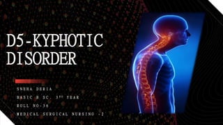
1. S N E H A D E R I A
B A S I C B S C . 3 R D Y E A R
R O L L N O - 3 6
M E D I C A L S U R G I C A L N U R S I N G - 2
D5-KYPHOTIC
DISORDER
2. • Kyphosis is a deformity of the spine as an increased roundness of the thoracic curve. It occurs
due to exaggeration or angulation of the normal posterior curve with convexity background and
forward curvatures of the shoulders.
• It may develop due to defective posture , rickets, congenital anomaly, diseased skin ( SYPHILIS,
TUBERCULOSIS ) malignancy , rheumatic arthritis , compression fractures or due to idiopathic
cause as in SCHEUREMANN disease.
• Management is performed by orthotic devices or orthopaedic after clinical and radiological
evaluation.
INTRODUCTION
6. CLASSIFICATION
There are three main types of KYPHOSIS
1. POSTURAL KYPHOSIS
2. SCHEUEMANN’S KYPHOSIS
3. CONGENITAL KYPHOSIS.
7. POSTURAL KYPHOSIS
• It is most common type of kyphosis.
• Most common in girls than in boys, which is
typically noticed in adolescent age.
• It is caused by poor posture and weakening
of the muscle , ligaments in the back.
• The vertebrae are typically placed in the
postural kyphosis.
• It progressively gets worse with time.
• These people will have symptoms of pain and
muscle failure.
8. SCHEUERMANN’S KYPHOSIS
• It is also first noticed during adolescent
period.
• It is result of structural deformity.
• It commonly develops with scoliosis.
• Reasons are not well understood.
9. CONGENITAL KYPHOSIS
• It is least common type of kyphosis .
• It is caused by abnormal development of
vertebrae prior to birth.
• It occurs due to fusion of many vertebrae
together.
11. RELATED ANATOMY AND PHYSIOLOGY
The spinal cord is a part of CNS, which lies within
the vertebral column.it begins as a continuation od
the medulla oblongata. It extends from the level of
FORAMEN MAGNUM to the lower border of L1
vertebra . It is approximately 45 cm long. In the
adult , it occupies only 2/3rd of the vertebral column.
12. External features -
• Spinal cord is a cylindrical structures that is
slightly flattened anteriorly and posteriorly.
• The cervical enlargements – the cervical spinal
nerves arising from here a plexus or network
called brachial plexus , which innervates the
muscles of the upper limb.
• The lumbosacral enlargements – innervates the
muscle of lower limb.
• The lowest part of spinal cord is conical and is
known as the CONUS MEDULLARIS . The conus
is a continuous below with a thin Filous cord ,
the FILUM TERMINALE.
13. COVERING OF SPINAL CORD
• The spinal cord is loosely surrounded by the three meninges as discussed – outer dura mater,
middle arachnoid and inner pia mater , which are continuous with the meninges of the brain.
14. Internal structures of spinal cord
• Transverse section of spinal cord shows that it is
compassed of grey and white mater. Gray mater
is placed centrally and white mater is placed
periphery. Gray mater is composed of nerve cells,
neuroglia and blood vessels and white mater is
composed of myelinated and non myelinated
nerve fibres.
16. Tracts of spinal cord
They are constituted by a group of sensory fibres ascending from the receptors to the central nervous
system . They include
ascending tracts
• Dorsal column
• Spinothalamic tracts
• Spinocerebellar tracts
• Spino olivary tracts
descending tracts
• Corticospinal tracts
• Extrapyramidal tracts
17. Spinal segments-
• The spinal cord gives attachment on the both
sides to a series of spinal nerves. the part of
the spinal cord giving origin to one pair of
spinal nerve called spinal segment. Spinal
cord is made up of spinal segments – 8
cervical , 12 thoracic, 5 lumbar , 5 sacral and
1 coccygeal.
19. Blood supply -
The anterior supply of the cord derived
from following –
• anterior spinal artery
• Two posterior spinal arteries
• The radicular arteries.
20. ETIOLOGY
• Congenital abnormalities
• Improper vertebral development
• Certain endocrine disorder
• Connective tissue disorder
• Infection
• Muscular dystrophy
• Poliomyelitis
• tumours
ACCORDING TO BOOK
• Congenital abnormalities from birth
( no genetic history of neuro problems)
ACCORDING TO PATIENT
21. RISK FACTORS OF KYPHOSIS
• Arthritis
• Cancerous tumors and cancer treatments
• Chronic disorders such as PAGET’S disease
• Genetic disease
• Injury to spine
• Infectious disease such as tuberculosis or polio
• Osteoporosis
• Spondyolothisthesis
23. CLINICAL MANIFESTATION
ACCORDING TO BOOK ACCORDING TO PATIENT
• Difficulty in breathing ( only in severe
cases)
• Fatigue
• Mild back pain
• Rounded back appearance
• Tenderness and stiffness of the spine
• Difficulty during walking
• Back pain
• Fatigue
• Tremor
• Muscle strain
• Difficulty in lower limb movement
25. INVESTIGATION
ACCORDING TO BOOK ACCORDING TO PATIENT
• History and physical
examination
• X- rays
• CT scan
• MRI
• MRI( MAGNETIC RESONANCE IMAGING)-
1. DATE- 25/01/2021
2. IMPRESSION- partial hypoplasia D5 vertebrae is seen with
anterior wedging likely developmental ?
kyphotic tilt of dorsal spine is seen. Indentation seen on dorsal
cord at D5 level which showing mild past compressive signal
changes .
• CYTOPATHOLOGY TEST -
1. DATE- 25/01/2021
2. IMPRESSION- CT guided FNAC from D5 vertebral lesion for
cytology. No acid fast bacilli is seen. No equivocal epithelioid
cell granuloma is seen
26. Contd..
ACCORDING TO PATIENT
• ELECTROCARDIOGRAPH TEST -
1. DATE – 23/01/2021
2. LVEF – 67%
3. LVFS- 60%
• C T SCAN -
1. DATE – 21/02/21
2. CT scan of dorso lumbar spine show– vertebral segmentation anomaly with hemivertebrae at D5
level causing acute kyphotic deformity at same level.
• COVID 19 TEST
1. DATE – 20/03/21
2. RAPID ANTIGEN ICT FOR COVID 19 - NAGETIVE
• GLASGO COMA SCALE – E4V5M6
28. MANAGEMENT
ACCORDING TO BOOK ACCORDING TO PATIENT
A. NON SURGICAL MANAGEMENT –
An initial programme of conservative treatment
that includes exercise and anti inflammatory
medications is recommended for patient with
SCHEUREMANN’S kyphosis. If the patient is still
growing, the doctor may prescribe a brace until
skeletal maturity is reached.
B. SURGICAL TREATMENT-
Surgery may be recommended if the kyphotic
curve exceeds 7.5 ,
A. MEDICAL MANAGEMENT-
1. Comfort devices are recommended
2. Medications –
Tab PCM 650 mg – BD
Tab Pan – 40 mg-ODAC
Tab Methyl cobalamin – 500
mg-OD
Tab non- Tryptiline -100 mg -OD
B. SURGICAL MANAGEMENT--
30. Contd..
• GOALS OF SURGERIES—
to reduce deformity
Reduce pain and neurological symptoms
To maintain the improvement over time.
NAME OF SURGERIES—
OSTEOTOMY
SPINAL INSTRUEMENTATION AND FUSION
BALLOON KYPHOPLASTY– A SPECIAL ORTHOPAEDIC BALLOON IS INSERTED INTO THE
COMPRESSED ( COLLAPSED) VERTEVRA.
31. COMPLICATIONS
ACCORDING TO BOOK ACCORDING TO PATIENT
• DECREASED LUNG CAPACITY
• DISABELING PAIN
• NEUROLOGICAL SYMPTOMS
INCLUDING LEG WEAKENING OR
PARALYSIIS
• ROUND BACK INFORMITY
• DIFFICULTIES IN WALKING
• TREMOR IN HANDS
• PAIN IN BACK
32. PROGRESS NOTE
CRITERIA DAY 1 DAY 2 DAY 3
TEMPERATURE (℉) 98.1 97.9 97.8
PULSE (b/m) 86 84 84
RESPIRATION RATE
(br/m)
22 20 24
BLOOD PRESSURE (
mm hg)
130/70 140/70 126/82
PAIN IN BACK
(pain scale)
present present Present
DIETARY INPUT balanced balanced Balanced
OUTPUT balanced balanced balanced
33. PROGNOSIS
• The progress rate of the patient is initially good. He is planned for surgery. So his pre operative
prognosis is good.
34. NURSING MANAGEMENT
• NURSING DIAGNOSIS 1 –
Impaired physical mobility related to neuro muscular impairment as evidenced by patient’s
verbalization.
• Goal –
To reduce impairment during physical mobility
• Intervention –
1. Continuously assessment of motor function of the patient should be done.
2. He should be assisted with full range of motion exercises in all extremities and joints using slow
movements.
3. Comfortable position should be given.
4. Lower extremities should be elevated in intervals in chair or raise foot or bed when permitted in
individual situation.
5. Neck collar should be provided if prescribed
35. Nursing diagnosis – 2
Pain at upper back related to spinal anomaly as evidenced by patients' verbalization
Goal –
To reduce pain
Interventions-
1. Degree of pain should be assessed by pain scale
2. Comfortable position should be given
3. Analgesic medication should be given as prescribed
4. Relaxation techniques should be provided as per patient’s comfort.2
36. Nursing diagnosis -3
Impaired skin integrity related to physical immobility as evidenced by patient’s verbalization and physical
examination.
Goal –
To maintain skin hygiene
Interventions-
1. Skin should be inspected
2. Pressure points should be assessed
3. Skin care should be given
4. Comfortable position change should be done at regular interval.
37. Nursing diagnosis -4
Anxiety related to physical immobility and lack of knowledge regarding anatomical deformities as evidenced by
patient’s facial expression and verbalization.
Goal-
To reduce anxiety
Interventions-
1. Psychological support should be given.
2. Orientation and good therapeutic relationship should be established.
3. Health talk and knowledge regarding KYPHOSIS should be given.
4. Spiritual support should be given.
5. Relaxation should be provided by different techniques like music , calm environment , muscle relaxation
technique etc.
Day 2
38. Nursing diagnosis – 5
Disturbed sleeping pattern related to physical discomfort as evidenced by patient’s verbalization
Goal-
To maintain normal sleeping pattern
Interventions-
1. Provide measures to take before bed time to assist in sleep .
2. Keep environment quiet and comfortable
3. Encourage minimum activities within limits during day time.
4. Teach relaxation techniques
5. Encourage patient to avoid coffee and caffeinated food and liquids
6. Try to maintain patient’s regular sleeping time and pattern.
7. Give mental support to the patient.
39. Nursing diagnosis 6
Risk for injury related to physical immobility
Goal-
To reduce risk of injury
Interventions-
1. Proper orientation of the ward should be done
2. Precautions like side rails, padded bed, low position of bed should be done
3. Provide adequate light
4. Avoid sedative drugs to avoid medications induces confusion
5. Remove any obstacles in floor, proper dry floor should be maintained
6. Calling bell should be provided at bed side for emergency
7. Reduce unnecessary movement of patient
40. Nursing diagnosis 7-
Self care deficit due to physical limitations as evidenced by physical examination
Goal-
To maintain self hygiene
Interventions-
1. Encourage patient to perform self car to maximum of ability to promote sense of self independency
2. Encourage patient in planning schedule of daily activities
3. We should anticipate hygiene needs
4. Consult with psychotherapist or occupational therapist
5. Provide ROM exercise on a regular basis to prevent muscle spasm.
Day 3
41. Nursing diagnosis 8-
Fatigue related to physical immobility as evidenced by patient’s verbalization and facial expression
Goal—
To reduce fatigue and encourage patient
Interventions—
1. Nurse should identify the factors affecting activities
2. Plan care with consistent rest periods in between activates
3. Avoid overheating and infection
4. Encourage patient in energy saving techniques
5. Try to maintain relaxation and encouraging activities like reading books, watching TVs , listening music
etc.
42. Nursing diagnosis 9—
Risk for urinary retention or incontinence
Goal—
To reduce risk for urinary retention
Interventions—
1. Assess the sign and symptoms of UTI
2. Encourage to take adequate amount of water and liquids
3. Provide prescribed drugs
4. Encourage minimum amount 0f exercise
5. Intake output chart should b maintained and informed to the physician if abnormalities occurs
43. I T I S E X A G G E R A T I O N O F P O S T E R I O R S P I N A L C U R V E
L O C A L I Z E D T O D O R S A L S P I N E . T H I S A B N O R M A L I T Y A F F E C T S
B O T H C L I N I C A L A N D Q U A N T I T A T I V E A S S E S S M E N T ,
A S S O C I A T E D W I T H D I M I N I S H E D F U N C T I O N A N D M O B I L I T Y
T A S K . T H E O S T E O P O R O S I S P R E V E N T I O N A N D T R E A T M E N T
C O U N S E L L I N G S H O U L D B E D O N E W I T H C O N S C I O U S M E D I C A L
G U I D E A N D N U R S I N G S U P E R V I S I O N .
CONCLUSION
44. BIBLIOGRAPHY
• Lewis ‘s medical surgical nursing, volume –II
Second south Asia edition
ELSVIER publication
• Brunner and Suddarth ‘s Textbook of medical surgical nursing
Janice l. Hinkle, Kerry h. Cheever
14th edition
• Textbook of anatomy and physiology for nurses
PR Ashalatha and G Deepa
third edition
JAYPEE publishers
• kenhub.com
• Spineuniverse.com
• Aurorahealthcare.org