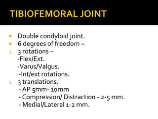
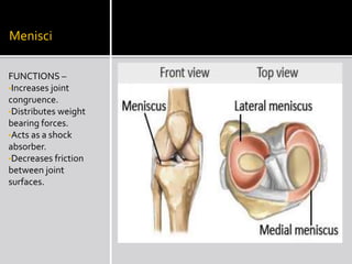
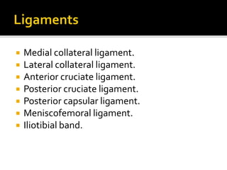
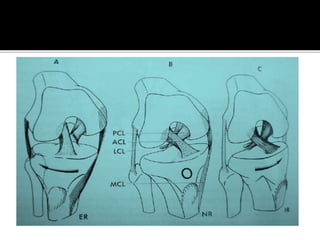
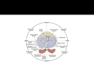
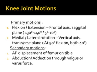
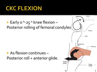
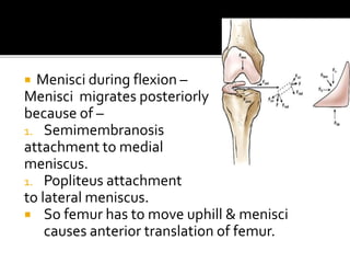
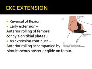
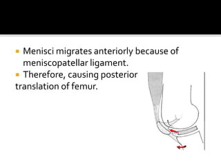
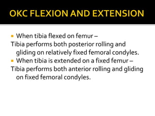
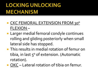
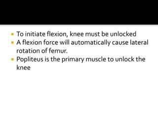
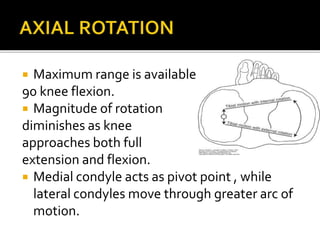
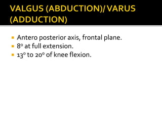
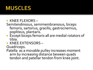
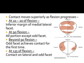
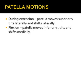
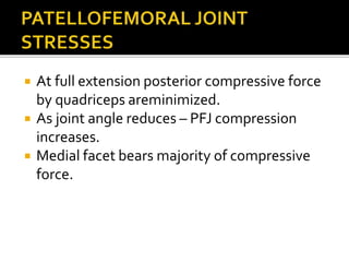
AKHILA NATESAN
 Double condyloid joint.
 6 degrees of freedom –
1. 3 rotations –
-Flex/Ext.
-Varus/Valgus.
-Int/ext rotations.
1. 3 translations.
- AP 5mm- 10mm
- Compression/ Distraction - 2-5 mm.
- Medial/Lateral 1-2 mm.
Menisci
FUNCTIONS –
•Increases joint
congruence.
•Distributes weight
bearing forces.
•Acts as a shock
absorber.
•Decreases friction
between joint
surfaces.
 Medial collateral ligament.
 Lateral collateral ligament.
 Anterior cruciate ligament.
 Posterior cruciate ligament.
 Posterior capsular ligament.
 Meniscofemoral ligament.
 Iliotibial band.
Primary motions :
1. Flexion / Extension – Frontal axis, saggital
plane ( 1300-1400 / 50-100)
2. Medial / Lateral rotation –Vertical axis,
transverse plane ( At 900 flexion, both 400)
Secondary motions :
1. AP displacement of femur on tibia.
2. Abduction/Adduction through valgus or
varus force.
 Early 0 0-25 0 knee flexion –
Posterior rolling of femoral condyles
 As flexion continues –
Posterior roll + anterior glide.
 Menisci during flexion –
Menisci migrates posteriorly
because of –
1. Semimembranosis
attachment to medial
meniscus.
1. Popliteus attachment
to lateral meniscus.
 So femur has to move uphill & menisci
causes anterior translation of femur.
 Reversal of flexion.
 Early extension –
Anterior rolling of femoral
condyle on tibial plateau.
 As extension continues –
Anterior rolling accompanied by
simultaneous posterior glide on femur.
 Menisci migrates anteriorly because of
meniscopatellar ligament.
 Therefore, causing posterior
translation of femur.
 When tibia flexed on femur –
Tibia performs both posterior rolling and
gliding on relatively fixed femoral condyles.
 When tibia is extended on a fixed femur –
Tibia performs both anterior rolling and gliding
on fixed femoral condyles.
 CKC FEMORAL EXTENSION FROM 300
FLEXION –
 Larger medial femoral condyle continues
rolling and gliding posteriorly when small
lateral side has stopped.
 This results in medial rotation of femur on
tibia, in last 50 of extension. (Automatic
rotation).
 OKC – Lateral rotation of tibia on femur.
 To initiate flexion, knee must be unlocked
 A flexion force will automatically cause lateral
rotation of femur.
 Popliteus is the primary muscle to unlock the
knee
 Maximum range is available at
90 knee flexion.
 Magnitude of rotation
diminishes as knee
approaches both full
extension and flexion.
 Medial condyle acts as pivot point , while
lateral condyles move through greater arc of
motion.
 Antero posterior axis, frontal plane.
 80 at full extension.
 130 to 200 of knee flexion.
 KNEE FLEXORS –
Semitendinosus, semimembranosus, biceps
femoris, sartorius, gracilis, gastrocnemius,
popliteus, plantaris.
 Except biceps femoris all are medial rotators of
tibia.
 KNEE EXTENSORS –
Quadriceps.
Patella as a movable pulley increases moment
arm by increasing distance beween quads
tendon and patellar tendon from knee joint.
 One of the most incongruent joints.
 Vertical position of patella in femoral sulcus is
related to length of patellar tendon – 1:1 – insall
salvati index.
 If patellar tendon longer- patella rests more
superiorly- patella alta – Larger knee ROM and
instability - more chances of subluxation.
 At fully extended knee, only inferior pole in
contact.
 Contact moves superiorly as flexion progresses –
 At 10 – 20 of flexion –
Inferior margin of medial lateral
facet.
 At 90 flexion –
All portion except odd facet.
 Beyond 90 flexion –
Odd facet achieves contact for
the first time.
 At 135 of flexion –
Contact on lateral and odd facet
 During extension – patella moves superiorly
tilts laterally and shifts laterally.
 Flexion – patella moves inferiorly , tilts and
shifts medially.
 At full extension posterior compressive force
by quadriceps areminimized.
 As joint angle reduces – PFJ compression
increases.
 Medial facet bears majority of compressive
force.
 PFJ is under permanent control of 2
restraining mechanism across each other at
right angles –
 Transverse group –
1. Medial lateral retinaculum.
2. Vatus medialis and lateralis.
 Longitudinal stabilizers –
1. Patellar tendon – inferiorly
2. Quadriceps tendon - superiorly.
 Kinesiology mechanics and pathomechanics;
2nd edition. Carol. A. Oatis.
 Joint Structure and function; 5th edition.
Cynthia norkins
Knee joint complex biomechanics

Knee joint complex biomechanics

  • 1.
  • 2.
     Double condyloidjoint.  6 degrees of freedom – 1. 3 rotations – -Flex/Ext. -Varus/Valgus. -Int/ext rotations. 1. 3 translations. - AP 5mm- 10mm - Compression/ Distraction - 2-5 mm. - Medial/Lateral 1-2 mm.
  • 3.
    Menisci FUNCTIONS – •Increases joint congruence. •Distributesweight bearing forces. •Acts as a shock absorber. •Decreases friction between joint surfaces.
  • 4.
     Medial collateralligament.  Lateral collateral ligament.  Anterior cruciate ligament.  Posterior cruciate ligament.  Posterior capsular ligament.  Meniscofemoral ligament.  Iliotibial band.
  • 7.
    Primary motions : 1.Flexion / Extension – Frontal axis, saggital plane ( 1300-1400 / 50-100) 2. Medial / Lateral rotation –Vertical axis, transverse plane ( At 900 flexion, both 400) Secondary motions : 1. AP displacement of femur on tibia. 2. Abduction/Adduction through valgus or varus force.
  • 8.
     Early 00-25 0 knee flexion – Posterior rolling of femoral condyles  As flexion continues – Posterior roll + anterior glide.
  • 9.
     Menisci duringflexion – Menisci migrates posteriorly because of – 1. Semimembranosis attachment to medial meniscus. 1. Popliteus attachment to lateral meniscus.  So femur has to move uphill & menisci causes anterior translation of femur.
  • 10.
     Reversal offlexion.  Early extension – Anterior rolling of femoral condyle on tibial plateau.  As extension continues – Anterior rolling accompanied by simultaneous posterior glide on femur.
  • 11.
     Menisci migratesanteriorly because of meniscopatellar ligament.  Therefore, causing posterior translation of femur.
  • 12.
     When tibiaflexed on femur – Tibia performs both posterior rolling and gliding on relatively fixed femoral condyles.  When tibia is extended on a fixed femur – Tibia performs both anterior rolling and gliding on fixed femoral condyles.
  • 13.
     CKC FEMORALEXTENSION FROM 300 FLEXION –  Larger medial femoral condyle continues rolling and gliding posteriorly when small lateral side has stopped.  This results in medial rotation of femur on tibia, in last 50 of extension. (Automatic rotation).  OKC – Lateral rotation of tibia on femur.
  • 14.
     To initiateflexion, knee must be unlocked  A flexion force will automatically cause lateral rotation of femur.  Popliteus is the primary muscle to unlock the knee
  • 15.
     Maximum rangeis available at 90 knee flexion.  Magnitude of rotation diminishes as knee approaches both full extension and flexion.  Medial condyle acts as pivot point , while lateral condyles move through greater arc of motion.
  • 16.
     Antero posterioraxis, frontal plane.  80 at full extension.  130 to 200 of knee flexion.
  • 17.
     KNEE FLEXORS– Semitendinosus, semimembranosus, biceps femoris, sartorius, gracilis, gastrocnemius, popliteus, plantaris.  Except biceps femoris all are medial rotators of tibia.  KNEE EXTENSORS – Quadriceps. Patella as a movable pulley increases moment arm by increasing distance beween quads tendon and patellar tendon from knee joint.
  • 18.
     One ofthe most incongruent joints.  Vertical position of patella in femoral sulcus is related to length of patellar tendon – 1:1 – insall salvati index.  If patellar tendon longer- patella rests more superiorly- patella alta – Larger knee ROM and instability - more chances of subluxation.  At fully extended knee, only inferior pole in contact.
  • 19.
     Contact movessuperiorly as flexion progresses –  At 10 – 20 of flexion – Inferior margin of medial lateral facet.  At 90 flexion – All portion except odd facet.  Beyond 90 flexion – Odd facet achieves contact for the first time.  At 135 of flexion – Contact on lateral and odd facet
  • 20.
     During extension– patella moves superiorly tilts laterally and shifts laterally.  Flexion – patella moves inferiorly , tilts and shifts medially.
  • 21.
     At fullextension posterior compressive force by quadriceps areminimized.  As joint angle reduces – PFJ compression increases.  Medial facet bears majority of compressive force.
  • 22.
     PFJ isunder permanent control of 2 restraining mechanism across each other at right angles –  Transverse group – 1. Medial lateral retinaculum. 2. Vatus medialis and lateralis.  Longitudinal stabilizers – 1. Patellar tendon – inferiorly 2. Quadriceps tendon - superiorly.
  • 23.
     Kinesiology mechanicsand pathomechanics; 2nd edition. Carol. A. Oatis.  Joint Structure and function; 5th edition. Cynthia norkins