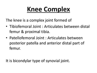
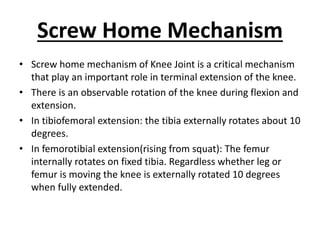
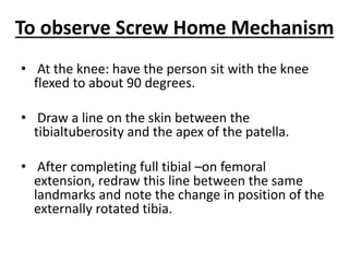
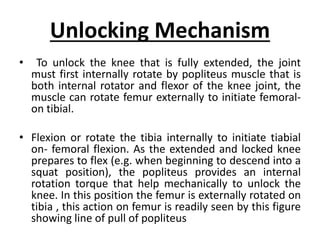
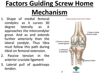
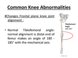
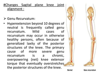
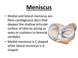
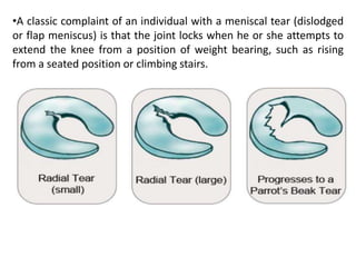
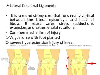
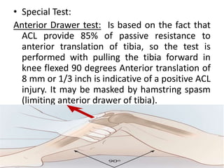
Pathomechanics
of Knee Joint
Dr. Venkat Singh (PT)
MPT (ORTHO)
Knee Complex
The knee is a complex joint formed of
• Tibiofemoral Joint : Articulates between distal
femur & proximal tibia.
• Patellofemoral Joint : Articulates between
posterior patella and anterior distal part of
femur.
It is bicondylar type of synovial joint.
What does Pathomechanics
means?
• It is defined as the mechanics of living systems
in motion resulting in, or leading to,
dysfunction or injury.
Brown, L. P., & Yavorsky, P. (1987). Locomotor biomechanics and pathomechanics: a
review. The Journal of orthopaedic and sports physical therapy, 9(1), 3–10.
https://doi.org/10.2519/jospt.1987.9.1.3
Screw Home Mechanism
• Screw home mechanism of Knee Joint is a critical mechanism
that play an important role in terminal extension of the knee.
• There is an observable rotation of the knee during flexion and
extension.
• In tibiofemoral extension: the tibia externally rotates about 10
degrees.
• In femorotibial extension(rising from squat): The femur
internally rotates on fixed tibia. Regardless whether leg or
femur is moving the knee is externally rotated 10 degrees
when fully extended.
To observe Screw Home Mechanism
• At the knee: have the person sit with the knee
flexed to about 90 degrees.
• Draw a line on the skin between the
tibialtuberosity and the apex of the patella.
• After completing full tibial –on femoral
extension, redraw this line between the same
landmarks and note the change in position of the
externally rotated tibia.
Unlocking Mechanism
• To unlock the knee that is fully extended, the joint
must first internally rotate by popliteus muscle that is
both internal rotator and flexor of the knee joint, the
muscle can rotate femur externally to initiate femoral-
on tibial.
• Flexion or rotate the tibia internally to initiate tiabial
on- femoral flexion. As the extended and locked knee
prepares to flex (e.g. when beginning to descend into a
squat position), the popliteus provides an internal
rotation torque that help mechanically to unlock the
knee. In this position the femur is externally rotated on
tibia , this action on femur is readily seen by this figure
showing line of pull of popliteus
Factors Guiding Screw Home
Mechanism
1. Shape of medial femoral
condyles: as it curves 30
degree laterally as it
approaches the intercondylar
grove. And as and extends
further anteriorly than the
lateral condyle. Thus tibia
must follow this path during
tibial on femoral extension.
2. Passive tension in the
anterior cruciate ligament
3. Lateral pull of quadriceps
tendon.
Common Knee Abnormalities
#Changes Frontal plane knee joint
alignment :
• Normal Tibiofemoral angle:
normal alignment is distal end of
femur makes an angle of 180 -
185° with the mechanical axis.
Tibiofemoral
Angle
180-185°
Genu Valgum :
• The medial condyle is enlarged
and the lateral condyle is
diminished and flattened.
• Effects of Genu valgum: -The
weight bearing line is placed
on the outer side of the knee
effects -Strain on medial
collateral ligament. -
Osteoarthritis in later stages.
Angle more
than 185°
Genu Varum:
• When angle is 175° or less
• There is deflection in the axis of
tibia in varus and inward in relation
to the femur.
• Effects of genu varum:
-The subtalar joint goes into inversion
to adapt the sole on the ground.
-Strain on the lateral collateral
ligament
-Osteoarthritis in later stages.
#Changes Sagital plane knee joint
alignment :
 Genu Recurvatum:
• Hyperextension beyond 10 degrees of
neutral is frequently called genu
recurvatum. Mild cases of
recurvatum may occur in otherwise
healthy persons, often because of
generalized laxity of the posterior
structures of the knee. The primary
cause of more severe genu
recurvatum is a chronic,
overpowering (net) knee extensor
torque that eventually overstretches
the posterior structures of the knee.
Meniscus
• Medial and lateral meniscus are
fibro-cartilaginous discs that
deepen the shallow articular
surface of tibia by acting as
seats or cushions to femoral
condyles.
• Medial meniscus is C shaped
while lateral meniscus is 0
shaped.
Function of Meniscus
1-Reduce the huge compressive stress at the
tibio-femoral joint that may reach 2-3 times
body weight during routine knee activity.
2- Stabilizing the joint during motion.
3-Lubricating the articular cartilage.
4-Reducing friction.
Attachment of Meniscus:
1- Each other anteriorly,
2- Tibial plateau,
3- Femoral condyles,
4- Joint capsule,
5- Quadriceps, semimembrenosus.
Additionally medial meniscus is
attached to medial collateral ligament,
lateral meniscus is attached to
popliteus muscle.
Medial Meniscus:
• The medial meniscus is more frequently injured
more than lateral meniscus because its
attachment to the medial collateral ligaments.
• As medial meniscus movements are more limited
than lateral meniscus movements
Common Mechanism of meniscus tear:
• Tears of menisci often occurs by forceful
horizontal plane rotations of the femoral
condyles over a partially weight bearing flexed
knee.
•A classic complaint of an individual with a meniscal tear (dislodged
or flap meniscus) is that the joint locks when he or she attempts to
extend the knee from a position of weight bearing, such as rising
from a seated position or climbing stairs.
Collateral Ligament Injury
 The medial collateral ligament
(MCL):
• It is a flat, board structure that
spans the medial side of the joint.
• It resist valgus stress (abduction),
extension, and extreme axial
rotations.
• Common Mechanism of injury:
1-Valgus force with foot planted.
2- severe hyperextension injury of
knee.
Lateral Collateral Ligament:
• It is a round strong cord that runs nearly vertical
between the lateral epicondyle and head of
fibula. It resist varus stress (adduction),
extension, and extreme axial rotations.
• Common mechanism of injury :
1-Valgus force with foot planted
2- severe hyperextension injury of knee.
Cruciate Ligament Injury
 Anterior cruciate ligaments:
• ACL attaches along anterior condylar area of the tibial plateau, to attach
on medial side of the lateral femoral condyle.
• Most of its fibers are taught when knee approaches full extension .
• Functions :
1- Most fibers resist excessive anterior translation of the tibia or excessive
posterior translation of the femur.
2- Limit full knee extension.
3- Resist extremes of varus, valgus and axial rotations.
• Mechanism of injury:
1- Hyperextension of knee.
2- Large valgus force with foot planted on ground.
3-Either of the above combined with large internal axial rotation torque e.g.
the femur forcefully externally rotates over a fixed tibia.
• Special Test:
Anterior Drawer test: Is based on the fact that
ACL provide 85% of passive resistance to
anterior translation of tibia, so the test is
performed with pulling the tibia forward in
knee flexed 90 degrees Anterior translation of
8 mm or 1/3 inch is indicative of a positive ACL
injury. It may be masked by hamstring spasm
(limiting anterior drawer of tibia).
Relation Between muscular
contraction and ACL injury
• A contraction of quadriceps muscle extends the
knee and slides the tibia anteriorly relative to the
femur thus increase the tension in most fibers of
the ACL.
• While contraction of the hamstring muscle, in
contrast, causes a posterior translation of the
tibia that slackens most fibers of the ACL.
• Thus in rehabilitation following injuries over
strengthening of hamstrings is an essential
component
Posterior Cruciate Ligament
• PCL attaches from the posterior intercondylar area of tibia
to the lateral side of the femoral medial condyle.
• Become taught in extreme flexion, pulled taught by
hamstring contraction and subsequent posterior slide of
tibia.
1- Most fibers resist excessive posterior translation of the tibia
or excessive anterior translation of the femur.
2- Most fibers taught in full knee flexion.
3- Some fibers are taught at extremes of varus, valgus and
axial rotations
• Mechanisms of injury of PCL: 1-Hypeflexion of knee 2-
Large valgus or varus force with foot planted . 3- Any of the
above, combined with large axial rotation.
• Posterior drawer test: Pushing tibia posteriorly with
knee flexed 90 degree. Normally the PCL provide 95%
degrees of passive resistance to posterior translation of
tibia.
 Relation between hamstrings and PCL:
• Contraction of hamstrings muscles flexes the knee and
slides the tibia posteriorly relative to the femur thus
stressing the posterior cruciate ligaments.
• A contraction of quadriceps muscle extends the knee
and slides the tibia anteriorly relative to the femur thus
decreases the tension in most fibers of the PCL.
• Thus in rehabilitation following injuries over
strengthening of hamstrings is an essential component
Patellofemoral Disorder
• The patellofemoral joint is the interface between the
articular side of the patella and the intercondylar groove on
the femur.
A. Painful Patellofemoral Joint:
• Patellofemoral joint pain syndrome is a common
conditions, cases may be mild involving only a generalized
aching about the anterior knee, or they may be severe
and involve recurrent dislocation or sublaxation of the
patella from the intercondylar groove. Overtime, some of
them develop degenerative changes in the joint surfaces
(chondromalacia patellae).
B. Chondromalaciapatellae is charecterized by:
1- Excessive cartilage degeneration on the
posterior side of the patella.
2-Those with this condition often experience
retro-patellar pain and crepitus, especially
while squatting or climbing steep stairs or
after sitting a prolonged period.
3-The cartilage becomes soft, pitted, and
fragmented. Note the irregular surfaces and
marked degeneration on the cartilage of the
femur and patella
C. Quadriceps Weakness (Extensor lag):
• Inability to fully extend knee in open kinematic in
the last 15 to 20, Although the knee can be fully
extended passively; commonly seen in persons
with moderate weakness in the quadriceps.
• Biomechanical causes includes:
1- In moderate weakness as the knee approaches
terminal extension, the maximal internal torque
potential of the quadriceps is least while the
opposing external (flexor) torque is greatest.
2- Swelling or effusion of the knee increases the
likelihood of an extensor lag. Swelling increases
intra articular pressure, which ca physically
impede full knee extension, by reflexively inhibit
the neural activation of the quadriceps muscle.
D. Patellectomy:
• It is a condition of removal of the patella.
• In this case the patellar tendon lies closer to the
center of the tibiofemoral joint than in a normal
knee joint so the quadriceps muscle will act with
a shorter lever arm, leading to increasing in the
force requirements for the muscle. The
quadriceps muscle has to develop more tension
(about 30%) than is normally required.
• So in cases of patellectomy, the patient must be
exercised to increase the strength of the
quadriceps muscle by about 30% than normal
(especially before operation).
Reference
1. Curwin S (2011). Joint structure and
function. Levangie P.K., & Norkin C.C.(Eds.), Joint
Structure and Function: A Comprehensive Analysis, 5e.
McGraw
Hill. https://fadavispt.mhmedical.com/Content.aspx?
bookid=1862&sectionid=136083890
2. Brown, L. P., & Yavorsky, P. (1987). Locomotor
biomechanics and pathomechanics: a review. The
Journal of orthopaedic and sports physical
therapy, 9(1), 3–10.
https://doi.org/10.2519/jospt.1987.9.1.3
Pathomechanics Knee.pptx

Pathomechanics Knee.pptx

  • 1.
    Pathomechanics of Knee Joint Dr.Venkat Singh (PT) MPT (ORTHO)
  • 2.
    Knee Complex The kneeis a complex joint formed of • Tibiofemoral Joint : Articulates between distal femur & proximal tibia. • Patellofemoral Joint : Articulates between posterior patella and anterior distal part of femur. It is bicondylar type of synovial joint.
  • 6.
    What does Pathomechanics means? •It is defined as the mechanics of living systems in motion resulting in, or leading to, dysfunction or injury. Brown, L. P., & Yavorsky, P. (1987). Locomotor biomechanics and pathomechanics: a review. The Journal of orthopaedic and sports physical therapy, 9(1), 3–10. https://doi.org/10.2519/jospt.1987.9.1.3
  • 7.
    Screw Home Mechanism •Screw home mechanism of Knee Joint is a critical mechanism that play an important role in terminal extension of the knee. • There is an observable rotation of the knee during flexion and extension. • In tibiofemoral extension: the tibia externally rotates about 10 degrees. • In femorotibial extension(rising from squat): The femur internally rotates on fixed tibia. Regardless whether leg or femur is moving the knee is externally rotated 10 degrees when fully extended.
  • 9.
    To observe ScrewHome Mechanism • At the knee: have the person sit with the knee flexed to about 90 degrees. • Draw a line on the skin between the tibialtuberosity and the apex of the patella. • After completing full tibial –on femoral extension, redraw this line between the same landmarks and note the change in position of the externally rotated tibia.
  • 10.
    Unlocking Mechanism • Tounlock the knee that is fully extended, the joint must first internally rotate by popliteus muscle that is both internal rotator and flexor of the knee joint, the muscle can rotate femur externally to initiate femoral- on tibial. • Flexion or rotate the tibia internally to initiate tiabial on- femoral flexion. As the extended and locked knee prepares to flex (e.g. when beginning to descend into a squat position), the popliteus provides an internal rotation torque that help mechanically to unlock the knee. In this position the femur is externally rotated on tibia , this action on femur is readily seen by this figure showing line of pull of popliteus
  • 11.
    Factors Guiding ScrewHome Mechanism 1. Shape of medial femoral condyles: as it curves 30 degree laterally as it approaches the intercondylar grove. And as and extends further anteriorly than the lateral condyle. Thus tibia must follow this path during tibial on femoral extension. 2. Passive tension in the anterior cruciate ligament 3. Lateral pull of quadriceps tendon.
  • 12.
    Common Knee Abnormalities #ChangesFrontal plane knee joint alignment : • Normal Tibiofemoral angle: normal alignment is distal end of femur makes an angle of 180 - 185° with the mechanical axis. Tibiofemoral Angle 180-185°
  • 13.
    Genu Valgum : •The medial condyle is enlarged and the lateral condyle is diminished and flattened. • Effects of Genu valgum: -The weight bearing line is placed on the outer side of the knee effects -Strain on medial collateral ligament. - Osteoarthritis in later stages. Angle more than 185°
  • 14.
    Genu Varum: • Whenangle is 175° or less • There is deflection in the axis of tibia in varus and inward in relation to the femur. • Effects of genu varum: -The subtalar joint goes into inversion to adapt the sole on the ground. -Strain on the lateral collateral ligament -Osteoarthritis in later stages.
  • 15.
    #Changes Sagital planeknee joint alignment :  Genu Recurvatum: • Hyperextension beyond 10 degrees of neutral is frequently called genu recurvatum. Mild cases of recurvatum may occur in otherwise healthy persons, often because of generalized laxity of the posterior structures of the knee. The primary cause of more severe genu recurvatum is a chronic, overpowering (net) knee extensor torque that eventually overstretches the posterior structures of the knee.
  • 16.
    Meniscus • Medial andlateral meniscus are fibro-cartilaginous discs that deepen the shallow articular surface of tibia by acting as seats or cushions to femoral condyles. • Medial meniscus is C shaped while lateral meniscus is 0 shaped.
  • 17.
    Function of Meniscus 1-Reducethe huge compressive stress at the tibio-femoral joint that may reach 2-3 times body weight during routine knee activity. 2- Stabilizing the joint during motion. 3-Lubricating the articular cartilage. 4-Reducing friction.
  • 18.
    Attachment of Meniscus: 1-Each other anteriorly, 2- Tibial plateau, 3- Femoral condyles, 4- Joint capsule, 5- Quadriceps, semimembrenosus. Additionally medial meniscus is attached to medial collateral ligament, lateral meniscus is attached to popliteus muscle.
  • 19.
    Medial Meniscus: • Themedial meniscus is more frequently injured more than lateral meniscus because its attachment to the medial collateral ligaments. • As medial meniscus movements are more limited than lateral meniscus movements Common Mechanism of meniscus tear: • Tears of menisci often occurs by forceful horizontal plane rotations of the femoral condyles over a partially weight bearing flexed knee.
  • 20.
    •A classic complaintof an individual with a meniscal tear (dislodged or flap meniscus) is that the joint locks when he or she attempts to extend the knee from a position of weight bearing, such as rising from a seated position or climbing stairs.
  • 21.
    Collateral Ligament Injury The medial collateral ligament (MCL): • It is a flat, board structure that spans the medial side of the joint. • It resist valgus stress (abduction), extension, and extreme axial rotations. • Common Mechanism of injury: 1-Valgus force with foot planted. 2- severe hyperextension injury of knee.
  • 23.
    Lateral Collateral Ligament: •It is a round strong cord that runs nearly vertical between the lateral epicondyle and head of fibula. It resist varus stress (adduction), extension, and extreme axial rotations. • Common mechanism of injury : 1-Valgus force with foot planted 2- severe hyperextension injury of knee.
  • 24.
    Cruciate Ligament Injury Anterior cruciate ligaments: • ACL attaches along anterior condylar area of the tibial plateau, to attach on medial side of the lateral femoral condyle. • Most of its fibers are taught when knee approaches full extension . • Functions : 1- Most fibers resist excessive anterior translation of the tibia or excessive posterior translation of the femur. 2- Limit full knee extension. 3- Resist extremes of varus, valgus and axial rotations. • Mechanism of injury: 1- Hyperextension of knee. 2- Large valgus force with foot planted on ground. 3-Either of the above combined with large internal axial rotation torque e.g. the femur forcefully externally rotates over a fixed tibia.
  • 25.
    • Special Test: AnteriorDrawer test: Is based on the fact that ACL provide 85% of passive resistance to anterior translation of tibia, so the test is performed with pulling the tibia forward in knee flexed 90 degrees Anterior translation of 8 mm or 1/3 inch is indicative of a positive ACL injury. It may be masked by hamstring spasm (limiting anterior drawer of tibia).
  • 26.
    Relation Between muscular contractionand ACL injury • A contraction of quadriceps muscle extends the knee and slides the tibia anteriorly relative to the femur thus increase the tension in most fibers of the ACL. • While contraction of the hamstring muscle, in contrast, causes a posterior translation of the tibia that slackens most fibers of the ACL. • Thus in rehabilitation following injuries over strengthening of hamstrings is an essential component
  • 27.
    Posterior Cruciate Ligament •PCL attaches from the posterior intercondylar area of tibia to the lateral side of the femoral medial condyle. • Become taught in extreme flexion, pulled taught by hamstring contraction and subsequent posterior slide of tibia. 1- Most fibers resist excessive posterior translation of the tibia or excessive anterior translation of the femur. 2- Most fibers taught in full knee flexion. 3- Some fibers are taught at extremes of varus, valgus and axial rotations • Mechanisms of injury of PCL: 1-Hypeflexion of knee 2- Large valgus or varus force with foot planted . 3- Any of the above, combined with large axial rotation.
  • 28.
    • Posterior drawertest: Pushing tibia posteriorly with knee flexed 90 degree. Normally the PCL provide 95% degrees of passive resistance to posterior translation of tibia.  Relation between hamstrings and PCL: • Contraction of hamstrings muscles flexes the knee and slides the tibia posteriorly relative to the femur thus stressing the posterior cruciate ligaments. • A contraction of quadriceps muscle extends the knee and slides the tibia anteriorly relative to the femur thus decreases the tension in most fibers of the PCL. • Thus in rehabilitation following injuries over strengthening of hamstrings is an essential component
  • 29.
    Patellofemoral Disorder • Thepatellofemoral joint is the interface between the articular side of the patella and the intercondylar groove on the femur. A. Painful Patellofemoral Joint: • Patellofemoral joint pain syndrome is a common conditions, cases may be mild involving only a generalized aching about the anterior knee, or they may be severe and involve recurrent dislocation or sublaxation of the patella from the intercondylar groove. Overtime, some of them develop degenerative changes in the joint surfaces (chondromalacia patellae).
  • 30.
    B. Chondromalaciapatellae ischarecterized by: 1- Excessive cartilage degeneration on the posterior side of the patella. 2-Those with this condition often experience retro-patellar pain and crepitus, especially while squatting or climbing steep stairs or after sitting a prolonged period. 3-The cartilage becomes soft, pitted, and fragmented. Note the irregular surfaces and marked degeneration on the cartilage of the femur and patella
  • 31.
    C. Quadriceps Weakness(Extensor lag): • Inability to fully extend knee in open kinematic in the last 15 to 20, Although the knee can be fully extended passively; commonly seen in persons with moderate weakness in the quadriceps. • Biomechanical causes includes: 1- In moderate weakness as the knee approaches terminal extension, the maximal internal torque potential of the quadriceps is least while the opposing external (flexor) torque is greatest. 2- Swelling or effusion of the knee increases the likelihood of an extensor lag. Swelling increases intra articular pressure, which ca physically impede full knee extension, by reflexively inhibit the neural activation of the quadriceps muscle.
  • 32.
    D. Patellectomy: • Itis a condition of removal of the patella. • In this case the patellar tendon lies closer to the center of the tibiofemoral joint than in a normal knee joint so the quadriceps muscle will act with a shorter lever arm, leading to increasing in the force requirements for the muscle. The quadriceps muscle has to develop more tension (about 30%) than is normally required. • So in cases of patellectomy, the patient must be exercised to increase the strength of the quadriceps muscle by about 30% than normal (especially before operation).
  • 33.
    Reference 1. Curwin S(2011). Joint structure and function. Levangie P.K., & Norkin C.C.(Eds.), Joint Structure and Function: A Comprehensive Analysis, 5e. McGraw Hill. https://fadavispt.mhmedical.com/Content.aspx? bookid=1862&sectionid=136083890 2. Brown, L. P., & Yavorsky, P. (1987). Locomotor biomechanics and pathomechanics: a review. The Journal of orthopaedic and sports physical therapy, 9(1), 3–10. https://doi.org/10.2519/jospt.1987.9.1.3