Isabel Miguel: Quadriceps muscle anatomy Cadaver study - PRP
•
11 likes•3,524 views

Isabel Miguel MD PhD. Human anatomy Lecture at Unit of Human Anatomy and Embryology, Spain PRP for Quadriceps Muscles Injuries 8th MuscleTech Network Workshop 3rd October, Barcelona

Recommended
More Related Content
What's hot
What's hot (20)
Viewers also liked
Viewers also liked (20)
Similar to Isabel Miguel: Quadriceps muscle anatomy Cadaver study - PRP
Similar to Isabel Miguel: Quadriceps muscle anatomy Cadaver study - PRP (20)
More from MuscleTech Network
More from MuscleTech Network (13)
Recently uploaded
Recently uploaded (20)
Isabel Miguel: Quadriceps muscle anatomy Cadaver study - PRP
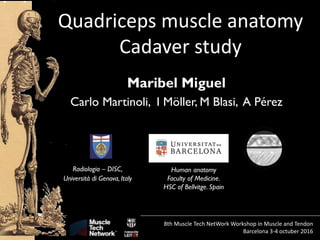
- 1. Quadriceps muscle anatomy Cadaver study Maribel Miguel Carlo Martinoli, I Möller, M Blasi, A Pérez Human anatomy Faculty of Medicine. HSC of Bellvitge. Spain Radiologia – DISC, Università di Genova, Italy 8th Muscle Tech NetWork Workshop in Muscle and Tendon Barcelona 3-4 octuber 2016
- 2. Quadriceps muscle anatomy Cadaver study Maribel Miguel Carlo Martinoli, I Möller, M Blasi, A Pérez 8th Muscle Tech NetWork Workshop in Muscle and Tendon Barcelona 3-4 octuber 2016 R Balius, G Rodes, J de la Fuente, X Alomar, C Pedret…
- 3. Quadriceps femoris muscle It is formed for several parts: Rectus femoris Vastus lateralis Vastus medialis Vastus intermedius
- 4. All these muscles are located at the anterior compartment of the thigh, and involved by the deep fascia, called the fascia lata Quadriceps femoris muscle
- 5. All these muscles are located at the anterior compartment of the thigh, and involved by the deep fascia, called the fascia lata Quadriceps femoris muscle
- 6. All these muscles are located at the anterior compartment of the thigh, and involved by the deep fascia, called the fascia lata Quadriceps femoris muscle Bottoni et cols, 2009
- 7. Quadriceps femoris muscle Vastus lateralis Vastus medialis Vastus intermedius Classicaly we know that they have their origen in the aspera line in the posterior part of the femur and other points as the major trochantis and they go to the anterior part of the thigh to find the rectus femoris muscle
- 8. Quadriceps femoris muscle A fifth muscle of the anterior thigh, the tensor of the vastus intermedius (TVI), has been described recently. This muscle belly has a separate aponeurotic tendon, closely associated with the aponeurosis of the VI, which joins the quadriceps tendon distally Grob et cols 2016
- 9. Quadriceps femoris muscle A fifth muscle of the anterior thigh, the tensor of the vastus intermedius (TVI), has been described recently. This muscle belly has a separate aponeurotic tendon, closely associated with the aponeurosis of the VI, which joins the quadriceps tendon distally Grob et cols 2016
- 10. Quadriceps femoris muscle Rectus femoris Vastus lateralis Vastus medialis Vastus intermedius All of them converge in a comun tendon, patellar tendon, in the more distal part of the thigh that goes to insert in the patella and finally in the tibial tuberosity as the patellar ligament
- 11. Quadriceps femoris muscle Rectus femoris Vastus lateralis Vastus medialis Vastus intermedius All of them converge in a comun tendon in the more distal part of the thigh that goes to insert in the patella and finally in the tibial tuberosity Remember that the muscular fibers from vastus medialis extends longer than the vastus lateralis and makes more force in the medial side and the presence of the iliotibialis tract in the lateral side to compensate it.
- 12. Quadriceps femoris muscle Rectus femoris Vastus lateralis Vastus medialis Vastus intermedius
- 14. Rectus femoris The most commonly injured muscle of the anterior thigh Quadriceps femoris muscle
- 15. Rectus femoris The most commonly injured muscle of the anterior thigh Three distinctive patterns have been described according to the location of the injury: • At the entesis • Within the tendon • Musculotendinous junction Pesquert et cols 2016 Quadriceps femoris muscle
- 16. Rectus femoris muscle Its origin is tendinous and consists of : Direct or straight head arises from the anteroinferior iliac spine (Bony footprint of 13 mm short axis and 26 mm long axis) Continues down as the aponeurosis fascia of the muscle Martinolli et al 2007 ; Ryan et al 2014
- 17. Rectus femoris muscle Its origin is tendinous and consists of : Direct or straight head arises from the anteroinferior iliac spine (Bony footprint of 13 mm short axis and 26 mm long axis) Indirect or reflected head arises from the superior acetabular ridge in a more lateral position (Bony footprint of 17 mm short axis and 48 mm long axis Continues down as the anterior aponeurosis of the muscle And form a sagitally and central aponeurosis, located inside the proximal muscle belly and courses until two-thirds of the muscle belly and forms most of the posterior component of the conjoined tendon Martinolli et al 2007 ; Ryan et al 2014
- 18. Rectus femoris muscle Direct head Indirect head
- 19. Rectus femoris muscle Indirect head
- 20. Rectus femoris muscle Indirect head
- 21. Rectus femoris muscle Direct head Indirect head
- 23. Rectus femoris muscle Direct head Indirect head
- 24. Rectus femoris muscle Direct head Indirect head
- 25. Rectus femoris muscle Direct head Indirect head Both differents origins give differents musculars fibbers in these special position that so clear explains Dr Martinoli: Direct musculars fibbers arises until the more posterior position in the posterior aponeurosis Indirect muculars fibbers arises also to the posterior aponeurosis but since the intramuscular tendon
- 29. Rectus femoris muscle But we want to add some different origin, sometimes no so clear and that they can be confused by aponeurosis of the others anteriors muscles Reflected portion is a small tendon. It is directed more laterally to merge with the anterior capsule of the hip joint.
- 30. Rectus femoris muscle Reflected portion As Tubb et al (2006) confirm this tendon attach to the anterior aspect of the greater trochanter in an inferolateral direction, superficially with the tendon of the gluteus minimus
- 31. Rectus femoris muscle But we want to add some different origin, sometimes no so clear. They can be confused by aponeurosis of the others anteriors muscles Inguinal tendon
- 32. Rectus femoris muscle But we want to add some different origin, sometimes no so clear. They can be confused by aponeurosis of the others anteriors muscles
- 33. Thank you for your attention