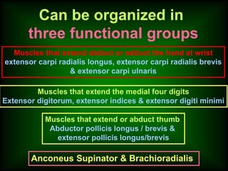
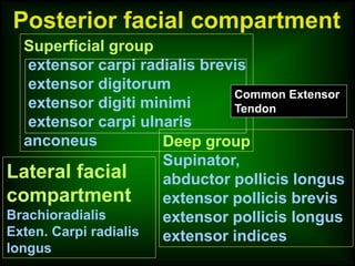
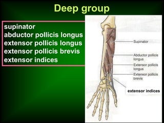
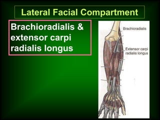
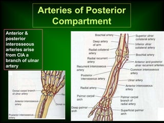
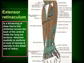
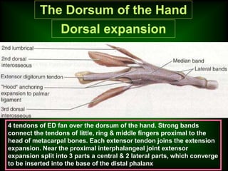
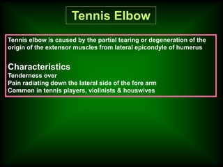
Anconeus
Common
Extensor
Tendon
Objectives
Fascial Compartmentalization
Organization of Muscles
according to their action
Grouping of muscles
into superficial & deep
Vessels & nerve
Muscles
Name them posterior extensor supinator
All are innervated by radial nerve
Can be organized in
three functional groups
Muscles that extend abduct or adduct the hand at wrist
extensor carpi radialis longus, extensor carpi radialis brevis
& extensor carpi ulnaris
Muscles that extend the medial four digits
Extensor digitorum, extensor indices & extensor digiti minimi
Muscles that extend or abduct thumb
Abductor pollicis longus / brevis &
extensor pollicis longus/brevis
Anconeus Supinator & Brachioradialis
Posterior facial compartment
Superficial group
extensor carpi radialis brevis
extensor digitorum
extensor digiti minimi
extensor carpi ulnaris
anconeus Deep group
Supinator,
abductor pollicis longus
extensor pollicis brevis
extensor pollicis longus
extensor indices
Common Extensor
Tendon
Lateral facial
compartment
Brachioradialis
Exten. Carpi radialis
longus
extensor carpi radialis brevis
extensor digitorum,
extensor digiti minimi,
extensor carpi ulnaris
anconeus
Superficial group
Anconeus
1
2
3
4
5
Common
Extensor
Tendon
Deep group
supinator
abductor pollicis longus
extensor pollicis longus
extensor pollicis brevis
extensor indices
extensor indices
Lateral Facial Compartment
Brachioradialis &
extensor carpi
radialis longus
Arteries of Posterior
Compartment
Anterior &
posterior
interosseous
arteries arise
from CIA a
branch of ulnar
artery
Deep branch arises from
radial nerve in front of the
lateral epicondyle of
humerus pierces the
supinator & hinds around
the neck of the radius, runs
b/w the superficial & deep
group.
Gives muscular & articular
branches
Nerves of Posterior
Compartment
Structures pass superficial to the extensor
retinaculum from medial to lateral
Dorsal cutaneous br of ulnar
nerve
Basilic vein
Cephalic vein
Superficial br of radial nerve
Structures pass deep to the extensor retinaculum
from medial to lateral
The tendons of:
Ext carpi ulnaris
Ext digiti minimi
Ext digitorum
Ext indicis
Ext pollicis longus
Ext carpi radialis longus
Abductor pollicis longus
Ext pollicis brevis
Extensor
retinaculum
Is a thickening of
deep fascia that
stretches across the
back of the wrist &
holds the long ext
tendons. Attached
medially to pisiform
& hook of hamate &
laterally to the distal
end of radius.
1
2
3
4
5
6
Synovial tendon
sheath
Synovial tendon sheath/cyst
The Dorsum of the Hand
Dorsal expansion
4 tendons of ED fan over the dorsum of the hand. Strong bands
connect the tendons of little, ring & middle fingers proximal to the
head of metacarpal bones. Each extensor tendon joins the extension
expansion. Near the proximal interphalangeal joint extensor
expansion split into 3 parts a central & 2 lateral parts, which converge
to be inserted into the base of the distal phalanx
Arterial supply
Dorsal Venous Arch
Nerve supply of
dorsum of the hand
Anatomical snuff box
Tennis Elbow
Tennis elbow is caused by the partial tearing or degeneration of the
origin of the extensor muscles from lateral epicondyle of humerus
Characteristics
Tenderness over
Pain radiating down the lateral side of the fore arm
Common in tennis players, violinists & houswives
Baseball fracture
11-Extensor Mus forearm.pptx

11-Extensor Mus forearm.pptx

  • 2.
  • 3.
    Objectives Fascial Compartmentalization Organization ofMuscles according to their action Grouping of muscles into superficial & deep Vessels & nerve
  • 4.
    Muscles Name them posteriorextensor supinator All are innervated by radial nerve
  • 5.
    Can be organizedin three functional groups Muscles that extend abduct or adduct the hand at wrist extensor carpi radialis longus, extensor carpi radialis brevis & extensor carpi ulnaris Muscles that extend the medial four digits Extensor digitorum, extensor indices & extensor digiti minimi Muscles that extend or abduct thumb Abductor pollicis longus / brevis & extensor pollicis longus/brevis Anconeus Supinator & Brachioradialis
  • 6.
    Posterior facial compartment Superficialgroup extensor carpi radialis brevis extensor digitorum extensor digiti minimi extensor carpi ulnaris anconeus Deep group Supinator, abductor pollicis longus extensor pollicis brevis extensor pollicis longus extensor indices Common Extensor Tendon Lateral facial compartment Brachioradialis Exten. Carpi radialis longus
  • 7.
    extensor carpi radialisbrevis extensor digitorum, extensor digiti minimi, extensor carpi ulnaris anconeus Superficial group Anconeus 1 2 3 4 5 Common Extensor Tendon
  • 8.
    Deep group supinator abductor pollicislongus extensor pollicis longus extensor pollicis brevis extensor indices extensor indices
  • 9.
    Lateral Facial Compartment Brachioradialis& extensor carpi radialis longus
  • 10.
    Arteries of Posterior Compartment Anterior& posterior interosseous arteries arise from CIA a branch of ulnar artery
  • 11.
    Deep branch arisesfrom radial nerve in front of the lateral epicondyle of humerus pierces the supinator & hinds around the neck of the radius, runs b/w the superficial & deep group. Gives muscular & articular branches Nerves of Posterior Compartment
  • 12.
    Structures pass superficialto the extensor retinaculum from medial to lateral Dorsal cutaneous br of ulnar nerve Basilic vein Cephalic vein Superficial br of radial nerve Structures pass deep to the extensor retinaculum from medial to lateral The tendons of: Ext carpi ulnaris Ext digiti minimi Ext digitorum Ext indicis Ext pollicis longus Ext carpi radialis longus Abductor pollicis longus Ext pollicis brevis
  • 13.
    Extensor retinaculum Is a thickeningof deep fascia that stretches across the back of the wrist & holds the long ext tendons. Attached medially to pisiform & hook of hamate & laterally to the distal end of radius.
  • 14.
  • 15.
  • 17.
    The Dorsum ofthe Hand Dorsal expansion 4 tendons of ED fan over the dorsum of the hand. Strong bands connect the tendons of little, ring & middle fingers proximal to the head of metacarpal bones. Each extensor tendon joins the extension expansion. Near the proximal interphalangeal joint extensor expansion split into 3 parts a central & 2 lateral parts, which converge to be inserted into the base of the distal phalanx
  • 18.
  • 19.
  • 20.
  • 21.
  • 22.
    Tennis Elbow Tennis elbowis caused by the partial tearing or degeneration of the origin of the extensor muscles from lateral epicondyle of humerus Characteristics Tenderness over Pain radiating down the lateral side of the fore arm Common in tennis players, violinists & houswives
  • 23.