GONIOSCOPY
Presented by Dr. Kabir Singh
INTRODUCTION
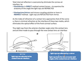
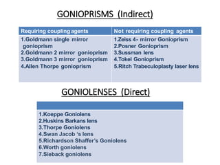
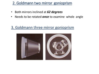
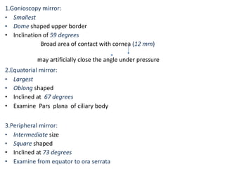
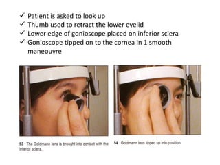
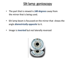
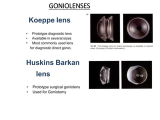
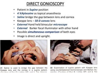
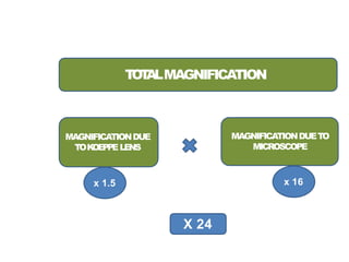
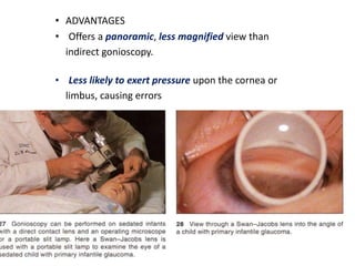
Gonioscopy
allows visualization of
the anterior chamber angle by using
direct or indirect contact lenses to overcome
the
Total Internal Reflection
 Enables glaucomas to be classified by assessing
whether the angle is open or closed
 Various Pathologies involving angle structures can
be observed
HISTORICALASPECTS
 Trantas (1907)
• 1st visualized the anterior chamber angle in a
patient with keratoglobus by indenting the limbus
• coined the term gonioscopy.
 Koeppe (1919)
• designed an improved steeper lens.
 Goldmann (1938)
• introduced the gonioprism
 Barkan
• established the use of gonioscopy in the management of
glaucoma
PRINCIPLES ANDOPTICS
•Critical angle for the cornea-air interface is approximately 46
degrees.
•Total internal reflection prevents direct visualization of ang le in
nearly all eyes.
•The incident angle of light- reflected from angle is greater
than the critical angle at the cornea–air interface.
Li < Lr
lightraysarerefractedatthe
contact lens-air interface
lightraysarereflectedbya mirror
lightraysleavethelensatnearlyright
anglesatthecontactlens-air interface
Total internal reflection is overcome by eliminate the cornea-air
interface by
Goniolenses in DIRECT method contact lenses –-to examine the
anatomy of the angle the light rays are REFRACTED
Gonioprisms/mirror and viscous coupling solution or tears in
INDIRECT method . Light rays are REFLECTED from mirror.
As the index of refraction of a contact lens approaches that of the corne
a, there is minimal refraction at the interface of these two media, which
eliminates the optical effect of the front corneal surface.
Thus light rays from the anterior chamber angle enter the contact lens
and are then made to pass through the new contact lens-air interface
GONIOPRISMS (Indirect)
GONIOLENSES (Direct)
Requiring coupling agents Not requiring coupling agents
1.Goldmann single mirror 1.Zeiss 4- mirror Gonioprism
gonioprism 2.Posner Gonioprism
2.Goldmann 2 mirror gonioprism 3.Sussman lens
3.Goldmann 3 mirror gonioprism 4.Tokel Gonioprism
4.Allen Thorpe gonioprism 5.Ritch Trabeculoplasty laser lens
1.Koeppe Goniolens
2.Huskins Barkans lens
3.Thorpe Goniolens
4.Swan Jacob ‘s lens
5.Richardson Shaffer’s Goniolens
6.Worth goniolens
7.Sieback goniolens
GONIOPRISMS
Requiring coupling agents
1.Goldmann single mirror gonioprism
Prototype diagnostic gonioprism
• Mirror inclined at 62 degrees from plano front
surface
• Needs to be rotated 3times to examine the whole angle
• Mirror Height -12 mm
• Central well diameter -12 mm
• Posterior Radius of curvature -7.38 mm
2. Goldmann two mirror gonioprism
• Both mirrors inclined at 62 degrees
• Needs to be rotated once to examine whole angle
3. Goldmann three mirror gonioprism
1.Gonioscopy mirror:
• Smallest
• Dome shaped upper border
• Inclination of 59 degrees
Broad area of contact with cornea (12 mm)
may artificially close the angle under pressure
2.Equatorial mirror:
• Largest
• Oblong shaped
• Inclined at 67 degrees
• Examine Pars plana of ciliary body
3.Peripheral mirror:
• Intermediate size
• Square shaped
• Inclined at 73 degrees
• Examine from equator to ora serrata
4. Allen Thorpe gonioprism
• 4 prisms instead of mirrors
• Allows examination of whole angle without rotating
the prisms
• Suspended by a frame
Advantages of Goldmann gonioprisms
• Easy to use
• Excellent view
• Peripheral retina can be seen
• Stabilizes the globe
Disadvantages of Goldmann gonioprisms
• Only 1 mirror for gonio-has to be rotated by 360
degrees
• Cannot be used for indentation
• In case of 3 mirror lens, broad area of contact with
cornea may cause artefactual closure of angle
Curvature of lens > cornea
Coupling material required
Blurs vision andfundus
Field charting ,direct and indirect ophthalmosc
opy
cantbedone immediatelyafteruse
GONIOPRISMS
Not requiring coupling agents
1.Zeiss 4- mirror Gonioprism
• 4 identical mirrors angled at 64 degrees
• On an UNGER HOLDER
• Small area of contact with the cornea (9mm)
Indentation gonioscopy can be performed
Zeiss 4- mirror Gonioprism
ADVANTAGES DISADVANTAGES
• Easy to perform
• All 4 quadrants visible at sa
me time
• Rotation of 11 degrees
covers area between the
mirrors
• Indentation gonio
• Coupling material not
required,
thus fundus viewing and
photography possible
• Difficult to master
• Does not stabilise the
globe
• May open the angle
artefactually If
pressure is applied
2.Posner Gonioprism
• Similar to Zeiss
• Made of plastic instead of glass
• Has a fixed handle as compared to
zeiss.
3.Sussman lens
Similar to Zeiss but
has no handle
4.Tokel Gonioprism
• Single mirror prism
• Broader viewing area
than Goldmann single mirror lens
5. Ritch Trabeculoplasty laser lens
• 2 mirrors tilted at 59degrees to see inferior angle
• 2 mirrors at 64 degrees to view the superior angle
• A convex button in front of a 59 degree mirror and a 64
degree mirror for extra magnification and laser
treatment
INDIRECT GONIOSCOPY
PROCEDURE
 Dim illumination
 Eye anaesthetised with topical agent
 Appropriate positioning of the patient at slit lamp
The concave face of Goldmann lens should be filled with a
Methyl cellulose coupling fluid before its applied to the
eye.
Care should be taken to keep air bubbles out of the
solution
 Patient is asked to look up
 Thumb used to retract the lower eyelid
 Lower edge of gonioscope placed on inferior sclera
 Gonioscope tipped on to the cornea in 1 smooth
maneouvre
Slit lamp gonioscopy
• The part that is viewed is 180 degrees away from
the mirror that is being used.
• Slit lamp beam is focussed on the mirror that shows the
angle diametrically opposite to it.
• Image is inverted but not laterally reversed
Advantages of Indirect Gonioscopy
• Easier to learn
• Faster to perform, particularly with the Zeiss
four-mirror lenses and modified Goldmann-type
lenses, because no viscous bridge is required.
• Slitlamp provides better optics, variable
magnification and illumination
• Requires fewer additional instruments and
occupies less space than direct gonioscopy.
• Gonioprisms with a posterior radius of curvature closer
to that of the anterior corneal surface may also reduce
Corneal distortion.
• Gonioprisms with taller mirrors facilitate
visualization of narrow angles.
• The slit beam can create a corneal wedge to help to define
the structures of the angle.
• Because of its relatively small diameter of
corneal contact, the Zeiss four-mirror lens
can also be used in compressive gonioscopy.
• Indentation gonioscopy can be performed with the
Posner or Sussmann lens to distinguish
appositional from synechial angle closure.
• Magnified stereoscopic view of the optic disc can be
obtained
Disadvantages of Indirect
Gonioscopy
• Limited positioning of light rays
• Comparison not possible
• Difficult to perform in horizontal meridian
• Inverted Mirror image seen – confusing
• Excess pressure over the cornea will displace aqueous from
the center of the anterior chamber into the periphery,disloc
ating the iris posteriorly and falsely opening the anterior ch
amber angle.
• Posterior pressure may indent the sclera and falsely narrow
the angle.
• Goldmann lens requires an optical coupling
between the cornea and the lens.
( four-mirror Zeiss lens ,Posner, Sussman have a smaller
area of contact and have almost the same
radius of curvature as the cornea, which allows the tear
film to function as the optical coupling agent.)
GONIOLENSES
Koeppe lens
• Prototype diagnostic lens
• Available in several sizes
• Most commonly used lens
for diagnostic direct gonio.
Huskins Barkan
lens
• Prototype surgical goniolens
• Used for Goniotomy
Swanjacob’s
lens
• Surgical
goniolens
• used in children
Richardson Shaffer’s lens
Small lens for use in infants
Worst goniolens
It anchors to the cornea by partial vaccum
Sieback goniolens
Tiny goniolens which floats on the cornea
DIRECT GONIOSCOPY
• Patient in Supine position
• 4 %Xylocaine as topical anaesthesia
• Saline bridge the gap between lens and cornea
• Koeppe lens – 50 D convex lens
• External Hand held binocular microscope
• External Barker focal illuminator with other hand
• Possible simultaneous comparison of both eyes
• Image is direct and upright.
T
O
T
ALMAGNIFICATION
MAGNIFICATIONDUE
TOK
OEPPELENS
MAGNIFICATIONDUETO
MICROSCOPE
x 1.5 x 16
X 24
• ADVANTAGES
• Offers a panoramic, less magnified view than
indirect gonioscopy.
• Less likely to exert pressure upon the cornea or
limbus, causing errors
 Goniolens may cause less distortion of the anterior
chamber
 Using 2 lenses, both eyes can be
simultaneously examined
 Possible to vary the angle of visualization more
easily. Therefore, a narrow angle can be assessed to
see if it is a steep approach to an open angle or a
completely closed angle.
 Can be used for surgical procedures like goniotomy and
goniosynechialysis
 Can be used in sedated or anesthetized patient s, as in
the examination of children
Advantages of Direct
Gonioscopy
 The height of the observer may be changed to look
deeper into a narrow angle, whereas the gonioprism
is limited by the height of the mirror
 Angle becomes deeper in supine position –
easier to see angle
 Provides a straight-on view of the angle rather than
the mirror image given by the indirect lenses.
 Panoramic view, so 1 part of angle can be com
pared to other
Disdvantages of Direct Gonioscopy
 Inconvenient
 Annoying light reflexes from the cornea
 Timeconsuming
 Benefits of slit lamp not available
• With a narrow, short slit beam off axis, the quadrant o f the
angle to be assessed is first examined with the f our-mirror
lens, with
 no pressure on the cornea
 the patient looking sufficiently far in the direction of the
mirror that the examiner can see as deeply into the angl e as
possible.
• The inferior portion of the angle is typically the widest and
where the trabecular meshwork has the most pigment,
thus easiest to identify
structures and familiarize with patient ‘s anatomy
• Thus most clinicians apply lens so that mirror is at the top of
the eye, to allow inferior angle to be examined first.
• Then the goniolens is rotated to view other portions of the
angle
TYPES OF G0NIOSCOPY
• GONIOSCOPY IN SITU
• MANIPULATIVE
• INDENTATION
Occludable Angles
During Gonioscopy in situ
(No anglestructuresare visible)
Optical or Apparent
closure
Appositional closure
Synechial Closure
Look for abnormalities inangle
Manipulative gonioscopy
Indentation gonioscopy
Tolook for angle abnormalities
• Increase the room and slit lamp illumination
• Allow light to impinge on pupil
• Thereby opening up angle
Manipulative/Dynamic Gonioscopy
 In eyes with a steep iris configuration
 manipulate Goldmann lens to visualise over a steep iris
(OVER HILL VIEW)
 Ask patient to look in direction of mirror or
 Move mirror towards angle being viewed
Indentation Gonioscopy
 performed in a completely darkened room using the
smallest square of light for a slit beam to avoid stimulating
the pupillary light reflex.
 Effective with Zeiss , Posner, Sussman ,Allen Thorpe lens
whose areas of contact are smaller than the cornea(no
coupling media)
 Goldmann and Koeppe have larger areas of contact and
may make the angle shallower with indentation
• Bending of the cornea results in mechanical rotation of the
limbus, giving more direct view of the angle
• Permits examiner to look deep into angle recess for
iridodialysis, foreign bodies or cyclodialysis clefts
• By deliberately varying the amount of pressure
applied to the cornea- observe the effects on
angle width.
• Measures extent of angle closure
• Useful in convex iris configuration and Plateau configur
ation - (retain convex profile)
• Performed in all cases
• The ability to visualize angle structures by indentation-redu
ced in the presence of elevated intraocular pressure.
• Differentiate form appositional or synechial
closure
INDICATIONS
To differentiate b/w primary open angle glaucoma & primary angle
closure glaucoma.
To diagnose
• Congenital glaucomas.
• Secondary glaucomas
• Angle recession glaucoma(ARG)
• Uveitic glaucoma
• Neovascularization
• ICE (Iridocorneal endothelial) syndrome
• Tumors of anterior segment
• Cyclodialysis
• Ciliary body cysts
• Intraocular foreign body
• Early detection of KF ring
• Unusual cases of glaucoma
e.g a haptic of posterior chamber lens protruding through the peripheral
iridectomy.The resultant pseudophakic pigmentary glaucoma can only be
diagnosed by gonioscopy.
• To perform:
• - Argon laser trabeculoplasty
• -Laser iridoplasty
• -Laser cytophotocoagulation
• Follow up of patient who had undergone
• -Peripheral iridotomy
• -Trabeculectomy
• Indentation gonioscopy can be used to break an attack of acute
• angle closure glaucoma.
ARTIFACT AND AVOIDANCE
• Use thin slit lamp
• illumination
• Goldman type lens - avoid indentation -> cause ar
tificially narrowing of angle
• Zeiss - avoid pressure -> artificial widening of the angle
CONTRAINDICATIONS
• Perforated Globe
• Hyphaema
• Herpes Simplex
• Epidemic Keratoconjunctivitis
• Epithelial basement dystrophies
DISINFECTION OF GONIOSCOPIC LENSES
• Concave contact area should be wiped with alcohol sponge
• 1:10 household bleach that is sodium hypochlorite solution flled
in the contact area and left for 5 mins then rinsed
• 2% glutaraldehyde
• Glass lenses can be autoclaved.
Interpretation of gonioscopy findings
IRIS
• Configuration of the peripheral iris
contour of the iris, noting its flatness -deep anterior
chamber
convexity (or even bowing) -a shallow anterior cham
ber
peripheral concavity -high myopia or
signs of pigment dispersion
• Site of iris insertion
in reference to structures within the angle recess
 at the level of the upper trabecular meshwork and S
chwalbe’s line
 at the level of the filtering trabecular
meshwork
 just below the scleral spur
 below the spur in the ciliary body
 deep posteriorly in the ciliary band.
Anteriorly inserting irides, at the level of the spur or TM -
more common among Asians and in patients with
hyperopia.
• Angulation between the iris insertion and the slo
pe of the inner cornea in the angle, in approxima
te steps of 10°.
This systematic assessment of angle anatomy is the
basis of the most detailed gonioscopic gradin systems.
• Abnormalities such as neovascularization, hypop
lasia,atrophy, and polycoria should be noted.
CILIARY BODY BAND
• The ciliary body band appears as a densely pigmented band
just behind Scleral Spur
• dull-brown to slate grey band
• Width depends on position of iris insertion
(Narrower -- hyperopes
wider – myopes)
• If abnormally deep and not symmetrical with the other eye –
 angle recession
 Cyclodialysis
 unilateral high myopia
SCLERAL SPUR
• Site of attachment of longitudinal muscle of Ciliary Body
• Appears as narrow, dense, shiny white band
• Imp. Landmark (relatively consistent appearance)
• Blood in the Schlemm ‘s canal –lies anterior to spur
SCHLEMM’S CANAL
• Lies deep to posterior trabeculum
• Normally not visible
• Seen if blood is present in Increased Episcleral V
enous Pressure
– Gonio lens - pressure
– Carotid-cavernous fistula
– Sturge Weber syndrome
– Venous Compression
– Hypotony
TRABECULAR MESHWORK
• Pigmented band anterior to Scleral Spur
• Width - 600µm
• Gonioscopic appearance - Ground glass, irregularly
roughened due to large openings of uveal meshwork
• 2 parts
Anterior - non functional part (White)
Posterior - functional pigmented part
(greyish blue) primary site of aqueous outflow
• has no pigment at birth, but with age, color develops,
from f aint tan to dark brown, depending on the degree
of pigment dispersion in the anterior chamber.
• distribution of pigment may be homogeneous in some
and ir
regular in others.
SCHWALBE’S LINE
• Collagen condensation of descement membrane between
T.M. and
endothelium
• Thin translucent line or ridge like structure
• The corneal wedge-identifying the schwalbe’s line
• Using a narrow slit beam at an oblique angle
• 2 linear reflections identified from
 external surface of cornea and its junction with sclera
 Internal surface of cornea
Parallelopiped beam of light is seen , apex of which
corresponds to
Schwalbe s line.
IRIS PROCESSES
• Small extensions from anterior surface of iris to level of
Scleral Spur but sometimes as far anteriorly as
schwalbe’s line
• Lacy fenestrated
• Underlying angle structures visible between strands
• Seen in 1/3 rd of normal eyes –not pathological
• Prominent in myopes / brown eyes
• Common in nasal Quadrant
Iris Processes
 Lacy fenestrated
 Underlying angle
stru ctures visible
between strands
 Tend to
follow
recess
PAS
Iridocorneal adhesions
 Short ,stout projections
 May obscure the scleral
spur
 Bridge the recess
 Tether iris to angle
and interfere with
posterior motion of
the iris during
Indentation
Blood Vessels in theAngle
• Two types
Circumferential vessels
• found at the base of the iris or in the angle
recess.
• Appearance- of an undulating “sea serpent”
• with segments of blood vessel visible against
the ciliary body, punctuate d by areas where
the vessel dips posteriorly and out of view
• never seen attached to the angle anterior to
the scleral spur.
Radial iris vessels within the iris stroma - mimic
corkscrews
• Fine
• Crossthescleralspur
• Branch,arborizeinT
.M.
Normal angle vessel
• Broad
• Appearsinshort segment
• Not extendanteriorto S.S
pur
• Do not arborizeintheT
.
M
Pathological angle
vessels
• Fine
• Crossthescleralspur
• Branch,arborizeinT
.M.
Sampaolesi line
• Line of irregular pigmentation deposit anterior to
Schwalbe’s line
• Sampaolesi’s Line can be mistaken for trabecular
meshwork in narrow angle.
Sampaolesi’s Line
Salt , pepper
Dark granular
Discontinuous
Pigmentation T.M
Brown sugar
Fine
Continuous
INTERPRETATION OF GONIOSCOPIC FINDINGS
• Several grading systems- describe the width of the anterior
chamber angle a nd its potential for angle closure.
• Shaffer, Scheie, and Spaeth-three most commonly used
systems.
ANGLE GRADING SYSTEMS FOR
GONIOSCOPY
SHAFFER’S GRADING
SL to CB
SL to SS
SL to TM
SL only
• Spaeth also graded posterior pigmented
meshwork in the 12 o’clock angle on a scale Of
0 to 4+ and this grade is often assigned
separately at the end of the gonioscopic
description.
Schematic drawing of gonioscopic findings:
• Gonioscopy involves various systems of classifying the anterior chamber
angle but they stop short of giving information about other pathologies
seen.
Becker came out with a scheme of representing
the gonioscopic findings which involves:-
• Drawing a dark circle( depicting scleral spur)
• Drawing three lighter circles outside that for trabecular meshwork.
• Drawing three circles inside it, depicting various levels of insertion of the
iris.
• Drawing the pupil at the centre.
A)Neovascularisation B)Peripheral anterior synechiae
C)Level of insertion of iris
D)Peripheral iridectomy
RECORDINGGONIOSCOPICFINDINGS
Illumination methods
Diffuse
illumination
Focal illumination with
a broad beam
Focal illumination with a narrow beam
• Using a narrow slit beam at an oblique angle
• 2 linear reflections identified from
 external surface of cornea and its junction with sclera
 Internal surface of cornea
• They meet at Schwalbe s line.
• Slit of light appears above Schwalbe ‘s line as a 3D parallelepiped of
light.
• Used for identifying landmarks in patients with
 Closed angles
 Open angles with no trabecular meshwork pigmentation
• NEOVASCULARIZATION OF ANGLE:
• Vessels- erratic course and/or extend anteriorly past the
level of the scleral spur.
• Vascular retinal abnormalities such as
 diabetic retinopathy
 retinal venous or arterial occlusions
 ocular ischemic syndrome.
• accompaniedby PAS
• Heterochromic cyclitis-
 vessels are fewer, finer
 not accompanied by
peripheral anterior
synechiae.
• Healed cataract incision
PATHOLOGICAL FINDINGS
PLATEAU IRIS
 Unusual form of
primary angle closure
, not by pupillary
block.
 Angle closed by
prominent last roll of iris
and abnormal approach
of iris to angle
 A patent PI or iridotomy
must be present for
the diagnosis
 Ciliary processes
– abnormally
forward
 On indentation , central
iris is pushed back but
peripheral iris held up by
ciliary processes
PAS IN ANTERIOR UVEITIS
Pseudoexfoliation
Pigment dispersion syndrome
Traumatic Iridodialysis
ANGLE RECESSION
Iris Bombe
Iris Coloboma
Posterior Embryotoxon
Axenfeld anomaly
Malignant melanoma
Angle closure-post uveitis
Foreign body
Aniridia
Disinfection
• With all lenses the manufacturer's instructions for disinfection
should be followed to prevent damage to the lens.
• It is important to carefully remove the disinfectant from the contact
surface before the next use, because alcohol and hydrogen peroxide
each cause transient corneal defects.
• Most lenses can be gas-sterilized and some glass lenses can be autoc
laved.
• Most common method is inverting the contact lens and wiping the
surface with an alcohol sponge.
• lens can be inverted and the concave contact area filled with a
solution of 1: 10 household bleach, which is left for 5 min and then
rinsed off with water.
Adenov
irus
type 8
soaking the lens for 5 to 15 minutes in diluted sodium
hypochlo rite (1:10 household bleach), 3% hydrogen
peroxide, or 70% is opropyl alcohol, or by wiping with
alcohol,hydrogenperoxide,i odophor (povidone-iodine),
or 1:1000 Merthiolate
HSV Type1 swabbingthelenswith70%isopropyl alcohol
HBV T
enminutesof continuousrinsinginrunning tap water
HIV-1 Wipe with3%hydrogenperoxideor 70%isopropyl
alcoholswab s
REFERENCES
1.SHIELD ‘S TEXTBOOK OF GLAUCOMA 6th e , by R RAND AL
LINGHAM
2.BECKER –SHAFFER S DIAGNOSIS AND THERAPY OF GLAU
COMAS
3.THE GLAUCOMA BOOK , A PRACTICAL EVIDENCE BASED A
PPROACH TO PATIENT CARE by Paul N. Schacknow
4.HANDBOOK OF GLAUCOMA by Augusto Azuara- Blanco
5.THEORY AND PRACTICE OF OPTICS AND REFRACTION by
A.K. Khurana
6.COLOUR ATLAS OF GONIOSCOPY by Wallace L.M. Alward

kabir ppt gonio copy.pptx

  • 1.
  • 2.
    INTRODUCTION Gonioscopy allows visualization of theanterior chamber angle by using direct or indirect contact lenses to overcome the Total Internal Reflection  Enables glaucomas to be classified by assessing whether the angle is open or closed  Various Pathologies involving angle structures can be observed
  • 3.
    HISTORICALASPECTS  Trantas (1907) •1st visualized the anterior chamber angle in a patient with keratoglobus by indenting the limbus • coined the term gonioscopy.  Koeppe (1919) • designed an improved steeper lens.  Goldmann (1938) • introduced the gonioprism  Barkan • established the use of gonioscopy in the management of glaucoma
  • 4.
    PRINCIPLES ANDOPTICS •Critical anglefor the cornea-air interface is approximately 46 degrees. •Total internal reflection prevents direct visualization of ang le in nearly all eyes. •The incident angle of light- reflected from angle is greater than the critical angle at the cornea–air interface.
  • 5.
    Li < Lr lightraysarerefractedatthe contactlens-air interface lightraysarereflectedbya mirror lightraysleavethelensatnearlyright anglesatthecontactlens-air interface Total internal reflection is overcome by eliminate the cornea-air interface by Goniolenses in DIRECT method contact lenses –-to examine the anatomy of the angle the light rays are REFRACTED Gonioprisms/mirror and viscous coupling solution or tears in INDIRECT method . Light rays are REFLECTED from mirror. As the index of refraction of a contact lens approaches that of the corne a, there is minimal refraction at the interface of these two media, which eliminates the optical effect of the front corneal surface. Thus light rays from the anterior chamber angle enter the contact lens and are then made to pass through the new contact lens-air interface
  • 6.
    GONIOPRISMS (Indirect) GONIOLENSES (Direct) Requiringcoupling agents Not requiring coupling agents 1.Goldmann single mirror 1.Zeiss 4- mirror Gonioprism gonioprism 2.Posner Gonioprism 2.Goldmann 2 mirror gonioprism 3.Sussman lens 3.Goldmann 3 mirror gonioprism 4.Tokel Gonioprism 4.Allen Thorpe gonioprism 5.Ritch Trabeculoplasty laser lens 1.Koeppe Goniolens 2.Huskins Barkans lens 3.Thorpe Goniolens 4.Swan Jacob ‘s lens 5.Richardson Shaffer’s Goniolens 6.Worth goniolens 7.Sieback goniolens
  • 7.
    GONIOPRISMS Requiring coupling agents 1.Goldmannsingle mirror gonioprism Prototype diagnostic gonioprism • Mirror inclined at 62 degrees from plano front surface • Needs to be rotated 3times to examine the whole angle • Mirror Height -12 mm • Central well diameter -12 mm • Posterior Radius of curvature -7.38 mm
  • 8.
    2. Goldmann twomirror gonioprism • Both mirrors inclined at 62 degrees • Needs to be rotated once to examine whole angle 3. Goldmann three mirror gonioprism
  • 9.
    1.Gonioscopy mirror: • Smallest •Dome shaped upper border • Inclination of 59 degrees Broad area of contact with cornea (12 mm) may artificially close the angle under pressure 2.Equatorial mirror: • Largest • Oblong shaped • Inclined at 67 degrees • Examine Pars plana of ciliary body 3.Peripheral mirror: • Intermediate size • Square shaped • Inclined at 73 degrees • Examine from equator to ora serrata
  • 11.
    4. Allen Thorpegonioprism • 4 prisms instead of mirrors • Allows examination of whole angle without rotating the prisms • Suspended by a frame
  • 12.
    Advantages of Goldmanngonioprisms • Easy to use • Excellent view • Peripheral retina can be seen • Stabilizes the globe
  • 13.
    Disadvantages of Goldmanngonioprisms • Only 1 mirror for gonio-has to be rotated by 360 degrees • Cannot be used for indentation • In case of 3 mirror lens, broad area of contact with cornea may cause artefactual closure of angle Curvature of lens > cornea Coupling material required Blurs vision andfundus Field charting ,direct and indirect ophthalmosc opy cantbedone immediatelyafteruse
  • 14.
    GONIOPRISMS Not requiring couplingagents 1.Zeiss 4- mirror Gonioprism • 4 identical mirrors angled at 64 degrees • On an UNGER HOLDER • Small area of contact with the cornea (9mm) Indentation gonioscopy can be performed
  • 15.
    Zeiss 4- mirrorGonioprism ADVANTAGES DISADVANTAGES • Easy to perform • All 4 quadrants visible at sa me time • Rotation of 11 degrees covers area between the mirrors • Indentation gonio • Coupling material not required, thus fundus viewing and photography possible • Difficult to master • Does not stabilise the globe • May open the angle artefactually If pressure is applied
  • 16.
    2.Posner Gonioprism • Similarto Zeiss • Made of plastic instead of glass • Has a fixed handle as compared to zeiss.
  • 17.
    3.Sussman lens Similar toZeiss but has no handle 4.Tokel Gonioprism • Single mirror prism • Broader viewing area than Goldmann single mirror lens
  • 18.
    5. Ritch Trabeculoplastylaser lens • 2 mirrors tilted at 59degrees to see inferior angle • 2 mirrors at 64 degrees to view the superior angle • A convex button in front of a 59 degree mirror and a 64 degree mirror for extra magnification and laser treatment
  • 19.
    INDIRECT GONIOSCOPY PROCEDURE  Dimillumination  Eye anaesthetised with topical agent  Appropriate positioning of the patient at slit lamp
  • 20.
    The concave faceof Goldmann lens should be filled with a Methyl cellulose coupling fluid before its applied to the eye. Care should be taken to keep air bubbles out of the solution
  • 21.
     Patient isasked to look up  Thumb used to retract the lower eyelid  Lower edge of gonioscope placed on inferior sclera  Gonioscope tipped on to the cornea in 1 smooth maneouvre
  • 23.
    Slit lamp gonioscopy •The part that is viewed is 180 degrees away from the mirror that is being used. • Slit lamp beam is focussed on the mirror that shows the angle diametrically opposite to it. • Image is inverted but not laterally reversed
  • 24.
    Advantages of IndirectGonioscopy • Easier to learn • Faster to perform, particularly with the Zeiss four-mirror lenses and modified Goldmann-type lenses, because no viscous bridge is required. • Slitlamp provides better optics, variable magnification and illumination • Requires fewer additional instruments and occupies less space than direct gonioscopy. • Gonioprisms with a posterior radius of curvature closer to that of the anterior corneal surface may also reduce Corneal distortion.
  • 25.
    • Gonioprisms withtaller mirrors facilitate visualization of narrow angles. • The slit beam can create a corneal wedge to help to define the structures of the angle. • Because of its relatively small diameter of corneal contact, the Zeiss four-mirror lens can also be used in compressive gonioscopy. • Indentation gonioscopy can be performed with the Posner or Sussmann lens to distinguish appositional from synechial angle closure. • Magnified stereoscopic view of the optic disc can be obtained
  • 26.
    Disadvantages of Indirect Gonioscopy •Limited positioning of light rays • Comparison not possible • Difficult to perform in horizontal meridian • Inverted Mirror image seen – confusing • Excess pressure over the cornea will displace aqueous from the center of the anterior chamber into the periphery,disloc ating the iris posteriorly and falsely opening the anterior ch amber angle. • Posterior pressure may indent the sclera and falsely narrow the angle.
  • 27.
    • Goldmann lensrequires an optical coupling between the cornea and the lens. ( four-mirror Zeiss lens ,Posner, Sussman have a smaller area of contact and have almost the same radius of curvature as the cornea, which allows the tear film to function as the optical coupling agent.)
  • 28.
    GONIOLENSES Koeppe lens • Prototypediagnostic lens • Available in several sizes • Most commonly used lens for diagnostic direct gonio. Huskins Barkan lens • Prototype surgical goniolens • Used for Goniotomy
  • 29.
    Swanjacob’s lens • Surgical goniolens • usedin children Richardson Shaffer’s lens Small lens for use in infants Worst goniolens It anchors to the cornea by partial vaccum Sieback goniolens Tiny goniolens which floats on the cornea
  • 30.
    DIRECT GONIOSCOPY • Patientin Supine position • 4 %Xylocaine as topical anaesthesia • Saline bridge the gap between lens and cornea • Koeppe lens – 50 D convex lens • External Hand held binocular microscope • External Barker focal illuminator with other hand • Possible simultaneous comparison of both eyes • Image is direct and upright.
  • 31.
  • 32.
    • ADVANTAGES • Offersa panoramic, less magnified view than indirect gonioscopy. • Less likely to exert pressure upon the cornea or limbus, causing errors
  • 33.
     Goniolens maycause less distortion of the anterior chamber  Using 2 lenses, both eyes can be simultaneously examined  Possible to vary the angle of visualization more easily. Therefore, a narrow angle can be assessed to see if it is a steep approach to an open angle or a completely closed angle.  Can be used for surgical procedures like goniotomy and goniosynechialysis  Can be used in sedated or anesthetized patient s, as in the examination of children
  • 34.
    Advantages of Direct Gonioscopy The height of the observer may be changed to look deeper into a narrow angle, whereas the gonioprism is limited by the height of the mirror  Angle becomes deeper in supine position – easier to see angle  Provides a straight-on view of the angle rather than the mirror image given by the indirect lenses.  Panoramic view, so 1 part of angle can be com pared to other
  • 35.
    Disdvantages of DirectGonioscopy  Inconvenient  Annoying light reflexes from the cornea  Timeconsuming  Benefits of slit lamp not available
  • 36.
    • With anarrow, short slit beam off axis, the quadrant o f the angle to be assessed is first examined with the f our-mirror lens, with  no pressure on the cornea  the patient looking sufficiently far in the direction of the mirror that the examiner can see as deeply into the angl e as possible. • The inferior portion of the angle is typically the widest and where the trabecular meshwork has the most pigment, thus easiest to identify structures and familiarize with patient ‘s anatomy • Thus most clinicians apply lens so that mirror is at the top of the eye, to allow inferior angle to be examined first. • Then the goniolens is rotated to view other portions of the angle
  • 37.
    TYPES OF G0NIOSCOPY •GONIOSCOPY IN SITU • MANIPULATIVE • INDENTATION
  • 38.
    Occludable Angles During Gonioscopyin situ (No anglestructuresare visible) Optical or Apparent closure Appositional closure Synechial Closure Look for abnormalities inangle Manipulative gonioscopy Indentation gonioscopy
  • 39.
    Tolook for angleabnormalities • Increase the room and slit lamp illumination • Allow light to impinge on pupil • Thereby opening up angle
  • 40.
    Manipulative/Dynamic Gonioscopy  Ineyes with a steep iris configuration  manipulate Goldmann lens to visualise over a steep iris (OVER HILL VIEW)  Ask patient to look in direction of mirror or  Move mirror towards angle being viewed
  • 41.
    Indentation Gonioscopy  performedin a completely darkened room using the smallest square of light for a slit beam to avoid stimulating the pupillary light reflex.  Effective with Zeiss , Posner, Sussman ,Allen Thorpe lens whose areas of contact are smaller than the cornea(no coupling media)  Goldmann and Koeppe have larger areas of contact and may make the angle shallower with indentation
  • 43.
    • Bending ofthe cornea results in mechanical rotation of the limbus, giving more direct view of the angle • Permits examiner to look deep into angle recess for iridodialysis, foreign bodies or cyclodialysis clefts • By deliberately varying the amount of pressure applied to the cornea- observe the effects on angle width. • Measures extent of angle closure • Useful in convex iris configuration and Plateau configur ation - (retain convex profile) • Performed in all cases • The ability to visualize angle structures by indentation-redu ced in the presence of elevated intraocular pressure.
  • 44.
    • Differentiate formappositional or synechial closure
  • 46.
    INDICATIONS To differentiate b/wprimary open angle glaucoma & primary angle closure glaucoma. To diagnose • Congenital glaucomas. • Secondary glaucomas • Angle recession glaucoma(ARG) • Uveitic glaucoma • Neovascularization • ICE (Iridocorneal endothelial) syndrome • Tumors of anterior segment • Cyclodialysis • Ciliary body cysts • Intraocular foreign body • Early detection of KF ring • Unusual cases of glaucoma e.g a haptic of posterior chamber lens protruding through the peripheral iridectomy.The resultant pseudophakic pigmentary glaucoma can only be diagnosed by gonioscopy.
  • 47.
    • To perform: •- Argon laser trabeculoplasty • -Laser iridoplasty • -Laser cytophotocoagulation • Follow up of patient who had undergone • -Peripheral iridotomy • -Trabeculectomy • Indentation gonioscopy can be used to break an attack of acute • angle closure glaucoma.
  • 48.
    ARTIFACT AND AVOIDANCE •Use thin slit lamp • illumination • Goldman type lens - avoid indentation -> cause ar tificially narrowing of angle • Zeiss - avoid pressure -> artificial widening of the angle
  • 49.
    CONTRAINDICATIONS • Perforated Globe •Hyphaema • Herpes Simplex • Epidemic Keratoconjunctivitis • Epithelial basement dystrophies
  • 50.
    DISINFECTION OF GONIOSCOPICLENSES • Concave contact area should be wiped with alcohol sponge • 1:10 household bleach that is sodium hypochlorite solution flled in the contact area and left for 5 mins then rinsed • 2% glutaraldehyde • Glass lenses can be autoclaved.
  • 51.
  • 52.
    IRIS • Configuration ofthe peripheral iris contour of the iris, noting its flatness -deep anterior chamber convexity (or even bowing) -a shallow anterior cham ber peripheral concavity -high myopia or signs of pigment dispersion • Site of iris insertion in reference to structures within the angle recess  at the level of the upper trabecular meshwork and S chwalbe’s line  at the level of the filtering trabecular meshwork  just below the scleral spur  below the spur in the ciliary body  deep posteriorly in the ciliary band.
  • 53.
    Anteriorly inserting irides,at the level of the spur or TM - more common among Asians and in patients with hyperopia. • Angulation between the iris insertion and the slo pe of the inner cornea in the angle, in approxima te steps of 10°. This systematic assessment of angle anatomy is the basis of the most detailed gonioscopic gradin systems. • Abnormalities such as neovascularization, hypop lasia,atrophy, and polycoria should be noted.
  • 55.
    CILIARY BODY BAND •The ciliary body band appears as a densely pigmented band just behind Scleral Spur • dull-brown to slate grey band • Width depends on position of iris insertion (Narrower -- hyperopes wider – myopes) • If abnormally deep and not symmetrical with the other eye –  angle recession  Cyclodialysis  unilateral high myopia
  • 56.
    SCLERAL SPUR • Siteof attachment of longitudinal muscle of Ciliary Body • Appears as narrow, dense, shiny white band • Imp. Landmark (relatively consistent appearance) • Blood in the Schlemm ‘s canal –lies anterior to spur
  • 57.
    SCHLEMM’S CANAL • Liesdeep to posterior trabeculum • Normally not visible • Seen if blood is present in Increased Episcleral V enous Pressure – Gonio lens - pressure – Carotid-cavernous fistula – Sturge Weber syndrome – Venous Compression – Hypotony
  • 58.
    TRABECULAR MESHWORK • Pigmentedband anterior to Scleral Spur • Width - 600µm • Gonioscopic appearance - Ground glass, irregularly roughened due to large openings of uveal meshwork • 2 parts Anterior - non functional part (White) Posterior - functional pigmented part (greyish blue) primary site of aqueous outflow • has no pigment at birth, but with age, color develops, from f aint tan to dark brown, depending on the degree of pigment dispersion in the anterior chamber. • distribution of pigment may be homogeneous in some and ir regular in others.
  • 60.
    SCHWALBE’S LINE • Collagencondensation of descement membrane between T.M. and endothelium • Thin translucent line or ridge like structure • The corneal wedge-identifying the schwalbe’s line • Using a narrow slit beam at an oblique angle • 2 linear reflections identified from  external surface of cornea and its junction with sclera  Internal surface of cornea Parallelopiped beam of light is seen , apex of which corresponds to Schwalbe s line.
  • 61.
    IRIS PROCESSES • Smallextensions from anterior surface of iris to level of Scleral Spur but sometimes as far anteriorly as schwalbe’s line • Lacy fenestrated • Underlying angle structures visible between strands • Seen in 1/3 rd of normal eyes –not pathological • Prominent in myopes / brown eyes • Common in nasal Quadrant
  • 62.
    Iris Processes  Lacyfenestrated  Underlying angle stru ctures visible between strands  Tend to follow recess PAS Iridocorneal adhesions  Short ,stout projections  May obscure the scleral spur  Bridge the recess  Tether iris to angle and interfere with posterior motion of the iris during Indentation
  • 63.
    Blood Vessels intheAngle • Two types Circumferential vessels • found at the base of the iris or in the angle recess. • Appearance- of an undulating “sea serpent” • with segments of blood vessel visible against the ciliary body, punctuate d by areas where the vessel dips posteriorly and out of view • never seen attached to the angle anterior to the scleral spur. Radial iris vessels within the iris stroma - mimic corkscrews • Fine • Crossthescleralspur • Branch,arborizeinT .M.
  • 64.
    Normal angle vessel •Broad • Appearsinshort segment • Not extendanteriorto S.S pur • Do not arborizeintheT . M Pathological angle vessels • Fine • Crossthescleralspur • Branch,arborizeinT .M.
  • 65.
    Sampaolesi line • Lineof irregular pigmentation deposit anterior to Schwalbe’s line • Sampaolesi’s Line can be mistaken for trabecular meshwork in narrow angle. Sampaolesi’s Line Salt , pepper Dark granular Discontinuous Pigmentation T.M Brown sugar Fine Continuous
  • 66.
    INTERPRETATION OF GONIOSCOPICFINDINGS • Several grading systems- describe the width of the anterior chamber angle a nd its potential for angle closure. • Shaffer, Scheie, and Spaeth-three most commonly used systems.
  • 67.
    ANGLE GRADING SYSTEMSFOR GONIOSCOPY
  • 68.
    SHAFFER’S GRADING SL toCB SL to SS SL to TM SL only
  • 70.
    • Spaeth alsograded posterior pigmented meshwork in the 12 o’clock angle on a scale Of 0 to 4+ and this grade is often assigned separately at the end of the gonioscopic description.
  • 72.
    Schematic drawing ofgonioscopic findings: • Gonioscopy involves various systems of classifying the anterior chamber angle but they stop short of giving information about other pathologies seen. Becker came out with a scheme of representing the gonioscopic findings which involves:- • Drawing a dark circle( depicting scleral spur) • Drawing three lighter circles outside that for trabecular meshwork. • Drawing three circles inside it, depicting various levels of insertion of the iris. • Drawing the pupil at the centre.
  • 73.
    A)Neovascularisation B)Peripheral anteriorsynechiae C)Level of insertion of iris D)Peripheral iridectomy
  • 74.
  • 75.
  • 76.
  • 77.
    • Using anarrow slit beam at an oblique angle • 2 linear reflections identified from  external surface of cornea and its junction with sclera  Internal surface of cornea • They meet at Schwalbe s line. • Slit of light appears above Schwalbe ‘s line as a 3D parallelepiped of light. • Used for identifying landmarks in patients with  Closed angles  Open angles with no trabecular meshwork pigmentation
  • 79.
    • NEOVASCULARIZATION OFANGLE: • Vessels- erratic course and/or extend anteriorly past the level of the scleral spur. • Vascular retinal abnormalities such as  diabetic retinopathy  retinal venous or arterial occlusions  ocular ischemic syndrome. • accompaniedby PAS • Heterochromic cyclitis-  vessels are fewer, finer  not accompanied by peripheral anterior synechiae. • Healed cataract incision PATHOLOGICAL FINDINGS
  • 80.
    PLATEAU IRIS  Unusualform of primary angle closure , not by pupillary block.  Angle closed by prominent last roll of iris and abnormal approach of iris to angle  A patent PI or iridotomy must be present for the diagnosis  Ciliary processes – abnormally forward  On indentation , central iris is pushed back but peripheral iris held up by ciliary processes
  • 81.
  • 82.
  • 83.
  • 84.
  • 85.
  • 86.
  • 87.
  • 88.
  • 89.
  • 91.
  • 92.
  • 93.
  • 94.
  • 95.
    Disinfection • With alllenses the manufacturer's instructions for disinfection should be followed to prevent damage to the lens. • It is important to carefully remove the disinfectant from the contact surface before the next use, because alcohol and hydrogen peroxide each cause transient corneal defects. • Most lenses can be gas-sterilized and some glass lenses can be autoc laved. • Most common method is inverting the contact lens and wiping the surface with an alcohol sponge. • lens can be inverted and the concave contact area filled with a solution of 1: 10 household bleach, which is left for 5 min and then rinsed off with water. Adenov irus type 8 soaking the lens for 5 to 15 minutes in diluted sodium hypochlo rite (1:10 household bleach), 3% hydrogen peroxide, or 70% is opropyl alcohol, or by wiping with alcohol,hydrogenperoxide,i odophor (povidone-iodine), or 1:1000 Merthiolate HSV Type1 swabbingthelenswith70%isopropyl alcohol HBV T enminutesof continuousrinsinginrunning tap water HIV-1 Wipe with3%hydrogenperoxideor 70%isopropyl alcoholswab s
  • 96.
    REFERENCES 1.SHIELD ‘S TEXTBOOKOF GLAUCOMA 6th e , by R RAND AL LINGHAM 2.BECKER –SHAFFER S DIAGNOSIS AND THERAPY OF GLAU COMAS 3.THE GLAUCOMA BOOK , A PRACTICAL EVIDENCE BASED A PPROACH TO PATIENT CARE by Paul N. Schacknow 4.HANDBOOK OF GLAUCOMA by Augusto Azuara- Blanco 5.THEORY AND PRACTICE OF OPTICS AND REFRACTION by A.K. Khurana 6.COLOUR ATLAS OF GONIOSCOPY by Wallace L.M. Alward