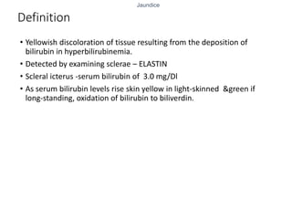
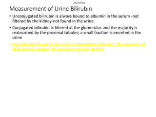
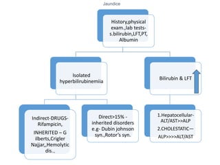
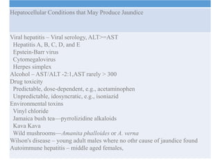
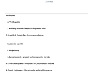
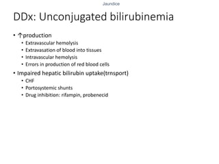
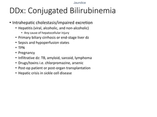
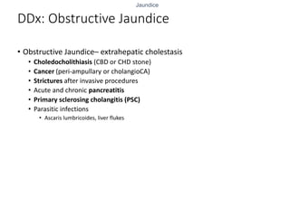
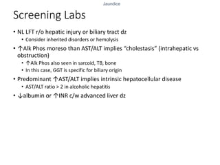
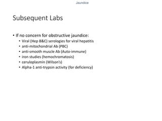
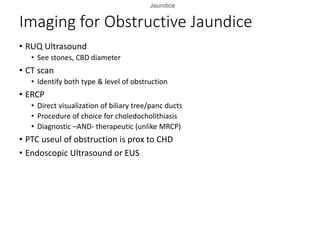
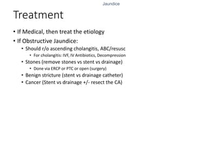
Jaundice
Jaundice
Jaundice
Overview
• Definition
• Production & metabolism
• Measurement of bilirubin – serum & urine
• Approach to the patient
• Broad Differential Diagnosis
• Work-up for “Medical” Jaundice
• Work-up if Obstructive Jaundice
Jaundice
Definition
• Yellowish discoloration of tissue resulting from the deposition of
bilirubin in hyperbilirubinemia.
• Detected by examining sclerae – ELASTIN
• Scleral icterus -serum bilirubin of 3.0 mg/Dl
• As serum bilirubin levels rise skin yellow in light-skinned &green if
long-standing, oxidation of bilirubin to biliverdin.
Jaundice
Differential diagnosis for yellowing of skin :
• Drugs – quinacrine, phenols.
• Carotenoderma -ingest excessive amounts of carotene. palms, soles,
forehead, and nasolabial folds. sparing of the sclerae.
Jaundice
Production and Metabolism of
Bilirubin:
• Bilirubin, a tetrapyrrole pigment.
Bilirubin
70-80%-senescent RBC
HEMOPROTIENS-
Myoglobin,cytochromes
Jaundice
Production steps:
Bilirubin formed in
RES
1.Opening of heme
ring
Formation of
Biliverdin by
Microsomal enzyme
2.Biliverdin
BILIRUBIN by
Cytosolic enz.
(Insoluble in plasma)
Jaundice
• To be transported in blood, bilirubin must be solubilized by
noncovalent binding to albumin.
• Bilirubin + albumin= unconjugated bilirubin transported to liver.
• Bilirubin minus albumin enters hepatocyte.
Jaundice
UNCONJ. Bilirubin Urobilinogen (80-90% excreted in feces)
10-20% portal circulation & reexcreted in liver
Jaundice
Measurement of Serum Bilirubin
• van den Bergh reaction- direct- and indirect
• the normal serum bilirubin is <1 mg/dL
• Up to 30% of the total may be direct-reacting (conjugated) bilirubin
Jaundice
Measurement of Urine Bilirubin
• Unconjugated bilirubin is always bound to albumin in the serum -not
filtered by the kidney-not found in the urine.
• Conjugated bilirubin is filtered at the glomerulus and the majority is
reabsorbed by the proximal tubules; a small fraction is excreted in the
urine
• Any bilirubin found in the urine is conjugated bilirubin. The presence of
bilirubinuria implies the presence of liver disease
Jaundice
Approach to the Patient: JAUNDICE
Jaundice
History,physical
exam.,lab tests-
s.bilirubin,LFT,PT,
Albumin
Isolated
hyperbilirubinemiia
Indirect-DRUGS-
Rifampicin,
INHERITED – G
ilberts,Crigler
Najjar,,Hemolytic
dis..,
Direct>15% -
inherited disorders
e.g- Dubin johnson
syn.,Rotor’s syn.
Bilirubin & LFT
1.Hepatocellular-
ALT/AST>>ALP
2.CHOLESTATIC—
ALP>>>>ALT/AST
Jaundice
Hepatocellular Conditions that May Produce Jaundice
Viral hepatitis – Viral serology, ALT>=AST
Hepatitis A, B, C, D, and E
Epstein-Barr virus
Cytomegalovirus
Herpes simplex
Alcohol – AST/ALT -2:1,AST rarely > 300
Drug toxicity
Predictable, dose-dependent, e.g., acetaminophen
Unpredictable, idosyncratic, e.g., isoniazid
Environmental toxins
Vinyl chloride
Jamaica bush tea—pyrrolizidine alkaloids
Kava Kava
Wild mushrooms—Amanita phalloides or A. verna
Wilson's disease – young adult males where no othr cause of jaundice found
Autoimmune hepatitis – middle aged females,
Jaundice
Initial Evaluation: History
• Jaundice, acholic stools, tea-colored urine
• Fever/chills, RUQ pain (cholangitis)
• Could lead to life-threatening septic shock
• Reasons to have hepatitis or cirrhosis?
• Alcohol, Viral, risk factors for viral hepatitis
• Exposure to toxins or offending drugs
• Inherited disorders or hemolytic conditions
• Recent blood transfusions or blood loss?
• Is patient septic or on TPN?
• Recent gallbladder surgery? (CBD injury)
Jaundice
• While ALT and AST values less than 8 times normal may be seen in
either hepatocellular or cholestatic liver disease, values 25 times
normal or higher are seen primarily in acute hepatocellular diseases.
Jaundice
Initial Evaluation: Physical Exam
• Signs of end stage liver disease (cirrhosis)
• Ascites, splenomegaly, spider angiomata, and gynecomastia
• Jaundice evident first underneath the tongue, also evident in sclerae
or skin
• Courvoisier’s sign = painless, but palpable or distended gallbladder
on exam
• Could indicate malignant obstruction
Jaundice
When the pattern of the liver tests suggests a cholestatic disorder, the next
step is to determine whether it is intra- or extrahepatic cholestasis
• Appropriate test is an ultrasound
• Biliary dilatation indicates extrahepatic cholestasis – SURGICAL
JAUNDICE
• Absence of biliary dilatation suggests intrahepatic cholestasis
Jaundice
Intrahepatic
A. Viral hepatitis
1. Fibrosing cholestatic hepatitis—hepatitis B and C
2. Hepatitis A, Epstein-Barr virus, cytomegalovirus
B. Alcoholic hepatitis
C. Drug toxicity
1. Pure cholestasis—anabolic and contraceptive steroids
2. Cholestatic hepatitis—chlorpromazine, erythromycin estolate
3. Chronic cholestasis—chlorpromazine and prochlorperazine
Jaundice
DDx: Unconjugated bilirubinemia
• ↑production
• Extravascular hemolysis
• Extravasation of blood into tissues
• Intravascular hemolysis
• Errors in production of red blood cells
• Impaired hepatic bilirubin uptake(trnsport)
• CHF
• Portosystemic shunts
• Drug inhibition: rifampin, probenecid
Jaundice
DDx: Unconjugated bilirubinemia
• Impaired bilirubin conjugation
• Gilbert’s disease
• Crigler-Najarr syndrome
• Neonatal jaundice (this is physiologic)
• Hyperthyroidism
• Estrogens
• Liver diseases
• chronic hepatitis, cirrhosis, Wilson’s disease
Jaundice
DDx: Conjugated Bilirubinemia
• Intrahepatic cholestasis/impaired excretion
• Hepatitis (viral, alcoholic, and non-alcoholic)
• Any cause of hepatocellular injury
• Primary biliary cirrhosis or end-stage liver dz
• Sepsis and hypoperfusion states
• TPN
• Pregnancy
• Infiltrative dz: TB, amyloid, sarcoid, lymphoma
• Drugs/toxins i.e. chlorpromazine, arsenic
• Post-op patient or post-organ transplantation
• Hepatic crisis in sickle cell disease
Jaundice
DDx: Obstructive Jaundice
• Obstructive Jaundice– extrahepatic cholestasis
• Choledocholithiasis (CBD or CHD stone)
• Cancer (peri-ampullary or cholangioCA)
• Strictures after invasive procedures
• Acute and chronic pancreatitis
• Primary sclerosing cholangitis (PSC)
• Parasitic infections
• Ascaris lumbricoides, liver flukes
Jaundice
Screening Labs
• NL LFT r/o hepatic injury or biliary tract dz
• Consider inherited disorders or hemolysis
• ↑Alk Phos moreso than AST/ALT implies “cholestasis” (intrahepatic vs
obstruction)
• ↑Alk Phos also seen in sarcoid, TB, bone
• In this case, GGT is specific for biliary origin
• Predominant ↑AST/ALT implies intrinsic hepatocellular disease
• AST/ALT ratio > 2 in alcoholic hepatitis
• ↓albumin or ↑INR c/w advanced liver dz
Jaundice
Subsequent Labs
• If no concern for obstructive jaundice:
• Viral (Hep B&C) serologies for viral hepatitis
• anti-mitochondrial Ab (PBC)
• anti-smooth muscle Ab (Auto-immune)
• iron studies (hemochromatosis)
• ceruloplasmin (Wilson’s)
• Alpha-1 anti-trypsin activity (for deficiency)
Jaundice
Imaging for Obstructive Jaundice
• RUQ Ultrasound
• See stones, CBD diameter
• CT scan
• Identify both type & level of obstruction
• ERCP
• Direct visualization of biliary tree/panc ducts
• Procedure of choice for choledocholithiasis
• Diagnostic –AND- therapeutic (unlike MRCP)
• PTC useul of obstruction is prox to CHD
• Endoscopic Ultrasound or EUS
Jaundice
Treatment
• If Medical, then treat the etiology
• If Obstructive Jaundice:
• Should r/o ascending cholangitis, ABC/resusc
• For cholangitis: IVF, IV Antibiotics, Decompression
• Stones (remove stones vs stent vs drainage)
• Done via ERCP or PTC or open (surgery)
• Benign stricture (stent vs drainage catheter)
• Cancer (Stent vs drainage +/- resect the CA)
Jaundice
Thanks

JAUNDICE.pptx

  • 1.
  • 2.
    Jaundice Overview • Definition • Production& metabolism • Measurement of bilirubin – serum & urine • Approach to the patient • Broad Differential Diagnosis • Work-up for “Medical” Jaundice • Work-up if Obstructive Jaundice
  • 3.
    Jaundice Definition • Yellowish discolorationof tissue resulting from the deposition of bilirubin in hyperbilirubinemia. • Detected by examining sclerae – ELASTIN • Scleral icterus -serum bilirubin of 3.0 mg/Dl • As serum bilirubin levels rise skin yellow in light-skinned &green if long-standing, oxidation of bilirubin to biliverdin.
  • 4.
    Jaundice Differential diagnosis foryellowing of skin : • Drugs – quinacrine, phenols. • Carotenoderma -ingest excessive amounts of carotene. palms, soles, forehead, and nasolabial folds. sparing of the sclerae.
  • 5.
    Jaundice Production and Metabolismof Bilirubin: • Bilirubin, a tetrapyrrole pigment. Bilirubin 70-80%-senescent RBC HEMOPROTIENS- Myoglobin,cytochromes
  • 6.
    Jaundice Production steps: Bilirubin formedin RES 1.Opening of heme ring Formation of Biliverdin by Microsomal enzyme 2.Biliverdin BILIRUBIN by Cytosolic enz. (Insoluble in plasma)
  • 7.
    Jaundice • To betransported in blood, bilirubin must be solubilized by noncovalent binding to albumin. • Bilirubin + albumin= unconjugated bilirubin transported to liver. • Bilirubin minus albumin enters hepatocyte.
  • 8.
    Jaundice UNCONJ. Bilirubin Urobilinogen(80-90% excreted in feces) 10-20% portal circulation & reexcreted in liver
  • 9.
    Jaundice Measurement of SerumBilirubin • van den Bergh reaction- direct- and indirect • the normal serum bilirubin is <1 mg/dL • Up to 30% of the total may be direct-reacting (conjugated) bilirubin
  • 10.
    Jaundice Measurement of UrineBilirubin • Unconjugated bilirubin is always bound to albumin in the serum -not filtered by the kidney-not found in the urine. • Conjugated bilirubin is filtered at the glomerulus and the majority is reabsorbed by the proximal tubules; a small fraction is excreted in the urine • Any bilirubin found in the urine is conjugated bilirubin. The presence of bilirubinuria implies the presence of liver disease
  • 11.
    Jaundice Approach to thePatient: JAUNDICE
  • 12.
    Jaundice History,physical exam.,lab tests- s.bilirubin,LFT,PT, Albumin Isolated hyperbilirubinemiia Indirect-DRUGS- Rifampicin, INHERITED –G ilberts,Crigler Najjar,,Hemolytic dis.., Direct>15% - inherited disorders e.g- Dubin johnson syn.,Rotor’s syn. Bilirubin & LFT 1.Hepatocellular- ALT/AST>>ALP 2.CHOLESTATIC— ALP>>>>ALT/AST
  • 13.
    Jaundice Hepatocellular Conditions thatMay Produce Jaundice Viral hepatitis – Viral serology, ALT>=AST Hepatitis A, B, C, D, and E Epstein-Barr virus Cytomegalovirus Herpes simplex Alcohol – AST/ALT -2:1,AST rarely > 300 Drug toxicity Predictable, dose-dependent, e.g., acetaminophen Unpredictable, idosyncratic, e.g., isoniazid Environmental toxins Vinyl chloride Jamaica bush tea—pyrrolizidine alkaloids Kava Kava Wild mushrooms—Amanita phalloides or A. verna Wilson's disease – young adult males where no othr cause of jaundice found Autoimmune hepatitis – middle aged females,
  • 14.
    Jaundice Initial Evaluation: History •Jaundice, acholic stools, tea-colored urine • Fever/chills, RUQ pain (cholangitis) • Could lead to life-threatening septic shock • Reasons to have hepatitis or cirrhosis? • Alcohol, Viral, risk factors for viral hepatitis • Exposure to toxins or offending drugs • Inherited disorders or hemolytic conditions • Recent blood transfusions or blood loss? • Is patient septic or on TPN? • Recent gallbladder surgery? (CBD injury)
  • 15.
    Jaundice • While ALTand AST values less than 8 times normal may be seen in either hepatocellular or cholestatic liver disease, values 25 times normal or higher are seen primarily in acute hepatocellular diseases.
  • 16.
    Jaundice Initial Evaluation: PhysicalExam • Signs of end stage liver disease (cirrhosis) • Ascites, splenomegaly, spider angiomata, and gynecomastia • Jaundice evident first underneath the tongue, also evident in sclerae or skin • Courvoisier’s sign = painless, but palpable or distended gallbladder on exam • Could indicate malignant obstruction
  • 17.
    Jaundice When the patternof the liver tests suggests a cholestatic disorder, the next step is to determine whether it is intra- or extrahepatic cholestasis • Appropriate test is an ultrasound • Biliary dilatation indicates extrahepatic cholestasis – SURGICAL JAUNDICE • Absence of biliary dilatation suggests intrahepatic cholestasis
  • 18.
    Jaundice Intrahepatic A. Viral hepatitis 1.Fibrosing cholestatic hepatitis—hepatitis B and C 2. Hepatitis A, Epstein-Barr virus, cytomegalovirus B. Alcoholic hepatitis C. Drug toxicity 1. Pure cholestasis—anabolic and contraceptive steroids 2. Cholestatic hepatitis—chlorpromazine, erythromycin estolate 3. Chronic cholestasis—chlorpromazine and prochlorperazine
  • 19.
    Jaundice DDx: Unconjugated bilirubinemia •↑production • Extravascular hemolysis • Extravasation of blood into tissues • Intravascular hemolysis • Errors in production of red blood cells • Impaired hepatic bilirubin uptake(trnsport) • CHF • Portosystemic shunts • Drug inhibition: rifampin, probenecid
  • 20.
    Jaundice DDx: Unconjugated bilirubinemia •Impaired bilirubin conjugation • Gilbert’s disease • Crigler-Najarr syndrome • Neonatal jaundice (this is physiologic) • Hyperthyroidism • Estrogens • Liver diseases • chronic hepatitis, cirrhosis, Wilson’s disease
  • 21.
    Jaundice DDx: Conjugated Bilirubinemia •Intrahepatic cholestasis/impaired excretion • Hepatitis (viral, alcoholic, and non-alcoholic) • Any cause of hepatocellular injury • Primary biliary cirrhosis or end-stage liver dz • Sepsis and hypoperfusion states • TPN • Pregnancy • Infiltrative dz: TB, amyloid, sarcoid, lymphoma • Drugs/toxins i.e. chlorpromazine, arsenic • Post-op patient or post-organ transplantation • Hepatic crisis in sickle cell disease
  • 22.
    Jaundice DDx: Obstructive Jaundice •Obstructive Jaundice– extrahepatic cholestasis • Choledocholithiasis (CBD or CHD stone) • Cancer (peri-ampullary or cholangioCA) • Strictures after invasive procedures • Acute and chronic pancreatitis • Primary sclerosing cholangitis (PSC) • Parasitic infections • Ascaris lumbricoides, liver flukes
  • 23.
    Jaundice Screening Labs • NLLFT r/o hepatic injury or biliary tract dz • Consider inherited disorders or hemolysis • ↑Alk Phos moreso than AST/ALT implies “cholestasis” (intrahepatic vs obstruction) • ↑Alk Phos also seen in sarcoid, TB, bone • In this case, GGT is specific for biliary origin • Predominant ↑AST/ALT implies intrinsic hepatocellular disease • AST/ALT ratio > 2 in alcoholic hepatitis • ↓albumin or ↑INR c/w advanced liver dz
  • 24.
    Jaundice Subsequent Labs • Ifno concern for obstructive jaundice: • Viral (Hep B&C) serologies for viral hepatitis • anti-mitochondrial Ab (PBC) • anti-smooth muscle Ab (Auto-immune) • iron studies (hemochromatosis) • ceruloplasmin (Wilson’s) • Alpha-1 anti-trypsin activity (for deficiency)
  • 25.
    Jaundice Imaging for ObstructiveJaundice • RUQ Ultrasound • See stones, CBD diameter • CT scan • Identify both type & level of obstruction • ERCP • Direct visualization of biliary tree/panc ducts • Procedure of choice for choledocholithiasis • Diagnostic –AND- therapeutic (unlike MRCP) • PTC useul of obstruction is prox to CHD • Endoscopic Ultrasound or EUS
  • 26.
    Jaundice Treatment • If Medical,then treat the etiology • If Obstructive Jaundice: • Should r/o ascending cholangitis, ABC/resusc • For cholangitis: IVF, IV Antibiotics, Decompression • Stones (remove stones vs stent vs drainage) • Done via ERCP or PTC or open (surgery) • Benign stricture (stent vs drainage catheter) • Cancer (Stent vs drainage +/- resect the CA)
  • 27.