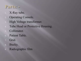
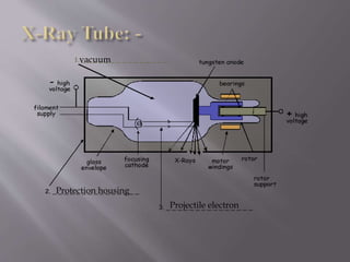
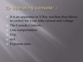
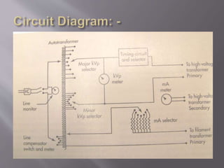
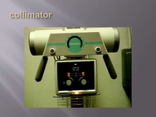
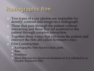
SlideShare a Scribd company logo
BY: Huzaifa Atique
Sir Syed University of Engineering and
Technology
1. X-Ray tube.
2. Operating Console.
3. High Voltage transformer.
4. Tube Head or Protective Housing.
5. Collimator.
6. Patient Table.
7. Grid.
8. Bucky.
9. Radiographic film
vacuum
Protection housing
Projectile electron
 It is an important component of x-ray machine
which is inaccessible as it is contained in a
protective housing.
 It is a vacuum tube.
 Early tubes were called Crook’s tube and were not
vacuum.
 Modern tubes are known as Coolidge tube and are
vacuumed.
 There are two primary parts.
 1) the cathode
 2) the Anode.
 It is an apparatus in X-Ray machine that allows
to control the x-ray tube current and voltage.
 The Console Controls: -
 Line compensation.
 kVp.
 mA.
 Exposure time.
 It have 5 further parts:
1. Line compensator.
2. Auto transformer.
3. Current source.
4. Voltage source.
5. Exposure time.
 Most machine are designed to operate at 220
volts while some will work with 110 volts or
440 volts
 The power company often cannot provide
exactly 220 volts at all times.
 Because of variations in power distribution to
the hospital and in power consumption by the
various sections of the hospital, the voltage to
the x-ray unit may vary by 5%, which will
result in large variations in x-ray output.
 The power supplied to
x-ray machine is
delivered to a special
transformer called an
Autotransformer.
 It works on the
principle of
electromagnetic
induction but is very
different from
conventional
transformer.
 It has only one winding and one core.
 The single winding has number of
connections, or electric taps.
 The purpose to use the Autotransformer
is to overcome induction losses.
 Its value ranges from 0 to 400V.
 X-ray console have adjustments labeled major
kVp and minor kVp.
 By the combinations of these two, we can get
the required kVp.
 The low voltage becomes input to the high
voltage step up transformer.
 The kVp meter is placed across the output
terminal of autotransformer.
 The tube current, the number of electrons
crossing from the cathode to anode per second
is measured in milli Amperes (mA).
 The quantity of electrons is determined by
filament temperature.
 The filament normally operates at currents
between 3 and 5 A.
 The Tube Current is controlled through a
separate circuit called the filament circuit.
 The transformer used for filament is step
down .
 It consists of a mechanical or electronic device
whose action is to make and break the high
voltage across the tube on the primary side of
the high voltage section.
 There are five types of timers:
 Mechanical Timers
 Synchronous Timers
 Electronic Timers
 mAs Timers
 Phototimers.
 The high voltage transformer is a step-up
transformer.
 There will be more winding on the secondary side
compared to the primary side.
 The ratio of windings is referred to as the turns
ratio.
 The only difference between the primary and
secondary waveforms is the amplitude.
 The turn ratio for most x-ray high voltage
transformers is between 500 and 1000.
 The primary voltage is measured in volts, and
secondary in kilovolts.
 X-ray tube is always mounted inside a lead-
lined protective housing that is designed to:
 Prevent excessive radiation exposure.
 Prevent electric shock to the patient and operator
(technologist)
 Incorporates specially designed high-voltage
receptacles.
 Provides mechanical support for the x-ray tube
and protects it from damage.
 Some tube housings contain oil in which the
tube is bathed.
 Some tube housings contain a cooling fan to
air-cool the tube.
 When properly designed, they reduce the level
of leakage radiation to less than 100 mR/hr
at 1 meter when operated at maximum
conditions.
 The Collimator is attached to the x-ray tube
below the glass window where the useful beam
is emitted.
 Lead shutters are used to restrict the beam.
 Its purpose is to minimize field of view, to
avoid un necessary exposure by using lead
plates.
 By virtue of function and material, collimator
and grid are same but they have different
location.
 It is made up of lead.
 It is located just after patient.
 It is used to destroy scattered radiation from
the body.
 Some of the X-Rays entering the body of a
patient are actually scattered and no longer
travel in a straight line, this scattering can
cause blurring of X-Ray image.
 The rays which are at 90’ can be passed .
 A Bucky is a component of x-ray units that
holds the x-ray film cassette and moves
the grid during x-ray exposure. The motion
keeps the lead strips from being seen on the x-
ray picture.
The name refers to Dr. Gustave Bucky who
invented the use of filter grids in 1913.
 Two types of x-ray photon are respnsible for
density, contrast and image on a radiograph.
 Those that pass through the patient without
interacting and those that are scattered in the
patient through compton interaction.
 Together these x-rays that exit from the patient and
intersect the film are called Remnant x-rays.
 Film Construction
 Radiographic Film has two basic parts.
 Base
 Emulsion
 Most film has two layers of emulsion so it is referred to as
Double Emulsion Film
• The emulsion is the heart of the film. The x-rays
or light from the intensifying screens interact
with the emulsion and transfer information to
the film.
 Thank you

More Related Content

What's hot

Tomographic equipment
Tomographic equipmentTomographic equipment
Tomographic equipment
Self
 
Viewing and storage of x ray film
Viewing and storage of x ray filmViewing and storage of x ray film
Viewing and storage of x ray film
Bangladesh Agricultural University,Mymemsingh
 
Multisection tomography and Transaxial Tomography
Multisection tomography and Transaxial TomographyMultisection tomography and Transaxial Tomography
Multisection tomography and Transaxial Tomography
Self
 
Rectifiers
RectifiersRectifiers
Rectifiers
Madhu Sudana
 
X ray generators
X ray generatorsX ray generators
X ray generators
kartheek guthikonda
 
Beam restricted device and filter used in x ray
Beam restricted device and filter used in x rayBeam restricted device and filter used in x ray
Beam restricted device and filter used in x ray
SushilPattar
 
Generations of CT scanner
Generations of  CT scannerGenerations of  CT scanner
Generations of CT scanner
sadhamhussain52
 
Portable n mobile unit
Portable n mobile unitPortable n mobile unit
Portable n mobile unit
Sudil Paudyal
 
Radiographic exposure and image quality
Radiographic exposure and image qualityRadiographic exposure and image quality
Radiographic exposure and image quality
Rad Tech
 
image intensifier.pptx
image intensifier.pptximage intensifier.pptx
image intensifier.pptx
GajananWattamwar1
 
Mobile & portable radiography
Mobile & portable radiographyMobile & portable radiography
Mobile & portable radiography
Priyanka Parimala
 
Computed radiography
Computed radiographyComputed radiography
Computed radiography
Illyasmk
 
Ct Generations
Ct  GenerationsCt  Generations
Ct Generations
Dr. Mohit Goel
 
X ray tube
X ray tubeX ray tube
Magnification(macro and micro radiography), distortion
Magnification(macro and micro radiography), distortionMagnification(macro and micro radiography), distortion
Magnification(macro and micro radiography), distortion
parthajyotidas11
 
Ward radiography.pptx
Ward radiography.pptxWard radiography.pptx
Ward radiography.pptx
Ritupanta1
 
The control of scattered radiation
The control of scattered radiationThe control of scattered radiation
The control of scattered radiation
Nitish Rajput
 
Radiographic film
Radiographic filmRadiographic film
Radiographic film
Rad Tech
 
Spectral matchin
Spectral matchinSpectral matchin
Spectral matchin
Shemiver Delino
 
Radiation protection & Personnel Monitoring Devices
Radiation protection & Personnel Monitoring DevicesRadiation protection & Personnel Monitoring Devices
Radiation protection & Personnel Monitoring Devices
Sheetal R Kashid
 

What's hot (20)

Tomographic equipment
Tomographic equipmentTomographic equipment
Tomographic equipment
 
Viewing and storage of x ray film
Viewing and storage of x ray filmViewing and storage of x ray film
Viewing and storage of x ray film
 
Multisection tomography and Transaxial Tomography
Multisection tomography and Transaxial TomographyMultisection tomography and Transaxial Tomography
Multisection tomography and Transaxial Tomography
 
Rectifiers
RectifiersRectifiers
Rectifiers
 
X ray generators
X ray generatorsX ray generators
X ray generators
 
Beam restricted device and filter used in x ray
Beam restricted device and filter used in x rayBeam restricted device and filter used in x ray
Beam restricted device and filter used in x ray
 
Generations of CT scanner
Generations of  CT scannerGenerations of  CT scanner
Generations of CT scanner
 
Portable n mobile unit
Portable n mobile unitPortable n mobile unit
Portable n mobile unit
 
Radiographic exposure and image quality
Radiographic exposure and image qualityRadiographic exposure and image quality
Radiographic exposure and image quality
 
image intensifier.pptx
image intensifier.pptximage intensifier.pptx
image intensifier.pptx
 
Mobile & portable radiography
Mobile & portable radiographyMobile & portable radiography
Mobile & portable radiography
 
Computed radiography
Computed radiographyComputed radiography
Computed radiography
 
Ct Generations
Ct  GenerationsCt  Generations
Ct Generations
 
X ray tube
X ray tubeX ray tube
X ray tube
 
Magnification(macro and micro radiography), distortion
Magnification(macro and micro radiography), distortionMagnification(macro and micro radiography), distortion
Magnification(macro and micro radiography), distortion
 
Ward radiography.pptx
Ward radiography.pptxWard radiography.pptx
Ward radiography.pptx
 
The control of scattered radiation
The control of scattered radiationThe control of scattered radiation
The control of scattered radiation
 
Radiographic film
Radiographic filmRadiographic film
Radiographic film
 
Spectral matchin
Spectral matchinSpectral matchin
Spectral matchin
 
Radiation protection & Personnel Monitoring Devices
Radiation protection & Personnel Monitoring DevicesRadiation protection & Personnel Monitoring Devices
Radiation protection & Personnel Monitoring Devices
 

Viewers also liked

Introduction to the parts of x ray machine
Introduction to the parts of x ray machineIntroduction to the parts of x ray machine
Introduction to the parts of x ray machine
Huzaifa Oxford
 
Introduction to X-Ray Machine
Introduction to X-Ray MachineIntroduction to X-Ray Machine
Introduction to X-Ray Machine
Syed Jamal
 
Components of X-Ray Machine
Components of X-Ray MachineComponents of X-Ray Machine
Components of X-Ray Machine
Syed Jamal
 
Basic of x ray production
Basic of x ray productionBasic of x ray production
Basic of x ray production
Khalis Karim
 
General awareness of diagnostic radiology
General awareness of diagnostic radiologyGeneral awareness of diagnostic radiology
General awareness of diagnostic radiology
Arun Kalpaka
 
AXI_X-Line 3D
AXI_X-Line 3DAXI_X-Line 3D
AXI_X-Line 3D
Dario Cordova
 
Cr & dr
Cr & drCr & dr
Moving from analog to digital 2.0
Moving from analog to digital 2.0Moving from analog to digital 2.0
Moving from analog to digital 2.0
stacypalmer
 
Specialised Techniques in Oral Radiology
Specialised Techniques in Oral RadiologySpecialised Techniques in Oral Radiology
Specialised Techniques in Oral Radiology
IAU Dent
 
Digital Radiography
Digital RadiographyDigital Radiography
Digital Radiography
ambar hazarika
 
Diagnostic imaging clinics
Diagnostic imaging clinicsDiagnostic imaging clinics
Diagnostic imaging clinics
Adenike Olatunji-Akioye
 
Intro to CT scan machine
Intro to CT scan machineIntro to CT scan machine
Intro to CT scan machine
Syed Jamal
 
ทีมหมอครอบครัว(Fct)พิมพ์ครั้งที่3
ทีมหมอครอบครัว(Fct)พิมพ์ครั้งที่3ทีมหมอครอบครัว(Fct)พิมพ์ครั้งที่3
ทีมหมอครอบครัว(Fct)พิมพ์ครั้งที่3
Auamporn Junthong
 
Spetrophotometer
SpetrophotometerSpetrophotometer
Spetrophotometer
Tapeshwar Yadav
 
L 4 ct physics
L 4  ct physicsL 4  ct physics
L 4 ct physics
Shahid Younas
 
Skm symps poster 2015 spect ct
Skm symps poster 2015 spect ctSkm symps poster 2015 spect ct
Skm symps poster 2015 spect ct
Shahid Younas
 
L 1 ct physics
L 1  ct physicsL 1  ct physics
L 1 ct physics
Shahid Younas
 
MRI
MRIMRI
L 6 ct physics
L 6 ct physicsL 6 ct physics
L 6 ct physics
Shahid Younas
 
Digital radiography-Avinesh Shrestha
Digital radiography-Avinesh ShresthaDigital radiography-Avinesh Shrestha
Digital radiography-Avinesh Shrestha
Avinesh Shrestha
 

Viewers also liked (20)

Introduction to the parts of x ray machine
Introduction to the parts of x ray machineIntroduction to the parts of x ray machine
Introduction to the parts of x ray machine
 
Introduction to X-Ray Machine
Introduction to X-Ray MachineIntroduction to X-Ray Machine
Introduction to X-Ray Machine
 
Components of X-Ray Machine
Components of X-Ray MachineComponents of X-Ray Machine
Components of X-Ray Machine
 
Basic of x ray production
Basic of x ray productionBasic of x ray production
Basic of x ray production
 
General awareness of diagnostic radiology
General awareness of diagnostic radiologyGeneral awareness of diagnostic radiology
General awareness of diagnostic radiology
 
AXI_X-Line 3D
AXI_X-Line 3DAXI_X-Line 3D
AXI_X-Line 3D
 
Cr & dr
Cr & drCr & dr
Cr & dr
 
Moving from analog to digital 2.0
Moving from analog to digital 2.0Moving from analog to digital 2.0
Moving from analog to digital 2.0
 
Specialised Techniques in Oral Radiology
Specialised Techniques in Oral RadiologySpecialised Techniques in Oral Radiology
Specialised Techniques in Oral Radiology
 
Digital Radiography
Digital RadiographyDigital Radiography
Digital Radiography
 
Diagnostic imaging clinics
Diagnostic imaging clinicsDiagnostic imaging clinics
Diagnostic imaging clinics
 
Intro to CT scan machine
Intro to CT scan machineIntro to CT scan machine
Intro to CT scan machine
 
ทีมหมอครอบครัว(Fct)พิมพ์ครั้งที่3
ทีมหมอครอบครัว(Fct)พิมพ์ครั้งที่3ทีมหมอครอบครัว(Fct)พิมพ์ครั้งที่3
ทีมหมอครอบครัว(Fct)พิมพ์ครั้งที่3
 
Spetrophotometer
SpetrophotometerSpetrophotometer
Spetrophotometer
 
L 4 ct physics
L 4  ct physicsL 4  ct physics
L 4 ct physics
 
Skm symps poster 2015 spect ct
Skm symps poster 2015 spect ctSkm symps poster 2015 spect ct
Skm symps poster 2015 spect ct
 
L 1 ct physics
L 1  ct physicsL 1  ct physics
L 1 ct physics
 
MRI
MRIMRI
MRI
 
L 6 ct physics
L 6 ct physicsL 6 ct physics
L 6 ct physics
 
Digital radiography-Avinesh Shrestha
Digital radiography-Avinesh ShresthaDigital radiography-Avinesh Shrestha
Digital radiography-Avinesh Shrestha
 

Similar to introductiontothepartsofx-raymachine-140825094645-phpapp02

X ray
X rayX ray
Production of x rays
Production of x raysProduction of x rays
X ray machine l
X ray machine lX ray machine l
X ray machine l
DiNa Amin
 
Bps xray mammo dsa
Bps xray mammo dsaBps xray mammo dsa
Bps xray mammo dsa
RMLIMS
 
X-Ray Tube complete-1.pptx
X-Ray Tube complete-1.pptxX-Ray Tube complete-1.pptx
X-Ray Tube complete-1.pptx
NMdcat2021
 
x ray machine ppt
x ray machine pptx ray machine ppt
x ray machine ppt
hunde7
 
Portable and mobile radiographic equipments [Autosaved].pptx
Portable and mobile radiographic equipments [Autosaved].pptxPortable and mobile radiographic equipments [Autosaved].pptx
Portable and mobile radiographic equipments [Autosaved].pptx
sarita gaire
 
Portable and mobile radiographic equipments [Autosaved].pptx
Portable and mobile radiographic equipments [Autosaved].pptxPortable and mobile radiographic equipments [Autosaved].pptx
Portable and mobile radiographic equipments [Autosaved].pptx
SaritaGaire1
 
Linear Accelerators.pptx
Linear Accelerators.pptxLinear Accelerators.pptx
Linear Accelerators.pptx
SaikatChakraborty80
 
basic of angiography physics and equipement.pdf
basic of angiography physics and equipement.pdfbasic of angiography physics and equipement.pdf
basic of angiography physics and equipement.pdf
naima SENHOU
 
Production of x rays
Production of x rays Production of x rays
Production of x rays
DrMeghaBahal
 
X ray tube, cassette and screens
X ray tube, cassette and screensX ray tube, cassette and screens
X ray tube, cassette and screens
Tarun Goyal
 
The x ray imaging system
The x ray imaging systemThe x ray imaging system
The x ray imaging system
Rad Tech
 
X ray generator, circuits
X  ray generator, circuitsX  ray generator, circuits
X ray generator, circuits
Anjan Dangal
 
X ray generator basic
X ray generator basicX ray generator basic
X ray generator basic
Md. Serajul islam bablu
 
X- Ray physics- X-Ray Tube, Transformer, Generator and Rectifiers by kajalsra...
X- Ray physics- X-Ray Tube, Transformer, Generator and Rectifiers by kajalsra...X- Ray physics- X-Ray Tube, Transformer, Generator and Rectifiers by kajalsra...
X- Ray physics- X-Ray Tube, Transformer, Generator and Rectifiers by kajalsra...
DrKajalLimbad
 
Linear accelerator.pptx
Linear accelerator.pptxLinear accelerator.pptx
Linear accelerator.pptx
ministry of health
 
X ray tube
X ray tubeX ray tube
X ray tube
Rad Tech
 
Auto speed control of solar car at specified locations like school zones
Auto speed control of solar car at specified locations like school zonesAuto speed control of solar car at specified locations like school zones
Auto speed control of solar car at specified locations like school zones
Vinod Mali
 
X ray Generator physics behind x ray generation.ppt
X ray Generator physics behind x ray generation.pptX ray Generator physics behind x ray generation.ppt
X ray Generator physics behind x ray generation.ppt
dypradio
 

Similar to introductiontothepartsofx-raymachine-140825094645-phpapp02 (20)

X ray
X rayX ray
X ray
 
Production of x rays
Production of x raysProduction of x rays
Production of x rays
 
X ray machine l
X ray machine lX ray machine l
X ray machine l
 
Bps xray mammo dsa
Bps xray mammo dsaBps xray mammo dsa
Bps xray mammo dsa
 
X-Ray Tube complete-1.pptx
X-Ray Tube complete-1.pptxX-Ray Tube complete-1.pptx
X-Ray Tube complete-1.pptx
 
x ray machine ppt
x ray machine pptx ray machine ppt
x ray machine ppt
 
Portable and mobile radiographic equipments [Autosaved].pptx
Portable and mobile radiographic equipments [Autosaved].pptxPortable and mobile radiographic equipments [Autosaved].pptx
Portable and mobile radiographic equipments [Autosaved].pptx
 
Portable and mobile radiographic equipments [Autosaved].pptx
Portable and mobile radiographic equipments [Autosaved].pptxPortable and mobile radiographic equipments [Autosaved].pptx
Portable and mobile radiographic equipments [Autosaved].pptx
 
Linear Accelerators.pptx
Linear Accelerators.pptxLinear Accelerators.pptx
Linear Accelerators.pptx
 
basic of angiography physics and equipement.pdf
basic of angiography physics and equipement.pdfbasic of angiography physics and equipement.pdf
basic of angiography physics and equipement.pdf
 
Production of x rays
Production of x rays Production of x rays
Production of x rays
 
X ray tube, cassette and screens
X ray tube, cassette and screensX ray tube, cassette and screens
X ray tube, cassette and screens
 
The x ray imaging system
The x ray imaging systemThe x ray imaging system
The x ray imaging system
 
X ray generator, circuits
X  ray generator, circuitsX  ray generator, circuits
X ray generator, circuits
 
X ray generator basic
X ray generator basicX ray generator basic
X ray generator basic
 
X- Ray physics- X-Ray Tube, Transformer, Generator and Rectifiers by kajalsra...
X- Ray physics- X-Ray Tube, Transformer, Generator and Rectifiers by kajalsra...X- Ray physics- X-Ray Tube, Transformer, Generator and Rectifiers by kajalsra...
X- Ray physics- X-Ray Tube, Transformer, Generator and Rectifiers by kajalsra...
 
Linear accelerator.pptx
Linear accelerator.pptxLinear accelerator.pptx
Linear accelerator.pptx
 
X ray tube
X ray tubeX ray tube
X ray tube
 
Auto speed control of solar car at specified locations like school zones
Auto speed control of solar car at specified locations like school zonesAuto speed control of solar car at specified locations like school zones
Auto speed control of solar car at specified locations like school zones
 
X ray Generator physics behind x ray generation.ppt
X ray Generator physics behind x ray generation.pptX ray Generator physics behind x ray generation.ppt
X ray Generator physics behind x ray generation.ppt
 

introductiontothepartsofx-raymachine-140825094645-phpapp02

  • 1. BY: Huzaifa Atique Sir Syed University of Engineering and Technology
  • 2. 1. X-Ray tube. 2. Operating Console. 3. High Voltage transformer. 4. Tube Head or Protective Housing. 5. Collimator. 6. Patient Table. 7. Grid. 8. Bucky. 9. Radiographic film
  • 4.  It is an important component of x-ray machine which is inaccessible as it is contained in a protective housing.  It is a vacuum tube.  Early tubes were called Crook’s tube and were not vacuum.  Modern tubes are known as Coolidge tube and are vacuumed.  There are two primary parts.  1) the cathode  2) the Anode.
  • 5.  It is an apparatus in X-Ray machine that allows to control the x-ray tube current and voltage.  The Console Controls: -  Line compensation.  kVp.  mA.  Exposure time.
  • 6.
  • 7.  It have 5 further parts: 1. Line compensator. 2. Auto transformer. 3. Current source. 4. Voltage source. 5. Exposure time.
  • 8.  Most machine are designed to operate at 220 volts while some will work with 110 volts or 440 volts  The power company often cannot provide exactly 220 volts at all times.  Because of variations in power distribution to the hospital and in power consumption by the various sections of the hospital, the voltage to the x-ray unit may vary by 5%, which will result in large variations in x-ray output.
  • 9.  The power supplied to x-ray machine is delivered to a special transformer called an Autotransformer.  It works on the principle of electromagnetic induction but is very different from conventional transformer.
  • 10.  It has only one winding and one core.  The single winding has number of connections, or electric taps.  The purpose to use the Autotransformer is to overcome induction losses.  Its value ranges from 0 to 400V.
  • 11.
  • 12.  X-ray console have adjustments labeled major kVp and minor kVp.  By the combinations of these two, we can get the required kVp.  The low voltage becomes input to the high voltage step up transformer.  The kVp meter is placed across the output terminal of autotransformer.
  • 13.
  • 14.  The tube current, the number of electrons crossing from the cathode to anode per second is measured in milli Amperes (mA).  The quantity of electrons is determined by filament temperature.  The filament normally operates at currents between 3 and 5 A.  The Tube Current is controlled through a separate circuit called the filament circuit.  The transformer used for filament is step down .
  • 15.  It consists of a mechanical or electronic device whose action is to make and break the high voltage across the tube on the primary side of the high voltage section.  There are five types of timers:  Mechanical Timers  Synchronous Timers  Electronic Timers  mAs Timers  Phototimers.
  • 16.  The high voltage transformer is a step-up transformer.  There will be more winding on the secondary side compared to the primary side.  The ratio of windings is referred to as the turns ratio.  The only difference between the primary and secondary waveforms is the amplitude.  The turn ratio for most x-ray high voltage transformers is between 500 and 1000.  The primary voltage is measured in volts, and secondary in kilovolts.
  • 17.
  • 18.  X-ray tube is always mounted inside a lead- lined protective housing that is designed to:  Prevent excessive radiation exposure.  Prevent electric shock to the patient and operator (technologist)  Incorporates specially designed high-voltage receptacles.  Provides mechanical support for the x-ray tube and protects it from damage.  Some tube housings contain oil in which the tube is bathed.  Some tube housings contain a cooling fan to air-cool the tube.
  • 19.  When properly designed, they reduce the level of leakage radiation to less than 100 mR/hr at 1 meter when operated at maximum conditions.
  • 20.
  • 21.  The Collimator is attached to the x-ray tube below the glass window where the useful beam is emitted.  Lead shutters are used to restrict the beam.  Its purpose is to minimize field of view, to avoid un necessary exposure by using lead plates.
  • 22.  By virtue of function and material, collimator and grid are same but they have different location.  It is made up of lead.  It is located just after patient.  It is used to destroy scattered radiation from the body.
  • 23.
  • 24.  Some of the X-Rays entering the body of a patient are actually scattered and no longer travel in a straight line, this scattering can cause blurring of X-Ray image.  The rays which are at 90’ can be passed .
  • 25.  A Bucky is a component of x-ray units that holds the x-ray film cassette and moves the grid during x-ray exposure. The motion keeps the lead strips from being seen on the x- ray picture. The name refers to Dr. Gustave Bucky who invented the use of filter grids in 1913.
  • 26.  Two types of x-ray photon are respnsible for density, contrast and image on a radiograph.  Those that pass through the patient without interacting and those that are scattered in the patient through compton interaction.  Together these x-rays that exit from the patient and intersect the film are called Remnant x-rays.  Film Construction  Radiographic Film has two basic parts.  Base  Emulsion  Most film has two layers of emulsion so it is referred to as Double Emulsion Film
  • 27. • The emulsion is the heart of the film. The x-rays or light from the intensifying screens interact with the emulsion and transfer information to the film.