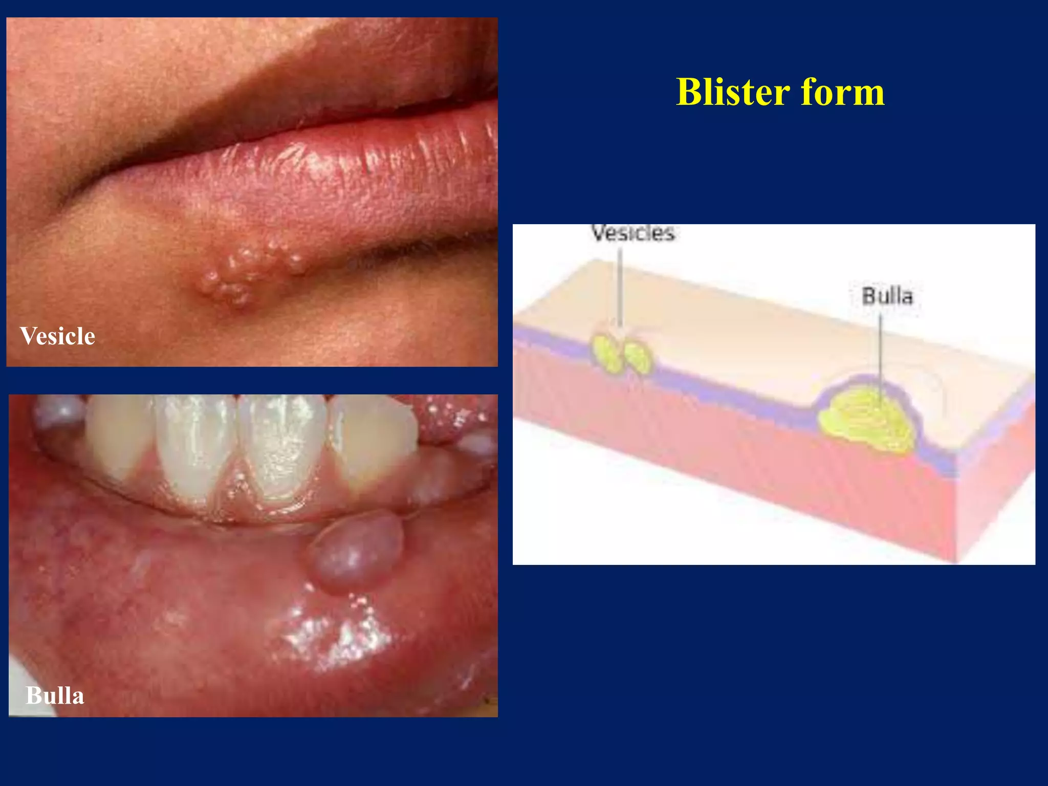
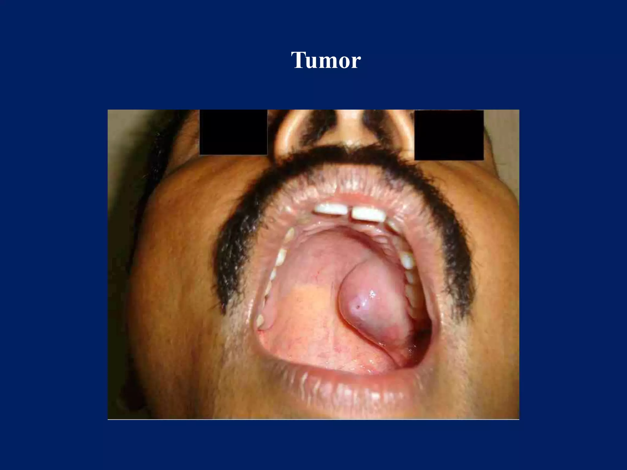
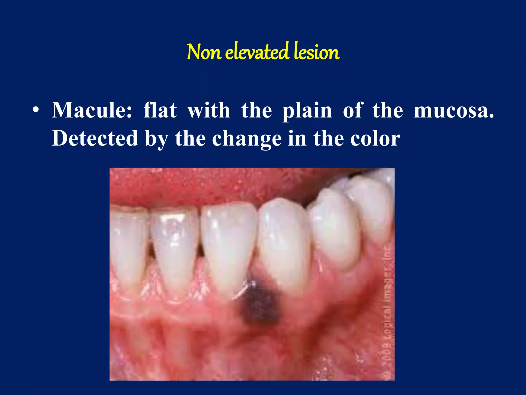
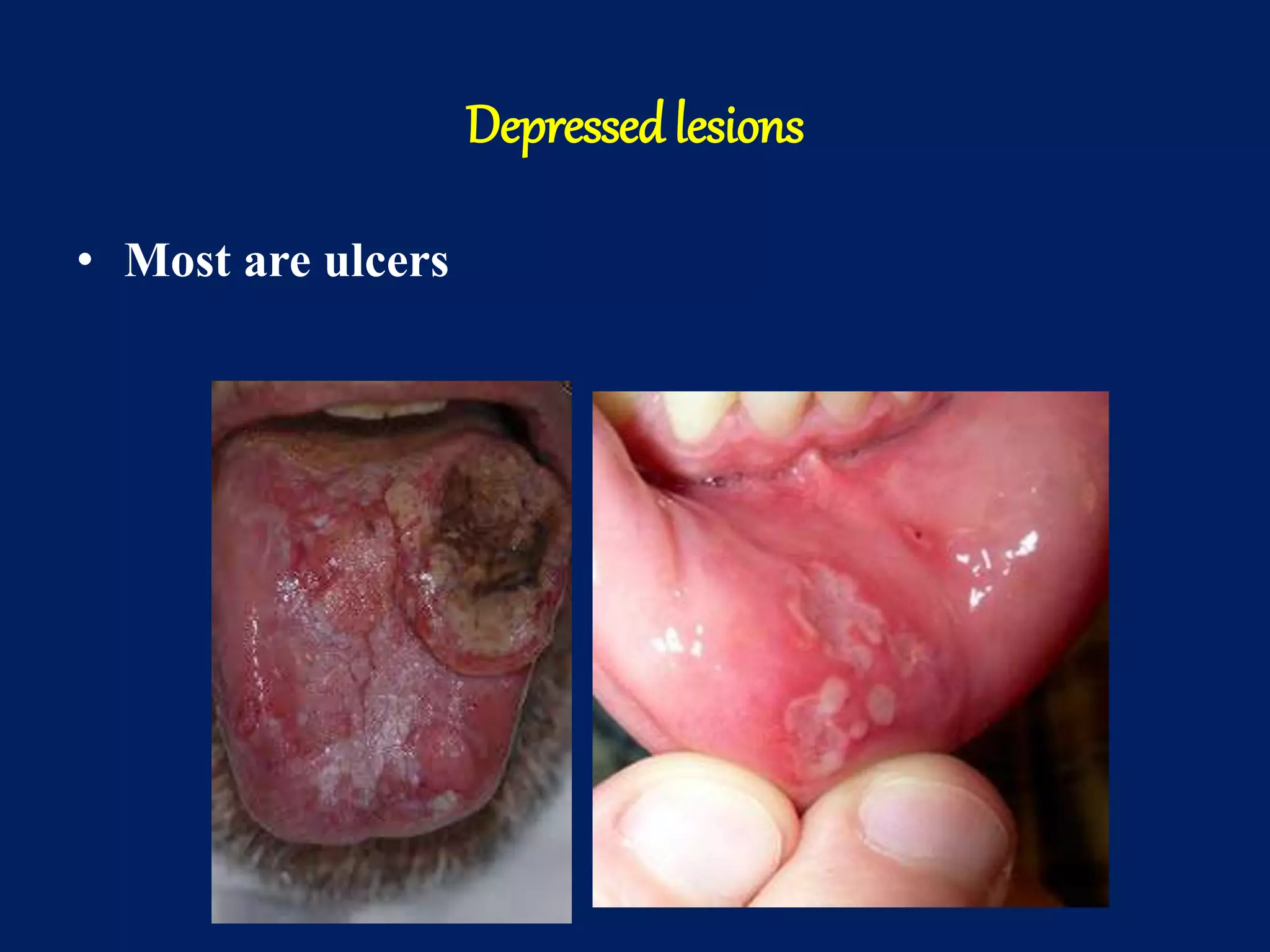
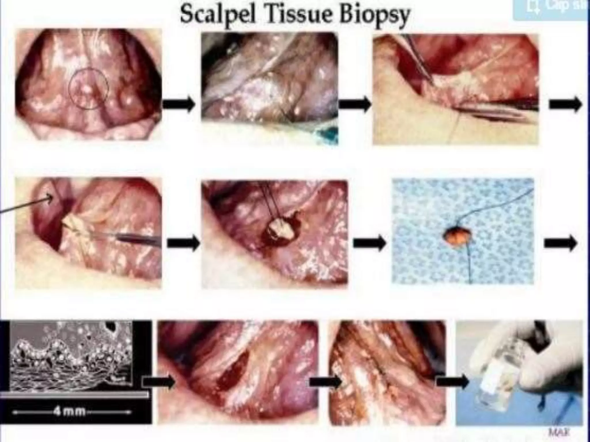
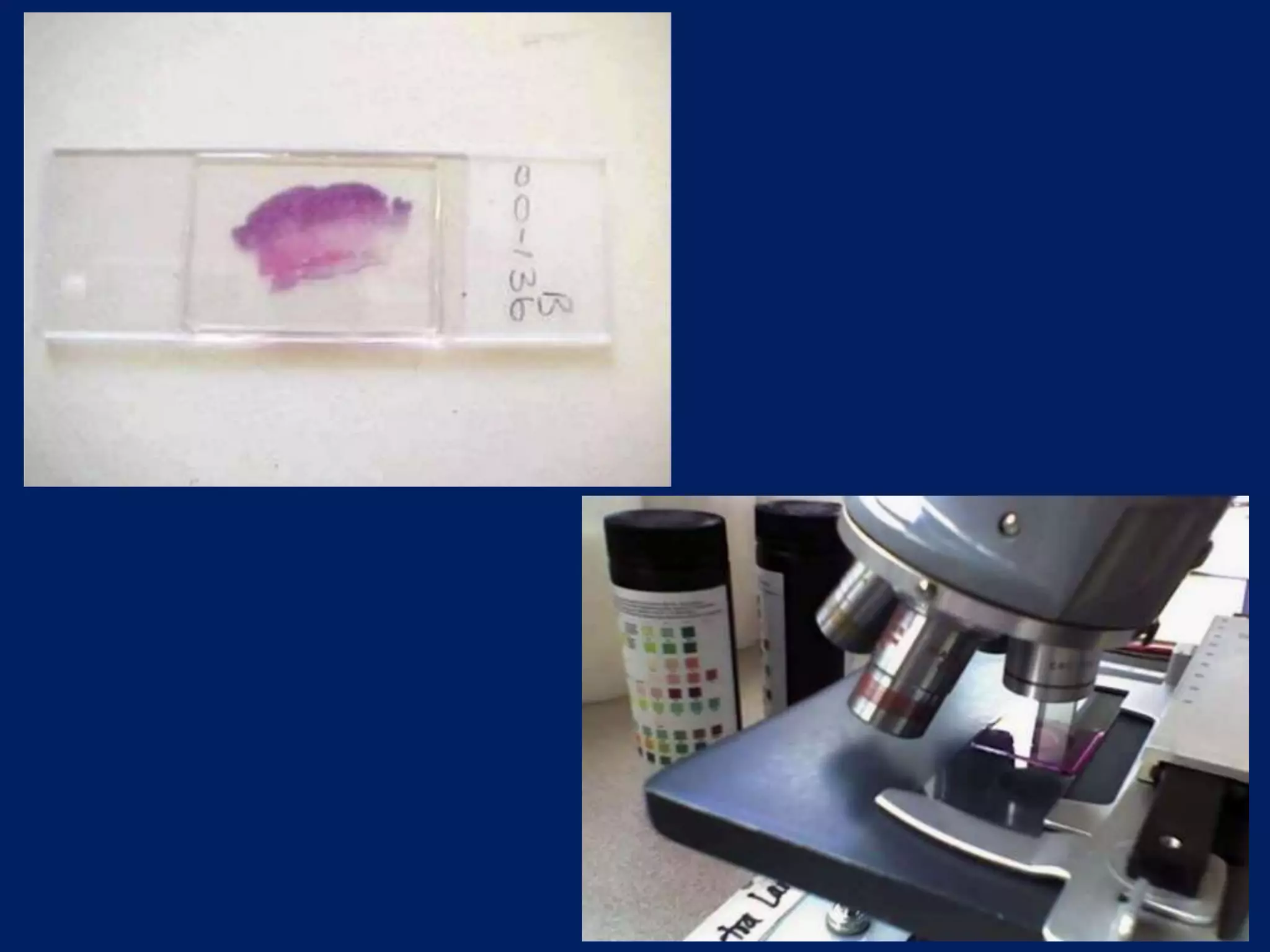
Introduction to oral pathology
Dr.Madhusudhan reddy
Introduction
• Oral and maxillofacial pathology is the specialty of
dentistry and pathology which deals with the
nature, identification, and management of diseases
affecting the oral and maxillofacial regions. It is
the science that investigates the causes, processes
and effects of these diseases.
• Practice of oral and maxillofacial pathology
includes research, diagnosis of diseases using
clinical, radiographic, microscopic, biochemical or
other examination and management of patients.
Classification of oral diseases
• According to the tissue affected
– The different oral and maxillofacial diseases
may affect different tissues which includes:
• Hard tissues (calcified) like teeth, jaw bones
and other bones in the maxillofacial regions
• Soft tissue in the oral and para-oral regions
Classification of oral soft tissue lesions
1. Epithelial in origin:
1. Lesions originated from odontogenic epithelium
2. Lesions originated from non odontogenic
epithelium
3. Lesions originated from glandular epithelium
2. Mesenchymal in origin
lesions originated from fibrous tissue
lesions originated from blood and lymphatic
vessels
lesions originated from the muscle
lesions originated from the nerve
lesions originated from adipose tissue
lesions originated from the bone
lesions originated from the cartilage
Diagnosis of the oral andmaxillofacial diseases
• Depends on the type of tissue affected:
• Disease affecting the hard tissue, the
diagnosis depend mainly on:
– Clinical features which includes the age, gender,
site, size and clinical effects.
– Radiographic features
– Microscopic features
Diagnosis of soft tissue lesions
• Clinical features.
– Site
– Morphology
• Elevate- above plain of mucosa
• Depressed- below the plain of mucosa
• Flat- with the plain of mucosa detected by the
change in colour
– Color
– size
• Microscopic features
Elevatedlesions
• Blister form (containing fluid)
– Vesicle: less than 5 mm
– Bulla: more than 5mm
– Pustule: filled with pus
• Non blisterform (not containing fluid)
– Papule: less than 5mm
– Nodule: more than 5mm and less than 2cm
– Tumor: more than 2cm
Blister form
Vesicle
Bulla
Blister form
Pustule
Non blisterform
Papule
Nodule
Tumor
Non elevated lesion
• Macule: flat with the plain of the mucosa.
Detected by the change in the color
Depressedlesions
• Most are ulcers
Intra bony lesions
– Inflammatory
– Developmental
– Tumor
Diagnostic Aids in Oral Pathology
• Exfoliative cytology
• Vital dyes
• Tissue biopsy
Tissue biopsy
• Surgical removal of the living tissue for
microscopic examination
– Indication
– Types of oral biopsy
• Excisional
• Incisional
histopathology
• Microscopic examination of biopsy or surgical
specimen by pathologist, after the specimen has
been processed and histological sections have been
placed onto glass slides.
Tissue processing
• Fixation: in 10% neutral buffered formalin
• Decalcification: for hard tissue
• Dehydration : to remove water (nprogressive
concentrations of ethanol are used)
• Clearing: xylene is used to remove alcohols
• Infiltration: paraffin wax is used
• Embedding
• Sectioning staining
Text books
• Shafers : text book of oral pathology : seventh or
eight edition
• Neville, Damm, Allen, Bouquot: Oral and
maxillofacial pathology text book: third edition

Introduction to oral pathology

  • 1.
    Introduction to oralpathology Dr.Madhusudhan reddy
  • 2.
    Introduction • Oral andmaxillofacial pathology is the specialty of dentistry and pathology which deals with the nature, identification, and management of diseases affecting the oral and maxillofacial regions. It is the science that investigates the causes, processes and effects of these diseases.
  • 3.
    • Practice oforal and maxillofacial pathology includes research, diagnosis of diseases using clinical, radiographic, microscopic, biochemical or other examination and management of patients.
  • 4.
    Classification of oraldiseases • According to the tissue affected – The different oral and maxillofacial diseases may affect different tissues which includes: • Hard tissues (calcified) like teeth, jaw bones and other bones in the maxillofacial regions • Soft tissue in the oral and para-oral regions
  • 5.
    Classification of oralsoft tissue lesions 1. Epithelial in origin: 1. Lesions originated from odontogenic epithelium 2. Lesions originated from non odontogenic epithelium 3. Lesions originated from glandular epithelium
  • 6.
    2. Mesenchymal inorigin lesions originated from fibrous tissue lesions originated from blood and lymphatic vessels lesions originated from the muscle lesions originated from the nerve lesions originated from adipose tissue lesions originated from the bone lesions originated from the cartilage
  • 7.
    Diagnosis of theoral andmaxillofacial diseases • Depends on the type of tissue affected: • Disease affecting the hard tissue, the diagnosis depend mainly on: – Clinical features which includes the age, gender, site, size and clinical effects. – Radiographic features – Microscopic features
  • 8.
    Diagnosis of softtissue lesions • Clinical features. – Site – Morphology • Elevate- above plain of mucosa • Depressed- below the plain of mucosa • Flat- with the plain of mucosa detected by the change in colour – Color – size • Microscopic features
  • 9.
    Elevatedlesions • Blister form(containing fluid) – Vesicle: less than 5 mm – Bulla: more than 5mm – Pustule: filled with pus • Non blisterform (not containing fluid) – Papule: less than 5mm – Nodule: more than 5mm and less than 2cm – Tumor: more than 2cm
  • 10.
  • 11.
  • 12.
  • 13.
  • 14.
    Non elevated lesion •Macule: flat with the plain of the mucosa. Detected by the change in the color
  • 15.
  • 16.
    Intra bony lesions –Inflammatory – Developmental – Tumor
  • 17.
    Diagnostic Aids inOral Pathology • Exfoliative cytology • Vital dyes • Tissue biopsy
  • 20.
    Tissue biopsy • Surgicalremoval of the living tissue for microscopic examination – Indication – Types of oral biopsy • Excisional • Incisional
  • 23.
    histopathology • Microscopic examinationof biopsy or surgical specimen by pathologist, after the specimen has been processed and histological sections have been placed onto glass slides.
  • 24.
    Tissue processing • Fixation:in 10% neutral buffered formalin • Decalcification: for hard tissue • Dehydration : to remove water (nprogressive concentrations of ethanol are used) • Clearing: xylene is used to remove alcohols • Infiltration: paraffin wax is used • Embedding • Sectioning staining
  • 28.
    Text books • Shafers: text book of oral pathology : seventh or eight edition • Neville, Damm, Allen, Bouquot: Oral and maxillofacial pathology text book: third edition