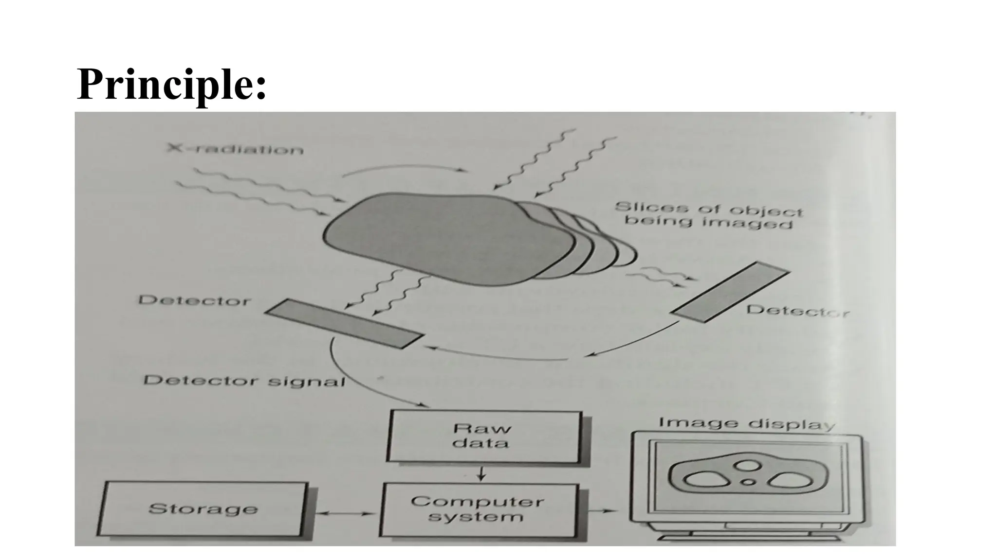
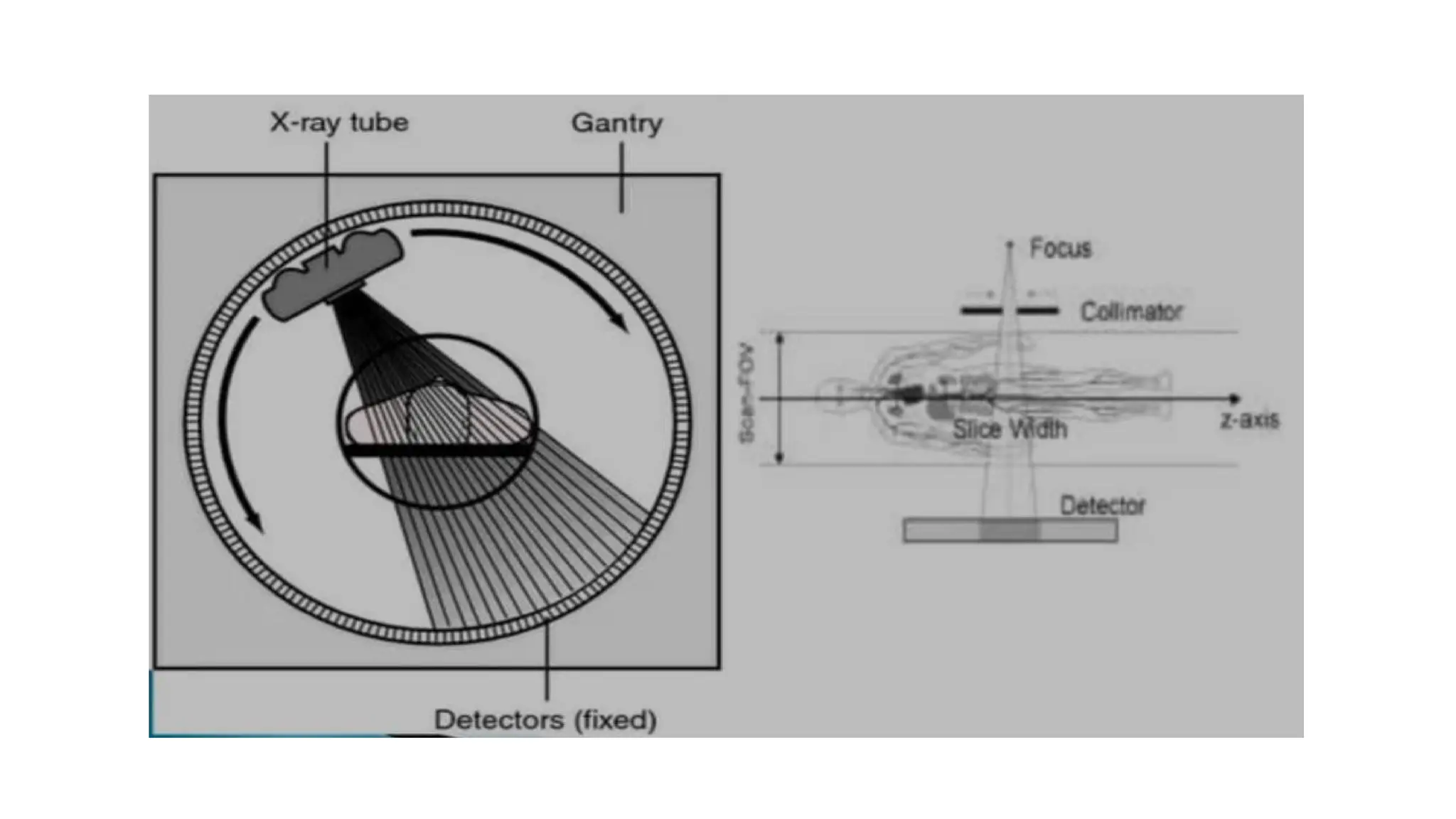
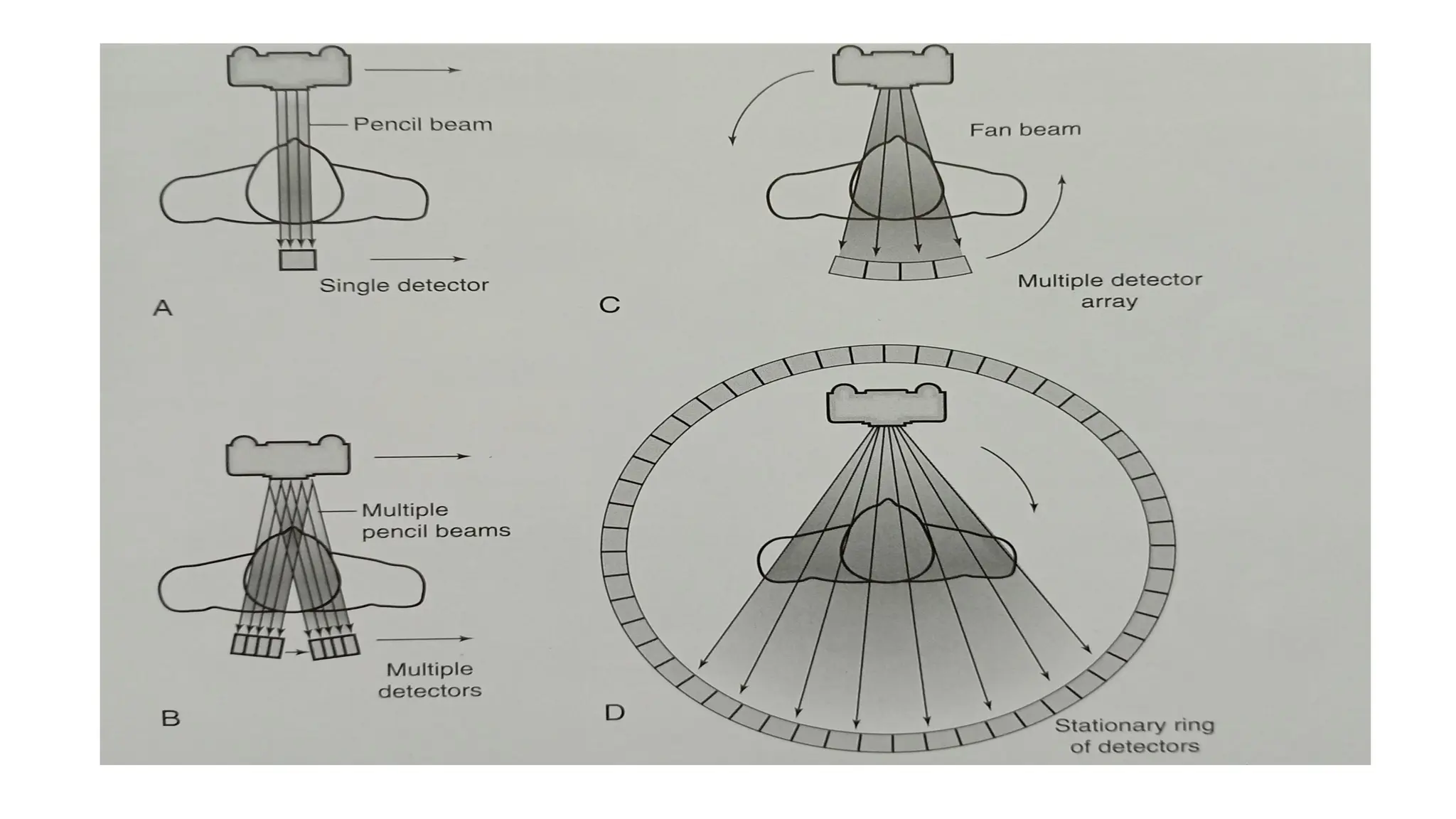
CT SCAN (Computed
Tomography)
Presented by,
J.SAROJINI
MPT (NEUROLOGY)
Introduction
Computed use of computer Tomography:
Greek word tomos means “slice”, graphy means “Write”.
Computed Tomography is the process of generating a two-dimensional
image of a slice or section through a 3-dimensional object (a
tomogram).
• Computed tomography (or computerized axial tomography) is an
examination that uses X-ray and computer to obtain a cross-sectional
image of the human body.
Parts of a CT scan machine
1. Gantry (scanner system)
2. Patient's table
3. Computer(operation control)
Scanner system:
1. X ray tube
2. Detector system
3. Collimators
4. Filters
Parts of Gantry:
• X-ray tube: generates x-ray beams
• Detector: detects the x-rays passing through the patient's body.
• Collimator: narrows the beams of x-rays
• Filters: these are used to filter some rays from entering the patient's
body that may be harmful.
Principle:
2 types:
• Conventional: scan is taken slice by slice and after slice the scan
stops.e.g.. from top of abdomen till the pelvis. This needs the patient
to hold their breath.
• Spiral scan: continuous scan taken in spiral fashion. It is quick.
Parameters:
• Scan range starting at base of the skull; ending at vertex of the skull
• Respiration: shallow breathing
• Positioning
• Supine
• Head immobilized
• Arms along the sides
Procedure:
• When X-rays pass through the human body, some of the rays are
absorbed and some pass through the body to produce an image.
• In plain X-ray imaging, the film directly absorbs penetrated X-rays.
• In CT scanning, an electronic device called a "detector array" absorbs
the penetrated X-rays, measures the X-ray amount, and transmits the
data to a computer system.
• A sophisticated computer system, in turn, calculates and analyzes data
from each detector in each level, and finally reconstructs multiple,
two- dimensional, cross-sectional images.
Process
• It involves 3 steps:
• Data Acquisition
• Image reconstruction
• Image display
Data Acquisition:
• The term data acquisition refers to the collection of x-ray
transmission measurements from the patient.
• Once x rays have passed through the patient, they fall onto special
electronic detectors that measure the transmission values.
Image Reconstruction:
• After enough transmission measurements have been collected by the
detectors, they are sent to the computer for processing.
• The computer uses special mathematical techniques to reconstruct
the CT image in a finite number of steps called image
reconstruction.
Image Display, Processing, Storage, computer can use Recording, and
Communications
• After the computer has performed the image reconstruction process,
the reconstructed image can be displayed and recorded for subsequent
view and stored for later analysis.
• The image is usually displayed on a cathode ray tube.
X-RAYS ARE ABSORBED TO DIFFERENT
DEGREES BY DIFFERENT TISSUES
• Always describe CT findings as densities- isodense/hypodense/hyperdense.
• Higher the density = whiter the appearance
• Lower the density = darker the appearance
• Higher density than brain-hyperdense ( skull is the best example)
• Anything darker (lower density) than brain= hypodense( CSF and air are
classical examples)
CT SCAN OF BRAIN:
• An imaging technique of the brain that reveal tumors, blood clots,
hemorrhages, or other abnormal anatomy.
• A series of computerized images of the brain at various levels are
taken to reveal normal anatomy or any abnormality.
• Routine scans of the brain usually begin at the base of the skull and
continue superiorly.
• Symmetry - Compare left and right side of the Midline
• Look for midline shift of cranium
• Cross-sectional anatomy - Review anatomical landmark for each
section.
• Brain tissue: gray matter, white matter, intracerebral lesions
• CSF space : ventricle (dilated or not)
• Skull and soft tissue: scalp swelling, fractures, sinuses, orbit
• Subdural windows: Look for blood collection adjacent to the skull
• Bone windows: Skull, orbit and sinuses, intracranial air
Normal scan:
• Ventricles are normal sized, the
grey versus white distinction is
clear.
• Midline is straight.
• Sulci are symmetrical on both sides.
• Skull is intact with no scalp edema.
USES:
• Evaluate acute cranial-facial trauma
• Determine acute stroke
• Evaluate suspected intracranial hemorrhage
• Evaluate headache
• Determine if there is abnormal development of the head and neck
• Tumors, clots
ADVANTAGES:
• Quick and painless
• Can detect the presence of more serious problems
• To Check if a previously treated disease has recurred
• No superimposition of images
DISADVANTAGES:
• Small risk of developing cancer in future from exposure to X-rays. Risk is greater for children
• Uses higher doses of radiation are used in chest, abdomen, so the risks are in general greater than other
imaging types
• Some procedures require anesthesia.
• Requires breath holding which some patients cannot manage.
• Ct scan of brain can affect nearby bone.
• There is also risk of childhood cancer and leukaemia in mothers who have imaging during pregnancy.
• Pregnant women are not allowed for this imaging
ISCHAEMIC STROKE
INTRACRANIAL HEMORRHAGE
Thank you...

COMPUTED TOMOGRAPHY (CT)simple way to learn

  • 1.
    CT SCAN (Computed Tomography) Presentedby, J.SAROJINI MPT (NEUROLOGY)
  • 2.
    Introduction Computed use ofcomputer Tomography: Greek word tomos means “slice”, graphy means “Write”. Computed Tomography is the process of generating a two-dimensional image of a slice or section through a 3-dimensional object (a tomogram). • Computed tomography (or computerized axial tomography) is an examination that uses X-ray and computer to obtain a cross-sectional image of the human body.
  • 3.
    Parts of aCT scan machine 1. Gantry (scanner system) 2. Patient's table 3. Computer(operation control) Scanner system: 1. X ray tube 2. Detector system 3. Collimators 4. Filters
  • 4.
    Parts of Gantry: •X-ray tube: generates x-ray beams • Detector: detects the x-rays passing through the patient's body. • Collimator: narrows the beams of x-rays • Filters: these are used to filter some rays from entering the patient's body that may be harmful.
  • 5.
  • 6.
    2 types: • Conventional:scan is taken slice by slice and after slice the scan stops.e.g.. from top of abdomen till the pelvis. This needs the patient to hold their breath. • Spiral scan: continuous scan taken in spiral fashion. It is quick.
  • 7.
    Parameters: • Scan rangestarting at base of the skull; ending at vertex of the skull • Respiration: shallow breathing • Positioning • Supine • Head immobilized • Arms along the sides
  • 8.
    Procedure: • When X-rayspass through the human body, some of the rays are absorbed and some pass through the body to produce an image. • In plain X-ray imaging, the film directly absorbs penetrated X-rays. • In CT scanning, an electronic device called a "detector array" absorbs the penetrated X-rays, measures the X-ray amount, and transmits the data to a computer system. • A sophisticated computer system, in turn, calculates and analyzes data from each detector in each level, and finally reconstructs multiple, two- dimensional, cross-sectional images.
  • 11.
    Process • It involves3 steps: • Data Acquisition • Image reconstruction • Image display
  • 12.
    Data Acquisition: • Theterm data acquisition refers to the collection of x-ray transmission measurements from the patient. • Once x rays have passed through the patient, they fall onto special electronic detectors that measure the transmission values.
  • 13.
    Image Reconstruction: • Afterenough transmission measurements have been collected by the detectors, they are sent to the computer for processing. • The computer uses special mathematical techniques to reconstruct the CT image in a finite number of steps called image reconstruction.
  • 14.
    Image Display, Processing,Storage, computer can use Recording, and Communications • After the computer has performed the image reconstruction process, the reconstructed image can be displayed and recorded for subsequent view and stored for later analysis. • The image is usually displayed on a cathode ray tube.
  • 15.
    X-RAYS ARE ABSORBEDTO DIFFERENT DEGREES BY DIFFERENT TISSUES • Always describe CT findings as densities- isodense/hypodense/hyperdense. • Higher the density = whiter the appearance • Lower the density = darker the appearance • Higher density than brain-hyperdense ( skull is the best example) • Anything darker (lower density) than brain= hypodense( CSF and air are classical examples)
  • 17.
    CT SCAN OFBRAIN: • An imaging technique of the brain that reveal tumors, blood clots, hemorrhages, or other abnormal anatomy. • A series of computerized images of the brain at various levels are taken to reveal normal anatomy or any abnormality. • Routine scans of the brain usually begin at the base of the skull and continue superiorly.
  • 18.
    • Symmetry -Compare left and right side of the Midline • Look for midline shift of cranium • Cross-sectional anatomy - Review anatomical landmark for each section. • Brain tissue: gray matter, white matter, intracerebral lesions • CSF space : ventricle (dilated or not) • Skull and soft tissue: scalp swelling, fractures, sinuses, orbit • Subdural windows: Look for blood collection adjacent to the skull • Bone windows: Skull, orbit and sinuses, intracranial air
  • 19.
    Normal scan: • Ventriclesare normal sized, the grey versus white distinction is clear. • Midline is straight. • Sulci are symmetrical on both sides. • Skull is intact with no scalp edema.
  • 22.
    USES: • Evaluate acutecranial-facial trauma • Determine acute stroke • Evaluate suspected intracranial hemorrhage • Evaluate headache • Determine if there is abnormal development of the head and neck • Tumors, clots
  • 23.
    ADVANTAGES: • Quick andpainless • Can detect the presence of more serious problems • To Check if a previously treated disease has recurred • No superimposition of images
  • 24.
    DISADVANTAGES: • Small riskof developing cancer in future from exposure to X-rays. Risk is greater for children • Uses higher doses of radiation are used in chest, abdomen, so the risks are in general greater than other imaging types • Some procedures require anesthesia. • Requires breath holding which some patients cannot manage. • Ct scan of brain can affect nearby bone. • There is also risk of childhood cancer and leukaemia in mothers who have imaging during pregnancy. • Pregnant women are not allowed for this imaging
  • 27.
  • 29.
  • 30.