Introduction
Def:
Microbiology
is the studyof living organisms/microbes
of microscopic size that cause infection.
It is the study of the structure, body
functions and physiological processes of
microorganism
The process of microbial inversion of the
body is called infection.
Historical dev’t ofmicro B
Some of the pioneers who are credited for the
development of microB include:
Antony Van Leeuwenhock (1675), the dutch
scientist who was the first person to see
micro-organism using a microscope. He was the
first man to build a microscope. He is credited
for coming up with a bacteria drawing that we
see today.
In 1796 Edward Jenner showed that small pox
could be prevented by inoculation with cowpox.
5.
Cont’d
Louis Pasteur (1864-1888)
Iscredited for the development of
modern microbiology. He demonstrated
that fermentation was caused by
growth of microorganisms.
Robert Koch (1877)
Described easy methods for
bacteriological exams and also devised
simple methods for isolating bacteria.
6.
Cont’d
Pasteur's work ledto an effective
sterilization method
He developed vaccines for diseases; anthrax,
fowl cholera and rabies
Beginning with Pasteur’s work, discoveries
included the relationship between microbes
and disease, immunity, and antimicrobial drugs
He postulated the Germ Theory of Disease
which states that microorganisms are the
causes of infectious diseases
7.
For an organismto be said to have caused a
particular disease then;
a) The micro-organism must be found in all
cases of disease
b) The micro-organism must be able to be
cultured outside the body for several
generations
c) The micro-organism must reproduce a
disease on inoculation into susceptible
animals
d) The antibody to the organism develops
during the course of infection
8.
Assignment
Explain Anthony vanLeuweenhoeks
contribution to the discovery of
microorganisms.
How did Louis Pasteur defeat the
theory of spontaneous generation
9.
Importance of micro-organismsto
man
• They cause decaying which is essential
for the organic cycle
• Causes disease to both plant and
animals
• Used in fermentation
Bacteria
Are unicellular/single celledorganisms
They lack membrane bound nucleus and
organelles.
Double stranded DNA lies loose in the
cytoplasm with arigid peptidoglycan cell
wall
Divide by binary fission
Some are harmful while others are
beneficial to man
Fungi
Are eukaryotic withrigid cell wall
May replicate asexually or sexually
They form spore like structure
Parasites
Comprise of worms, malaria parasite etc
14.
1. Bacteriology
This isthe study of bacteria.
Bacteria is a minute unicellular
organism.
Has rigid peptidoglycan cell-wall and
divides by binary fission
Cont’d
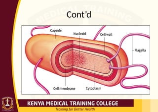
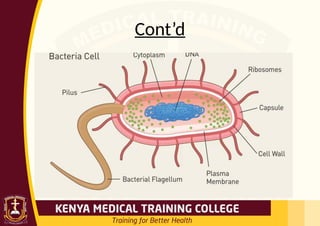
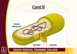
Capsule
Is amorphousmaterial which forms the
outermost layer of bacteria
Composed of polysaccharide and proteins
It inhibits phagocytosis and hence correlates
with bacterial virulence
Cell wall
Tough rigid structure strengthened by
peptidoglycan layers.
Protects the bacteria against osmotic changes.
21.
Cont’d
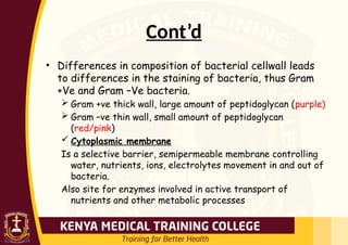
• Differences incomposition of bacterial cellwall leads
to differences in the staining of bacteria, thus Gram
+Ve and Gram –Ve bacteria.
Gram +ve thick wall, large amount of peptidoglycan (purple)
Gram –ve thin wall, small amount of peptidoglycan
(red/pink)
Cytoplasmic membrane
Is a selective barrier, semipermeable membrane controlling
water, nutrients, ions, electrolytes movement in and out of
bacteria.
Also site for enzymes involved in active transport of
nutrients and other metabolic processes
22.
Cont’d
Mesosome
Are convoluted invaginationsof cytoplasmic
membrane
Site for septum formation and involved in DNA
segregation during cell division
Nuclear material/nucleuos- for replication of
DNA and RNA
Ribosomes- site for protein synthesis
Flagella- long filaments structures for
bacterial motility
23.
Cont’d
Function of Flagella
•Bacterial propel themselves by rotating
their helical flagella.
Based on their location on the cell, flagella
may be polar or lateral.
(i) Polar: At one or both ends of bacterium.
(ii) Lateral: Along the sides of the
bacterium.
24.
Types of flagella
Monotrichous:A single polar flagellum.
Many that appears and function as monopolar
or bipolar flagella consists of bundles of 2 to
50 single units (polytrichous).
* Lophotrichous: A cluster of polar flagella.
* Amphitrichous: Flagella, either single or
clusters at both cell poles.
* Peritrichous: Cell surrounded by lateral
flagella.
Size
Bacteria are verysmall, 0.5 to 1.0 NM
in diameter.
Because of their small size they have
high surface area/volume ratio which
results in a high growth and
metabolism rate.
27.
Classification of bacteria
Bacteriaclassification is based on;
o Morphology
o Biochemical characteristics
o Lab identification
o Growth requirement/cultural
characteristics
28.
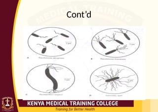
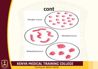
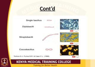
1. Morphological
Spherical-cocci egS. pneumonia
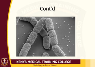
Cylindrical-bacilli or rods
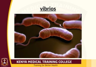
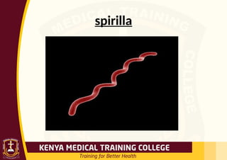
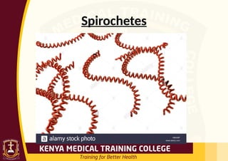
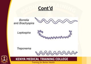
Helical- spirochaetes, flexible,coiled
and motile e.g Trepanoma pallidum
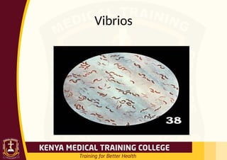
Vibrios-coma like
2. biochemical
• Coagulase-theenzyme that
clots/coagulates plasma and fibrin
• Hyalurodinase-breaks hyaluronic acid
the cement of connective tissue,
causing increased permeability
• Production of toxin-exotoxins and
endotoxins
38.
3. Lab identification
•Gram staining
• Giemsa staining-histopathological
diagnosis of malaria and other parasites
• Elisa (HIV) Ziehl Neelsen test- for acid
fast bacilli eg mycobacteria AFB
• Urinalysis- E. coli
• CSF analysis
• Field staining for malaria parasite
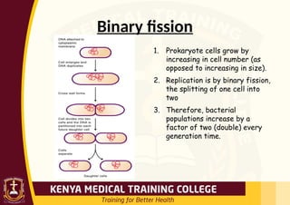
Binary fission
1. Prokaryotecells grow by
increasing in cell number (as
opposed to increasing in size).
2. Replication is by binary fission,
the splitting of one cell into
two
3. Therefore, bacterial
populations increase by a
factor of two (double) every
generation time.
41.
Generation time
• Thetime required to for a
population to double (doubling time)
in number. eg
• Ex. Escherichia coli (E. coli) double
every 20 minutes
• Ex. Mycobacterium tuberculosis
double every 12 to 24 hours
42.
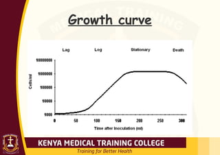
Growth in BatchCulture
• Bacteria growing in batch culture
produce a growth curve with up to
four distinct phases.
• Batch cultures are grown in tubes
or flasks and are closed systems
where no fresh nutrients are
added or waste products removed
43.
Phases of growth
1.Lag phase occurs when bacteria are adjusting to
them medium. For example, with a nutritionally poor
medium, several anabolic pathways need to be turned
on, resulting in a lag before active growth begins.
2. In log or exponential phase, the cells are growing as
fast as they can, limited only by growth conditions
and genetic potential. During this phase, almost all
cells are alive, they are most nearly identical, and
they are most affected by outside influences like
disinfectants.
44.
CONT’D
1. Due tonutrient depletion and/or
accumulation of toxic end products,
replication stops and cells enter a
stationary phase where there is no net
change in cell number.
2.Death phase occurs when cells can no
longer maintain viability and numbers
decrease as a proportion.
concept of infection
Objectives
Bythe end of this session, the learner will be
able to:
1. Describe the stages of development of an
infection
2.Define various terms related to infection
3.Describe the phases of an infectious d’se
49.
Infection
• is invasionby and multiplication of
pathogenic microorganisms in a body
part or tissue
• This invasion may produce subsequent
tissue injury and progress to disease
through a variety of toxic mechanisms.
50.
Stages in developmentof infection
1. Acquisition
2. Adhesion to host cells
3. Penetration of cells
4. Multiplication in tissues
5. Damage to tissues
6. Spread to other tissues
7. Resolution or death
51.
1. Acquisition, Adhesion& Penetration
• The microorganism must adhere to the cell,
then penetrate it.
• The microbe may use various methods to:
Special hairs on their surface,
Fimbriae (thin appendages used in attachment)
or Pilli (thin appendages used in genetic
exchange),
Secrete sticky substances (e.g. dextran)
Produce slime. (e.g. biofilm)
52.
4. Multiplication inthe host
• The microbe multiplies in the host cell
& overpowers the host’s defenses so as
to cause disease.
• The virus has the ability to switch the
metabolism of the cell to the
production of viral components.
53.
5. Tissue damage
•Pathogenic microorganisms cause
disease by damaging the host’s tissue.
• Damage occurs due to release of
enzymes or substances that destroy
cells or tissues in the local area.
54.
6. Spread toother tissues
• Some infections remain at the site of
invasion, causing symptoms related to
invasion of epithelial tissue at the
particular site.(e.g. Shigella invades the GIT)
• Some microorganisms spread once they
have been established in a particular site.
(e.g. salmonella typhi establishes itself in the GIT,
then spreads through blood and causes fever)
55.
7. Resolution ordeath
• The function of the cell may be disrupted
OR the cell may be destroyed when new
microbes are released.
• The effects depend on the particular
microbe and the location of the infected
cells. (e.g. Polio virus infects motor neuron cells and
shuts down protein synthesis causing the death of
neurons and paralysis of the muscles that they
innervate)
56.
Resolution or deathcont..
• The infection may resolve (death of
the pathogen) due to;
Improved host defenses
Antibiotic therapy
Pathogenicity
• Pathogens aremicrobial species that
invade and damage tissue to cause
disease.
• Pathogenicity is the capacity of
microorganism to cause disease.
59.
Pathogenicity cont’d….
• Somemicroorganisms cause a single
disease (e.g. clostridium tetani-
tetanus)
• Others cause a range of diseases:
e.g. Staphylococcus aureus can cause:
skin infections, wound infection,
pneumonia and Osteomyelitis.
60.
Pathogenicity cont’d….
• Somepathogens cause infections that
become severe in debilitated people.
• Opportunistic pathogens cause disease
in individuals with impaired defenses.
(e.g. Pneumocystis carinii, a normal flora in
healthy people but causes PCP in immune-
compromised people)
61.
Primary vs.Secondary Infection
•Primary Infection
– An infection that develops in an
otherwise healthy individual
• Secondary Infection
– An infection that develops in an
individual who is already infected with
a different pathogen
62.
Acute Vs. ChronicInfection
• Acute Infection
– An infection characterized by sudden
onset, rapid progression, and often with
severe symptoms
• Chronic Infection
– An infection characterized by delayed
onset and slow progression
63.
Localized vs. systemicinfection
• Localized Infection
– An infection that is restricted to a specific
location or region within the body of the
host
• Systemic Infection
– An infection that has spread to several
regions or areas in the body of the host
64.
Clinical vs. subclinicalinfection
• Clinical Infection
– An infection with obvious observable
or detectable symptoms
• Subclinical Infection
– An infection with few or no obvious
symptoms
65.
Opportunistic infection
• Aninfection caused by microorganisms
that are commonly found in the host’s
environment (normal flora)
66.
Nosocomial Infections
• Alsocalled Hospital Acquired
Infections (HAI’s)
• Infection which was neither present
nor incubating at the time of admission
• Infection that appears between 48
hours & 4 days following admission to a
hospital or other health-care facility.
67.
Nosocomial cont’d…
• 36%of these infections are preventable
through the adherence to strict guidelines by
health care workers when caring for patients
• Occurs in most modern hospitals
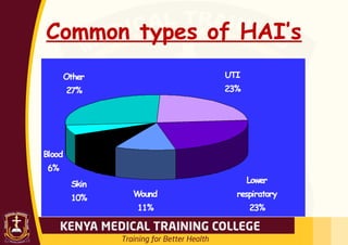
• Most common HAI’s:
Urinary tract infections (UTI’s).
Ventilator-associated pneumonia (VAP)
Surgical wound infections
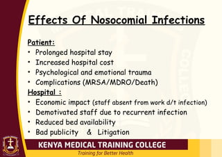
Effects Of NosocomialInfections
Patient:
• Prolonged hospital stay
• Increased hospital cost
• Psychological and emotional trauma
• Complications (MRSA/MDRO/Death)
Hospital :
• Economic impact (staff absent from work d/t infection)
• Demotivated staff due to recurrent infection
• Reduced bed availability
• Bad publicity & Litigation
72.
Phases of aninfectious disease
Incubation period
Refers to the time between infection and appearance of signs
and symptoms.
Prodromal phase
A stage in which the pathogens begin to invade tissues;
marked by early non specific symptoms.
Invasive phase
When the individual begins to experience the typical signs and
symptoms of the disease
73.
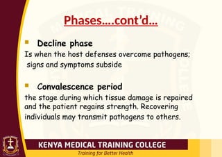
Phases….cont’d…
Decline phase
Iswhen the host defenses overcome pathogens;
signs and symptoms subside
Convalescence period
the stage during which tissue damage is repaired
and the patient regains strength. Recovering
individuals may transmit pathogens to others.
S & D
•Sterilization: Destruction of all forms
of microbial life including spores.
• Disinfection: Destruction of microbes
that cause disease; may not be
effective in killing spores.
• Antiseptic: chemical agent used to kill
organisms on the surface of the skin
and mucous membranes
76.
CONT’D
• Sterilizing anddisinfecting
agents are divided into two
groups;
1. Chemical methods
2. Physical methods
77.
1. Chemical methods
•Chemical agents destroy any type of
microbes without showing any form of
selectivity unlike antibiotics.
The efficacy of these agents depends on
the following factors.
1. Concentration of the agent -There is a
relationship between the concentration of
the agent and the time required to kill a
given fraction of the microbial population.
78.
CONT’D
2. Time ofexposure Microbes are killed with
a reasonable length of time with chemical
agents.
3. pH of the medium where action is to take
place Hydrogen ion concentration determines
degree of ionization of the chemical and
bacterial surface charge. The non-ionized
form passes through the bacterial cell
membrane more readily than the ionized
form
79.
CONT’D
4.Temperature Bactericidal potencyof
the chemical agent increases with an
increase in temperature. An increase in
100c doubles the bacterial death rate.
5. Nature of the organism . Species of the
bacteria . Growth phase of bacteria in
culture . Presence of capsule, spore and
other special structures . Number of
bacteria in test system
80.
CONT’D
6. Presence ofextraneous
materials Organic materials like
serum, blood or pus makes
chemicals inert that are highly
active in their absence.
81.
Classification of chemicalmethods of S
& D
1. Chemical agents that damage the cell
membrane .
-Surface active agents .
-Phenols .
-Organic solvents
2. Chemical agents that denature
proteins,
-Acids and Alkaline
82.
CONT’D
3. Chemical agentsthat modify
functional groups of proteins and
nucleic acids .
-Heavy metals .
-Oxidizing agents .
-Dyes .
-Alkylating agents
83.
2. Physical method
1.Heat: Denature proteins, membranes demage
or DNA cleavage;
A) Moist heat:-boiling
-Autoclaving (Steam under pressure)
121c (15-20mns)
-Pasteurization i.e 62c for 30mins
B) Dry heat;- open flames
- inceneration
-hot air oven
84.
CONT’D
2) Radiation:-x-rays, gammarays which
produces free radicals
3) filtration:-preferred for certain liquids
that heat sensitive like serum and enzymes
solution, membrane filters up to 0.22um
4) Freezing: Inactivation of living bacteria
by cold. It prevents active multiplication
of bacteria by decreasing the metabolic
activity of bacteria.
85.
Methods of controllingsterilization
1. Recording of temperature and time of
each sterilizing cycle.
2. Heat-sensitive autoclave tape fixed to the
outside of each pack. Color change of
autoclave tape from blue to brown-black
indicates complete sterilization.
3.Biological indicator : Use of paper strips
impregnated with spores of Bacillus stereo
thermophilus