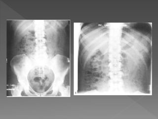
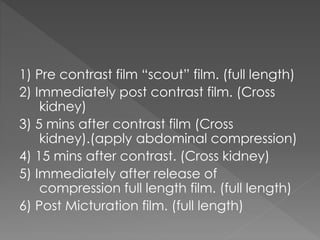
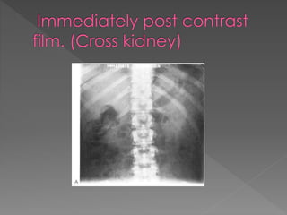
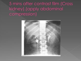
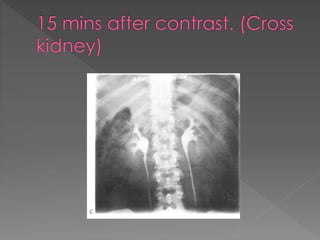
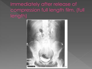
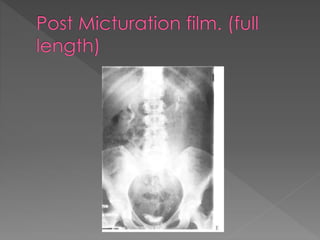
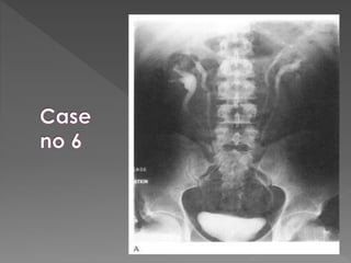
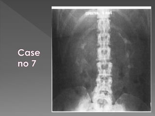
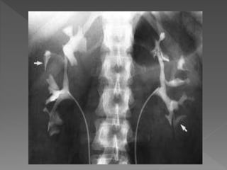
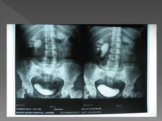
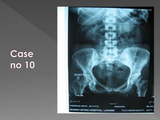
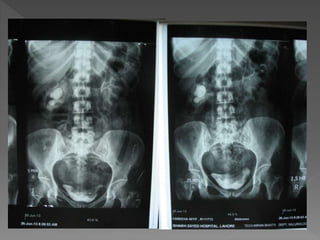
The document discusses the indications, technique, and findings of intravenous urography (IVU). It provides details on:
1) The traditional preparation of IVU including bowel preparation and fluid restriction to optimize contrast opacification of the urinary tract; however, recent recommendations state bowel preparation is unnecessary and fluid restriction should be avoided in high-risk patients.
2) The standard technique involves a series of films before and after intravenous contrast administration, including compression of the abdomen to distend the collecting system, with modifications for different clinical situations.
3) The expected timing of nephrogram, pyelogram, and other findings seen on IVU and their significance in evaluating the urinary tract anatomy and function.
Call Girls in Lucknow Just Call 👉👉8875999948 Top Class Call Girl Service Avai...
Intravenous urography
1.
2.
3. Indications
1) Persistent or frank hematuria.
2) Renal & ureteric calculi. (Especially prior to
endourological procedures)
3) Ureteric fistulas & strictures.
4) Complex urinary tract infections.
4. o Consists of series of film after IV
administration of contrast.
Traditional preperation
4 hours starvation & liquid deprivation along
with bowel purgation with laxatives.
5. Recent Recommendations
o Bowel preparation: unhelpful & is
unpleasant for patient.
Food should be avoided 2-4 hrs before
procedure.
o Fluid restriction: better opacification of
collecting system.
6. Fluid restriction increase risk of
nephrotoxicity
High risk conditions
› DM
› Multiple Myeloma
› Hyper urecemia
› Sickle cell disease
› Pre existing renal disease.
7. Risk of contrast induced injury to previously
healthy kidney is very low.
Fluid restriction should be avoided in high
risk patients
If dehydration is present it should be
corrected.
8.
9.
10. 1) Pre contrast film “scout” film. (full length)
2) Immediately post contrast film. (Cross
kidney)
3) 5 mins after contrast film (Cross
kidney).(apply abdominal compression)
4) 15 mins after contrast. (Cross kidney)
5) Immediately after release of
compression full length film. (full length)
6) Post Micturation film. (full length)
11.
12.
13.
14.
15.
16. Compression is omitted in :
Children.
Aortic aneurysm.
Tender abdomen.
Recent abdominal surgery.
Acute abdomen including renal colic.
If patient can not tolerate.
17. Modifications to deal with
different circumstances.
o To increase the sensitivity of the
procedure.
o To reduce the radiation dose.
18. Acute Obstruction:
There is delay in opacification of the
collecting system.
Delay may be considerable up to 24 hrs
or more.
It is then necessary to perform additional
films
19. Time interval b/w films is doubled
0.5 hrs/ 1 hr/ 2 hrs/ 4 hrs/ 16 hrs/ 24 hrs
as necessary.
To minimize the radiation exposure If there is
no opacification at 30 mins it is usually
unhelpful to perform next film before 4 hrs
20. In patient with strong suspicion of
ureteric calculi omit all films after
contrast until a full length film is
performed at 15 mins.
Patients with proven ureteric calculus for
follow up IVU there should be a full
length plain film & 15 mins post contrast
film
21. Pregnant female
Very rarely necessary to perform IVU.
If required minimize radiation exposure.
Single full length film pre contrast.
Solitary full length film at 30-45 mins.
22. Rapid sequencing IVU
Taken in cases of suspected renal
vascular hypertension to evaluate
differential rates of contrast uptake.
Several time-spaced films of kidneys
within 1st several minutes after contrast.
23. IV Frusemide
20 mg after 15 mins with a further film 15
mins later.
In suspected obstruction if no evidence
on 15 min film , it provokes
hydronephrosis & pain.
24.
25. Contrast takes 12-20 sec to reach the renal
arteries after iv injection.
Nephrogram : diffuse enhancement which
healthy kidneys show In the 1st min of IVU.
Renal size 3-4 vertebrae in length no > 4
vertebrae
Out lines best seen in 1st 30 sec.
26. Medullary or pyramidal blush:
Contrast may be visible as fine linear
opacities running along the medullary
pyramids
Pyelogram:
Excretion of contrast into the pelvis &
ureter
Compression impedes ureteric drainage
& distends PCS at12-15 mins
27. On releasing compression increase flow in
ureters making them prominent in post
release film.
Normal ureters have contractions & are not
seen entire length.
There are smoothly narrowed areas esp at
PUJ and as the ureters cross ilial vessels in
pelvis.