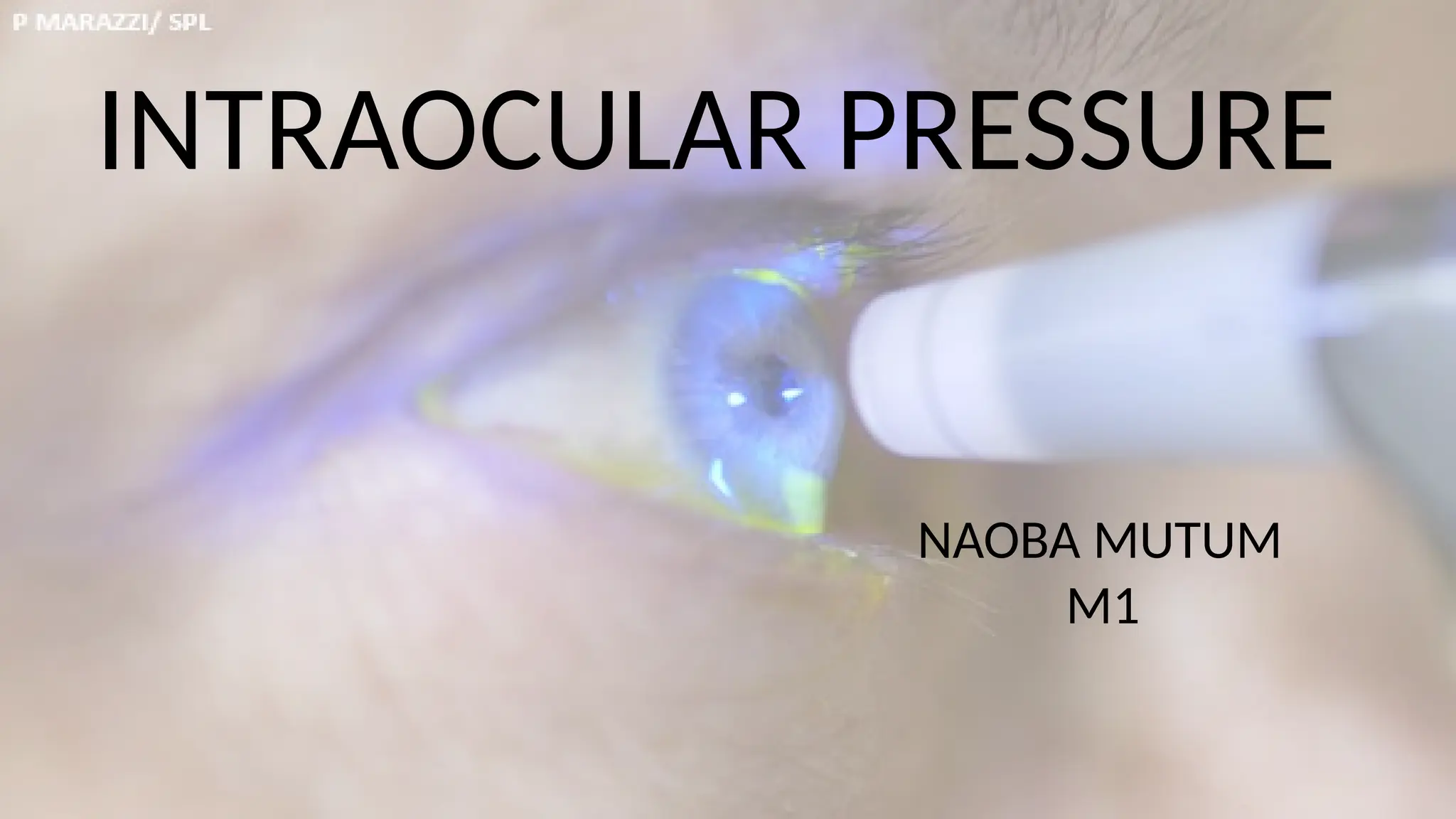
INTRAOCULAR PRESSURE
NAOBA MUTUM
M1
DEFINITION
The intraocular pressure (IOP) refers to the pressure
exerted by intraocular contents on the coats of the
eyeball. The intraocular pressure is important in
maintaining the shape of the eyeball and thus also
the optical integrity.
Normal IOP varies between 10.5 and 20.5 mm Hg with a mean
pressure of 15.5 ±2.57 mm Hg.
The intraocular pressure is created by aqueous formation which
has two components: first, a hydrostatic component from the
arterial blood pressure and ciliary body tissue pressure and,
second, an osmotic pressure induced by the active secretion of
sodium and other ions by the ciliary epithelium.
The IOP serves as the tissue pressure of the vascularized internal
structures of the eye and is thus much higher than the tissue
pressure elsewhere in the body (5 mm Hg).
FEATURES OF INTRAOCULAR PRESSURE
 Normal IOP is pulsatile, reflecting in part its vascular origin and the effects
of blood flow on the internal ocular structures.
 The IOP is a dynamic function. Any single measurement of IOP is just a
momentary sample and may or may not reflect the average pressure for
the patient in that hour, day or week
FREQUENCY DISTRIBUTION OF IOP IN THE POPULATION
Several population-based studies have been done to comment upon the
frequency distribution of the normal IOP. Despite the use of different
instruments and differing ethnic groups, the studies show remarkably close
correlation. The most frequently cited population study is that of Leydhecker
and associates,21 in which 10,000 persons with no known eye disease were
tested with Schiotz tonometer.
Conclusions drawn from the Leydhecker study regarding frequency
distribution of IOP in the population are as follows:
 The distribution of pressures observed resembled a Gaussian
curve, but was skewed towards the right. (Fig 1.)
Fig 1.
Distribution of intraocular pressure in non-glaucomatous (N) and
glaucomatous (Q) populations, showing overlap between the two
groups (dotted lines represent uncertainty of extreme value in both
populations.
 It has been assumed that perhaps two different population groups account
for the skewed distribution: a large ‘normal’ group (having a true Gaussian-
shaped curve) and a smaller group that was felt to be glaucomatous
without optic nerve head damage (causing a long tail on the right hand side
of the distribution curve).
 The mean IOP of normal group was 15.5 ± 2.57 mm Hg.
 95% of the population had an IOP between 10.5 and 20.5 mm Hg.
Some of the important conclusions drawn from the other population based
studies are as follows:
 A slight increase of mean IOP in each decade over 40 years.
 A slightly higher pressure exists in women than men in population above 40
years of age.
 IOP difference between right and left eye rarely exceeds 4 mm Hg.
 Level of IOP is inherited as a polygenic multifactorial trait.
FACTORS INFLUENCING THE INTRA – OCULAR PRESSURE
A. FACTORS CAUSING LONG TERM CHANGES IN IOP.
In addition to the glaucoma, following factors cause long-term influence on IOP:
1. Heredity: Relations of patients with primary open-angle glaucoma tend
to have higher IOP. Heredity influences IOP, possibly by multifactorial
modes.
2. Age: After the age of 40, there occurs a slight increase in the mean IOP
and standard deviation after each decade. This probably occurs due to
age-related reduction in aqueous outflow facility, despite a concomitant
reduction in the aqueous production.
3. Sex: IOP is equal between the sexes in ages of 20–40 years. In older age
group, increase in mean IOP with age is greater in females than males
4. Race: Though population-based studies in different ethnic groups do not
show significant difference in mean IOP, the race may occasionally influence IOP
distribution. For example, full blood Indians in a New Mexican tribe were found
to have significantly lower mean IOP than a control population.
5. Refractive error: Myopes tend to have slightly higher IOP as compared to
emmetropes
B. FACTORS CAUSING SHORT TERM CHANGES IN INTRAOCULAR PRESSURE
1. Arterial blood pressure. The IOP is generally not affected by physiological
changes in the arterial blood pressure; however, sudden large swings may affect
the IOP accordingly. For example, ligating one carotid can cause a fall in IOP on
the ipsilateral side (due to decreased blood flow) and an increase in IOP on the
contralateral side (due to increased blood flow).
2. Systemic venous pressure (SVP).
Changes in SVP can cause a profound effect on IOP by affecting
ipsilateral venous pressure,26 for about 1 mm Hg rise in episcleral venous
pressure (EVP) raises the IOP by 0.8 mm Hg.
 Common clinical conditions in which EVP is raised include external
pressure of jugular veins, compression of superior vena cava by tumour
(thoracic outlet syndrome), cavernous sinus thrombosis and neoplasms,
inflammatory or dysthyroid changes at the apex of the orbit.
 The Valsalva manoeuvre also increases the IOP by raising the EVP.
Postural variations in IOP when going from sitting or upright position to the
supine position and also the changes in IOP during coughing, straining are
related to changes in EVP.
3. Mechanical pressure on the globe from outside initially raises the IOP by
indentation but after some time due to acceleration of aqueous outflow, the
IOP returns to normal and by prolonged pressure decreases below the initial
levels.
This forms the basis of ocular massage to lower the IOP. Transient rise
due to external pressure on the globe is associated with conditions like forced
eyelid closure, mechanical pull on the restricted extraocular muscle and
simultaneous contraction of extraocular muscles in conditions like Duane’s
syndrome.
4. Plasma osmolarity.
Plasma osmolarity affects the IOP profoundly.
• When the total concentration of solute molecules in the blood exceeds,
the water from the eye (vitreous and perhaps aqueous also) is withdrawn,
lowering the IOP. This effect is used clinically to lower the IOP by use of
hyperosmotic agents like mannitol.
• Conversely, when the concentration of solutes in plasma is lower than the
ocular fluids, the water will enter the eye from plasma and raise the IOP.
This forms the basis of water drinking test used as provocative test for
glaucoma. A fall in IOP noted after prolonged exercise such as running or
bicycling has been attributed to increased serum osmolarity and metabolic
acidosis.
5. Blood pH.
Systemic acidosis lowers the IOP. Bietti has postulated that it is the
metabolic acidosis induced by carbonic anhydrase inhibition that is responsible
for their pressure lowering effect.
6. Diurnal variation in IOP.
Like many other biological parameters, the IOP also fluctuates
cyclically throughout the day.
There are four types of diurnal variation curves:
• Falling type: Maximal at 6–8 am followed by a continuous decline
• Rising type: Maximal at 4–6 pm
• Double variation type: With 2 peaks 9–11 am and 6pm
• Flat type of curve, with no variation
Most common pattern is falling type, i.e. the pressure is highest in the
early morning and lowest in the late evening( Fig 2.)
Fig 2.Diurnal variation of intraocular pressure
• The mean amplitude of daily fluctuation is usually less than 5 mm Hg
in normal individuals.
• A diurnal variation in IOP of more than 8 mm is considered
pathognomonic of glaucoma.
 Exact mechanism of diurnal IOP variation is uncertain, however, it has
been related to a diurnal variation in the level of plasma cortisol.
7. Seasonal variation in IOP has also been described with the highest pressure
recorded in Winter and lowest in Summer.
8. Systemic hyperthermia has been shown to cause an increased IOP in rabbits.
Similarly, a drop in body temperature will cause a decrease in the IOP, probably
by inhibition of aqueous secretion.
9. Effect of general anaesthesia on IOP has been widely studied.
 Intraocular pressure (IOP) may be affected under general anaesthesia by
premedication, induction agents, muscle relaxants, inhalation anaesthetics
and other drugs administered during preoperative period.
Apart from drugs, venous congestion or increased venous pressure,
hypertension and changes in blood gas tension also affect the IOP.
These changes in IOP basically occur due to alteration of physiological
determinants of intraocular pressure which include the following:
• Aqueous humour dynamics
• Vitreous volume
• Extraocular muscle tone, and
• Choroidal blood volume
Some facts about the effect of general anaesthesia on IOP are as
below:
a) Acute changes in the volume of blood present in the eye due to increase
in venous pressure as a result of coughing, retching, vomiting and bucking
on endotracheal tube cause a serious increase in IOP and is a factor of
cardinal importance to anaesthetist. Hypertension occurring during
laryngoscopy and intubation under general anaesthesia for the same
reason may cause a temporary increase in IOP.
b) Hypercapnia and hypoxia during general anaesthesia produce an increase
in IOP, while hypocapnia and hyperoxia produce a decrease in IOP due to
changes in volume of blood in the eye.
c). Drugs used in general anaesthesia produce a decrease in IOP provided
haemodynamic parameters and blood gases are kept in their normal limits.
 Premedicants such as diazepam, morphine, pethidine, pentazocine,
fentanyl, buprenorphine produce a decrease in IOP.
 Thiopentone sodium, an induction agent reduces IOP and so do most of
the gaseous and volatile anaesthetic agents with the possible exception of
nitrous oxide.
 Ketamine, a dissociative agent, has little effect on IOP when given
intramuscularly and may transiently raise the IOP when given intravenously.
 Parenteral use of atropine does not cause a significant rise in IOP, even in
the predisposed eye.
 Suxamethonium is a notorious drug and cause rise in IOP which can be
clinically important. The eyeball is compressed by the extraocular muscles.
The other possible mechanisms include a rise in arterial pressure, sharp
increase in venous pressure and rotation of eyes in divergent position.
d) In general, once the patient is under the effect of general anaesthesia, the
IOP is decreased. This effect occurs regardless of the type of anaesthetic.
Possible mechanisms for the decreased IOP include muscle relaxation,
decreased pressure, increased carbon dioxide levels in the blood, and/or
direct effect of anaesthetic agent.
10. Effects of drugs on IOP.
In addition to the well-established antiglaucoma drugs and effect of
general anaesthetic agents as discussed, many other drugs also affect the IOP.
Alcohol, heroin, and systemic vasodilators have been reported to lower the
IOP. While tobacco smoking, caffeine, LSD and corticosteroids may raise the
IOP.
CONTROL OF INTRAOCULAR PRESSURE
Although there are minor physiological variations of IOP, there
exists more or less a constant steady level of IOP, in spite of the fact that
there is continuous drainage of the aqueous humour or in other words
there is continuous leakage from the eye.
As already discussed, this steady level of IOP is the result of a
dynamic equilibrium between the aqueous humour formation, the normal
resistance offered by the drainage channels for the outflow of fluid from
the eye and the episcleral venous pressure
The homeostatic mechanisms locally help in maintaining the steady
state level of IOP. For example, when intraocular pressure rises, there
occurs a decrease in the aqueous inflow (pseudo-facility) maintaining the
equilibrium.
 It is now being assumed that there exists some central controlling
mechanisms that tend to maintain pressure homeostasis within the eye.
 The nature of location(s) of such a centre have still not been delineated.
However, the evidences have accumulated which suggest that the
controlling centre operates through the sympathetic, parasympathetic and
diencephalic influences.
The above conclusions are based on the following observations of various
workers:
CENTRAL CONTROLLING MECHANISM FOR IOP
a) Pressure-sensitive afferent fibres have been found in the long posterior
and short ciliary nervous.
b) Diencephalon’s stimulation has been reported to cause a fall or
rise in IOP, depending upon the area stimulated.
c) Cervical sympathetic ganglion’s stimulation produces a drop in IOP
due to diminished aqueous secretion.
d) Beta-adrenergic stimulation causes inhibition of aqueous secretion
and alpha-adrenergic stimulation causes an improvement in aqueous
outflow facility.
e) Parasympathetic system stimulation decreases IOP by an increase
in aqueous outflow facility.
MEASUREMENT OF INTRAOCULAR PRESSURE
1.MANOMETRY
Manometry is the only direct
measure of IOP. In this method, a
needle is introduced either into
the anterior chamber or into the
vitreous which is then connected
with a suitable mercury or water
manometer to measure the IOP.
(Fig3.)
Fig3. Manometry.
DISADVANTAGES OF MANOMETRY.
Not a practical method for routine in human beings. Needs general
anaesthesia which has its other effects on the IOP. Introduction of the
needle produces breakdown of blood-aqueous barrier and release of
prostaglandins which alter the IOP.
▪ Uses:
Manometer is of the greatest and perhaps of the only use for a
continuous measurements over time and recording the changes in IOP
induced by physiological and pharmacological manipulations in the
experiment research work on animal eyes.
2. TONOMETRY
 Tonometry is an indirect method of measuring the intraocular pressure
(IOP) with the help of specially designed instruments known as
tonometers.
 Tonometry has been over 180 years long journey. In late 19th
century, Donders designed the first instrument which displaced
intraocular fluid by contact with the sclera.
 The first commonly used mechanical tonometer designed by Schiotz in
early 1990’s soon became the new gold standard.
All clinical tonometers measure the IOP by relating a deformation of
the globe to the force responsible for the deformation.
They are broadly of two types, differing according to the shape of the
deformation (Fig.4.):
• The indentation or impression tonometers (truncated cone) and
• The applanation tonometers (simple flattening).
Fig4. Corneal deformation created by (A) indentation tonometers (a truncated cone) and
(B) applanation tonometers (simple flattening).
3. INDENTATION (IMPRESSION) TONOMETRY
Indentation (impression) tonometry is based on the fundamental fact
that a plunger will indent soft eye more than a hard eye. The indentation
tonometer in current use is that of Schiotz who devised it in 1905 and
continued to refine it through 1927.
Because of its simplicity, reliability, low price and relative accuracy, it is a
widely used tonometer in the world.
Indentation tonometers, other than Schiotz tonometer include:
• Herrington tonometer,
• Grants tonometer, and
• Maurice tonometer.
BASIC CONCEPT AND THEORY OF INDENTATION TONOMETRY
As the tonometer is placed on the cornea (Fig 5.),
the different forces come into play:
a. W, the weight of the tonometer, acts over an
area A and indents the cornea displacing a
volume Vc.
b. The tensile force T set up in the outer coats of
the eye at everywhere tangentially to the
corneal surface, with a component opposing W,
so that an additional force T is added to the
original baseline or resting intraocular pressure
(Po) which is artificially raised to a new value
(Pt). Fig 5. Basic principle of indentation tonometer.
SCHIOTZ TONOMETER
A Schiotz tonometer consists of the following parts (Fig.6):
• Handle for holding the instrument in vertical position on the
cornea.
• Footplate which rests on the cornea.
• Plunger which moves freely within a shaft in the footplate.
• A bent lever whose short arm rests on the upper end of the plunger
and a long arm which acts as a pointer needle. The degree to which
the plunger indents the cornea is indicated by the movement of this
needle on a scale.
• Weights: 5.5 g weight is permanently fixed to the plunger, which can
be increased by extra weight to 7.5,10 and 15 g.
Fig 6. Schiotz tonometer
TECHNIQUE OF SCHIOTZ TONOMETRY
 After anaesthetising the cornea with 2–4%
topical xylocaine, patient is made to lie
supine on a couch and instructed to fix at
a target on the ceiling.
 The examiner separates the lids with left
hand and gently rests the footplate of the
tonometer vertically on the centre of
cornea. The reading on scale is recorded
as soon as the needle becomes steady.
• It is customary to start with 5.5g weight.
However, if the scale reading is less than 3,
additional weight should be added to the
plunger to make it 7.5 g or 10 g as indicated; since with Schiotz tonometer the
greatest accuracy is attained when the deflection of the lever is between 3 and 4.
Fig 7. Friendenwald 1955 table for Schiotz tonometer.
ERRORS OF INDENTATION TONOMETRY
1. Errors inherent in the instrument.
These may be in the form of difference in the weight of different parts
of the tonometer, difference in the size and shape and curvature of the
footplate, friction arising in the working of plunger and differences in the
smoothness of gliding movement of the pointer on the scale.
The American Academy of Ophthalmology and Otolaryngology has
established a committee on tonometer standardization, which has got rigid
criteria for Schiotz tonometers.
2. Errors due to contraction of extraocular muscles.
When a tonometer is placed on the eye, always there occurs some reflex
contraction of the extraocular muscles, which tends to increase the intraocular
pressure.
3. Errors due to accommodation.
When the tonometer is brought near to the eye, there is a tendency to
look at the tonometer and thus inadvertently accommodation comes into play.
The contraction of ciliary muscle in accommodation also increases the facility
of aqueous outflow by pulling on the trabeculae and thus causes some
lowering of IOP.
4. Errors due to ocular rigidity.
Conversion tables of Schiotz tonometer are based on an average
coefficient of ocular rigidity (K). However, the ocular rigidity may differ
significantly in certain eyes and thus false values of IOP are obtained. Thus, the
deduction of IOP from tonometric readings will be higher in an eye with a
higher ocular rigidity and lower in an eye with an abnormal low ocular rigidity.
5. Errors due to variation in corneal curvature and thickness.
Either a steeper or thicker cornea will cause a greater displacement of
fluid during indentation tonometry which leads to a falsely high IOP readings.
Thus, errors may arise in cases of microphthalmos, buphthalmos, and high
myopia and corneal scars.
6. Errors in scale reading. An error of one scale reading on either side may occur
during observations. Thus, an actual IOP of 17.3 mm Hg (scale reading 5) may be
noted as 14.6 mm Hg (scale reading 6) or 20.6 mm Hg (scale reading 4).
7. Blood volume alteration. The variable expulsion of intraocular blood during
indentation tonometry may also influence the IOP measurement
8. Moses effect. At low scale readings, the cornea may mould into the space
between the plunger and hole, pushing the plunger up and leading to falsely
high pressure readings.
9. Repeated measurements lower the IOP.
Conclusion: Thus, from the above, it is quite clear that the results of
indentation tonometry are never absolutely accurate. An error to the tune of ±
2 mm Hg in normal range of IOP and of ± 4 mm Hg in higher range of IOP has
been reported.
STERILIZATION OF SCHIOTZ TONOMETER
Dissemble between each use and the barrel is cleaned with two pipe
cleaners, the first soaked in alcohol and the second dry. The footplate is
cleaned with alcohol swab. All surfaces must be dried before reassembling.
ELECTRONIC SCHIOTZ TONOMETER
It provides a continuous recording of IOP that is used for tonography. The
scale is also magnified which makes it easier to detect small changes in IOP. It
is limited to experimental studies in the present era.
4. APPLANATION TONOMETRY
The concept of applanation tonometry is
based on Imbert-Fick law which states that the
pressure inside a sphere (P) is equal to the force
(W) required to flatten its surface divided by the
area of flattening (A), i.e. P = W/A (Fig 8.) Modified
Imbert-Fick law: W + S = PA1 + B.
Therefore, one can determine the pressure by
measuring either the force necessary to flatten a
fixed area or by measuring the area flattened by a
fixed force. Applanation tonometers have been
designed using both the principles.
Fig 8. Basic principles of indentation
tonometry(Imbert-Fick law )
I. VARIABLE AREA (FIXED FORCE ) APPLANATION TONOMETERS
The types of tonometers measure the area of the cornea that is
flattened by a known force. These include:
Maklakov-Kalfa tonometer
Introduced in 1885, it consisted of a dumb-bell-shaped metal cylinder
with flat end plates of polished glass on either end with diameter of 10
mm. A set of four such instruments was available, weighing 5, 7.5,10 and
15 g.
Technique:
A thin coat of the dye is smeared on the anaesthetised cornea with
the patient supine, the flat bottom of the tonometer allowed to rest on
the cornea. In this way, the dye from the flattened part of cornea is
transferred to the tonometer bottom, which allows the measurement of the
area flattened by a fixed force. The IOP is then read from the conversion tables.
Presently, this tonometer is not much popular.
Other Variable Area Applanation Tonometers
Other variable area applanation tonometers developed are:
• Applanometer: Ceramic end plates .
• Tonomat: Disposable end plates.
• Halberg tonometer: Transparent end plate for direct reading
• Barraquer tonometer: Plastic tonometer for use in operating
room.
• Ocular tension indicator: Uses Goldmann prisms and standard
weight, for screening (measures above or below 21 mm Hg).
• Glaucotest: Screening tonometer with multiple end plates for
selecting different cutoff pressures.
II. VARIABLE FORCE (FIXED AREA) APPLANATION TONOMETERS
This type of tonometer measures the force that is required to applanate a
standard area of the corneal surface.
Fick in 1888 devised the first fixed area applanation tonometer.
However, it was Goldmann who in 1954 modernized this concept and
developed a tonometer which has revolutionized the use of applanation
tonometry all over the world.
This method of tonometry is far more accurate than indentation
tonometry since in this technique a very tiny area of the cornea is applanated
so the ocular rigidity does not interfere with the readings.
Some of the fixed area applanation tonometers are described below:
GOLDMANN APPLANATION TONOMETER (GAT):
Currently, Goldmann applanation tonometer (GAT) is the most popular
and accurate tonometer. It is mounted on the end of a lever hinged on a
standard slit lamp.
TECHNIQUE
 After anaesthetising the cornea with a drop of 2% xylocaine and staining
the tear film with fluorescein, patient is made to sit in front of a slit-lamp.
 The cornea and biprisms are illuminated with cobalt blue light from the slit-
lamp.
 The examiner views through the centre of a plastic biprism, which is used to
applanate the cornea. Biprism is then advanced until it just touches the apex
of cornea (Fig 9.)
Fig 9. Technique of applanation tonometry
At this point, two fluorescent semicircles
are viewed through the prism. Then, the
applanation force against cornea is
adjusted until the inner edges of the two
semicircles just touch (Fig10.) This is the
end point.
Fig10. Showing end point of applanation tonometry
A, too small; B, too large; C, end point.
 The prisms are caliberated in such a way that inner margins of semicircles
touch when 3.06 mm of cornea is applanated. The intraocular pressure is
determined by multiplying the dial reading with ten to obtain IOP in mm
Hg.
SOURCES OF ERROR
1. Fluorescein concentration affects the thickness of the mires leading to
overestimation (with thick mires) or underestimation (with thin mires) of IOP.
2. Inappropriate vertical alignment gives higher IOP readings.
3. Central corneal thickness. There is underestimation of IOP in thin corneas and
overestimation in thick corneas.
4. Corneal curvature. For every 3D increase in corneal curvature, IOP rises
approximately by 1 mm Hg as more fluid is displaced under steeper corneas
causing increase in ocular rigidity.
5. Corneal astigmatism.
With the rule (WTR) astigmatism underestimates while against the rule
(ATR) astigmatism overestimates the IOP.
To minimize this, tonometer biprism should be rotated so that axis of
least corneal curvature is opposite the red mark on the biprism. This results in a
separation of 43° and an applanation of an area of approximately 7.35 mm2.
The other method is to obtain measurements in both vertical and horizontal
meridians and average these readings.
6. Irregular corneal surface leads to distortion of mires.
EFFECTS OF CENTRAL CORNEAL THICKNESS
Variations in central corneal thickness (CCT) change the resistance of the
cornea to indentation so that this is no longer balanced entirely by the tear
film surface tension thus affecting the accuracy of IOP measurement.
A thinner cornea (physiological variation, pathological or
post-PRK/LASIK) would require less force to applanate it, leading to
underestimation of IOP and vice versa.
 Goldmann applanation tonometer was designed to give accurate reading when
the CCT was 520 μ. There occurs a change in applanation readings of 0.7 mm
Hg per 10 μ. Thus, a correction factor, as described by Dredsner (Fig 11), is
required to get accurate IOP levels with applanation tonometry.
Fig 11 : Dredsner correction table for IOP measured using GAT.
STERILIZATION
Infections likely to be transmitted by tonometry are:
• Adenovir epidemic keratoconjunctivitis (EKC)
• HSV—type 1
• HIV Hepatitis B
Methods used to sterilize applanation tonometer biprisms include:
• Soaking applanation tip for 5–15 minutes in diluted sodium hypochlorite
(1:10),
• hydrogen peroxide 3% or isopropyl alcohol 70%,
• or by wiping with alcohol, hydrogen peroxide, povidone iodine or
• 1 : 1000 merthiolate are some easy and handy methods to sterilize the
biprism of applanation tonometer.
• Ten minutes of rinsing in running tap zvater Soap and zvater zvash
• Disposable film covers for tips
• Exposure to UV lights.
Hand-held Goldmann type tonometers
I. Contact type
a. Perkin’s applanation tonometer (Fig. 12).
 It is a hand-held tonometer utilizing the same biprism as in the
Goldmann applanation tonometer.
 It is small, easy to carry and does not require slit-lamp.
 The light source is powered by a battery and the force is varied
manually.
 A counterbalance makes it possible to use the instrument in either the
vertical or horizontal position. It is not used commonly now.
Fig 12. Perkin’s hand-held applanation tonometer
b. Draeger applanation tonometer. Similar to the Perkin’s tonometer, but
uses a different biprism and has an electric motor that varies the force.
II. Non-contact type
a. Air-puff tonometer (Fig 13): It is a non-contact tonometer. In this,
central part of cornea is flattened by a jet of air. This tonometer is very
good for mass screening as there is no danger of cross-infection and
local anaesthetic is not required.
Fig. 13 air puff tonometer.
b. Pulse air tonometer (Fig. 14 ). It is a non-contact tonometer that can be
used with the patient in any position.
Fig 14. pulse air tonometer
III.TONO – PEN .
 It is the most commonly used MacKay-Marg type tonometer today.
 It is a hand-held instrument with a strain gauge that creates an
electrical signal as the footplate flattens the cornea.
 A built-in single-chip microprocessor senses the proper force curves
and averages 4 to 10 readings to give a final digital readout.
 It also provides the percentage of variability between the lowest
and highest acceptable readings from 5 to 20%.
Advantages of tonopen
• Used in cases of corneal epithelial irregularities.
• Measurement of IOP over bandage contact lens.
• Useful in oedematous and scarred corneas.
• Useful in patients with nystagmus and head tremors.
• Used in operation theatre Portable.
Fig 15. Tono pen
PNEUMOTONOMETER
In this, the cornea is applanated by
touching its apex by a silastic diaphragm
covering the sensing nozzle (which is
connected to a central chamber
containing pressurised air).
There is a pneumatic-to-electronic
transducer present which converts the air
pressure to a recording on a paper strip,
from where IOP is read. It gives
significantly higher IOP estimates.
Fig 16. Pneumotonometer
MISCELLENANEOUS ADVANCEMENTS IN TONOMETERS
1. Pascal dynamic contour tonometer
 It is a newer applanation
tonometer that uses principle of
contour matching to measure IOP
instead of applanation to eliminate
the systemic errors inherent in
previous tonometers.
Fig. 17 .Pascal dynamic contour tonometer
 It uses a miniature pressure sensor embedded within a tonometer tip
contour matched to the shape of the cornea.
 The tonometer tip rests on the cornea with a constant appositional
force of one gram.
 When the sensor is subjected to a change in pressure, the electrical
resistance is altered and the Pascal’s computer calculates a change in
pressure in concordance with the change in resistance.
 It repeatedly samples IOP 100 times per second in addition to ocular
pulse amplitude and the systemic pulse rate and provides a digital
output of the IOP and a graphic output of the ocular pressure pulse.
 A complete measurement cycle requires about eight seconds of contact
time. Audio feedback helps the clinician to ensure proper contact with the
cornea.
2. Diatom transpalpebral tonometer
 It is a hand-held device using a free falling thin rod on the
stretched upper eyelid.
 It is based on processing the rod acceleration time resulting from
its free fall from the constant height and the interaction with eye
through the eyelid.
 It requires no contact with the cornea, therefore, sterilization of
the device and anaesthesia are not required.
 It is of use especially in children, those with corneal pathology, or
those who have had corneal surgery.
3. Icare or rebound tonometer
Icare or Rebound tonometer consists of a pair of coils coaxial with
the probe shaft, a solenoid coil and a sensing coil.
A lightweight probe is propelled towards the cornea by the solenoid
and the sensing coil monitors the movement. The speed immediately
before impact, the deceleration during impact and the ratio of these
parameters are correlated to the IOP
Fig 18. Icare or rebound tonometer .
4. SmartLens
• It has a contact lens that incorporates an electronic pressure sensor,
which is able to measure IOP and ocular pulse amplitude continuously by
applanation of the central cornea.
• There is a Myler membrane covered hole at the centre of the lens.
The cavity behind the membrane is filled with silicone oil and is
connected with a piezoelectric pressure transducer.
• The measurement of IOP is achieved by the transmission of
pressure through the oil to the transducer when the applanation
membrane is displaced during its contact with the cornea. It is being
used mainly in experimental studies
5. Proview-pressue phosphene tonometer
It is a patient self-monitoring device that utilizes a psychophysical
technique based on entopic phenomenon of pressure phosphenes.
6. Implantable IOP sensors
• IOP-IOL probe. A sensor incorporated in the haptics of an IOL for continuous
monitoring of IOP.
• Contact lens sensor. It correlates the spherical deformation of eyeball with
the changes in IOP.
• Choroid-IOP sensor. Sensor lies in contact with choroid.
TONOMETRY IN SPECIAL CONDITIONS
Tonometry on irregular corneas
Pneumatic tonometers provide the most accurate result in eyes with
diseased or irregular corneas. There is overestimation of IOP with Tono-Pen in
such patients.
Other tonometers which can also be used are:
 Non-contact tonometers
 MacKay-Marg tonometer, and
 Tono-Pen
Tonometry over soft contact lens
Pneumotonometer and Tono-Pen measure IOP through bandage
contact lenses with reasonable accuracy.
Tonometry in presence of intraocular gases
In eyes after pars plana vitrectomy and gas fluid exchange, there occurs
alteration in scleral rigidity rendering indentation tonometry unsatisfactory.
Pneumatic tonometers underestimate Goldmann IOP measurement.
Tono-Pen measurements are comparable.
Tonometry in shallow anterior chamber
There is much variability in IOP measurements with GAT,
pneumotonometer and Tono-Pen.
Tonometry in eyes with keratoprosthesis
Due to rigid surface of keratoprosthesis, it is impossible to measure IOP
with indentation or applanation tonometers. Digital IOP assessment is the
only applicable method in such eyes.
Intraocular pressure presentation slideshare

Intraocular pressure presentation slideshare

  • 1.
  • 2.
    DEFINITION The intraocular pressure(IOP) refers to the pressure exerted by intraocular contents on the coats of the eyeball. The intraocular pressure is important in maintaining the shape of the eyeball and thus also the optical integrity.
  • 3.
    Normal IOP variesbetween 10.5 and 20.5 mm Hg with a mean pressure of 15.5 ±2.57 mm Hg. The intraocular pressure is created by aqueous formation which has two components: first, a hydrostatic component from the arterial blood pressure and ciliary body tissue pressure and, second, an osmotic pressure induced by the active secretion of sodium and other ions by the ciliary epithelium. The IOP serves as the tissue pressure of the vascularized internal structures of the eye and is thus much higher than the tissue pressure elsewhere in the body (5 mm Hg). FEATURES OF INTRAOCULAR PRESSURE
  • 4.
     Normal IOPis pulsatile, reflecting in part its vascular origin and the effects of blood flow on the internal ocular structures.  The IOP is a dynamic function. Any single measurement of IOP is just a momentary sample and may or may not reflect the average pressure for the patient in that hour, day or week
  • 5.
    FREQUENCY DISTRIBUTION OFIOP IN THE POPULATION Several population-based studies have been done to comment upon the frequency distribution of the normal IOP. Despite the use of different instruments and differing ethnic groups, the studies show remarkably close correlation. The most frequently cited population study is that of Leydhecker and associates,21 in which 10,000 persons with no known eye disease were tested with Schiotz tonometer. Conclusions drawn from the Leydhecker study regarding frequency distribution of IOP in the population are as follows:  The distribution of pressures observed resembled a Gaussian curve, but was skewed towards the right. (Fig 1.)
  • 6.
    Fig 1. Distribution ofintraocular pressure in non-glaucomatous (N) and glaucomatous (Q) populations, showing overlap between the two groups (dotted lines represent uncertainty of extreme value in both populations.
  • 7.
     It hasbeen assumed that perhaps two different population groups account for the skewed distribution: a large ‘normal’ group (having a true Gaussian- shaped curve) and a smaller group that was felt to be glaucomatous without optic nerve head damage (causing a long tail on the right hand side of the distribution curve).  The mean IOP of normal group was 15.5 ± 2.57 mm Hg.  95% of the population had an IOP between 10.5 and 20.5 mm Hg.
  • 8.
    Some of theimportant conclusions drawn from the other population based studies are as follows:  A slight increase of mean IOP in each decade over 40 years.  A slightly higher pressure exists in women than men in population above 40 years of age.  IOP difference between right and left eye rarely exceeds 4 mm Hg.  Level of IOP is inherited as a polygenic multifactorial trait.
  • 9.
    FACTORS INFLUENCING THEINTRA – OCULAR PRESSURE A. FACTORS CAUSING LONG TERM CHANGES IN IOP. In addition to the glaucoma, following factors cause long-term influence on IOP: 1. Heredity: Relations of patients with primary open-angle glaucoma tend to have higher IOP. Heredity influences IOP, possibly by multifactorial modes. 2. Age: After the age of 40, there occurs a slight increase in the mean IOP and standard deviation after each decade. This probably occurs due to age-related reduction in aqueous outflow facility, despite a concomitant reduction in the aqueous production. 3. Sex: IOP is equal between the sexes in ages of 20–40 years. In older age group, increase in mean IOP with age is greater in females than males
  • 10.
    4. Race: Thoughpopulation-based studies in different ethnic groups do not show significant difference in mean IOP, the race may occasionally influence IOP distribution. For example, full blood Indians in a New Mexican tribe were found to have significantly lower mean IOP than a control population. 5. Refractive error: Myopes tend to have slightly higher IOP as compared to emmetropes B. FACTORS CAUSING SHORT TERM CHANGES IN INTRAOCULAR PRESSURE 1. Arterial blood pressure. The IOP is generally not affected by physiological changes in the arterial blood pressure; however, sudden large swings may affect the IOP accordingly. For example, ligating one carotid can cause a fall in IOP on the ipsilateral side (due to decreased blood flow) and an increase in IOP on the contralateral side (due to increased blood flow).
  • 11.
    2. Systemic venouspressure (SVP). Changes in SVP can cause a profound effect on IOP by affecting ipsilateral venous pressure,26 for about 1 mm Hg rise in episcleral venous pressure (EVP) raises the IOP by 0.8 mm Hg.  Common clinical conditions in which EVP is raised include external pressure of jugular veins, compression of superior vena cava by tumour (thoracic outlet syndrome), cavernous sinus thrombosis and neoplasms, inflammatory or dysthyroid changes at the apex of the orbit.  The Valsalva manoeuvre also increases the IOP by raising the EVP. Postural variations in IOP when going from sitting or upright position to the supine position and also the changes in IOP during coughing, straining are related to changes in EVP.
  • 12.
    3. Mechanical pressureon the globe from outside initially raises the IOP by indentation but after some time due to acceleration of aqueous outflow, the IOP returns to normal and by prolonged pressure decreases below the initial levels. This forms the basis of ocular massage to lower the IOP. Transient rise due to external pressure on the globe is associated with conditions like forced eyelid closure, mechanical pull on the restricted extraocular muscle and simultaneous contraction of extraocular muscles in conditions like Duane’s syndrome.
  • 13.
    4. Plasma osmolarity. Plasmaosmolarity affects the IOP profoundly. • When the total concentration of solute molecules in the blood exceeds, the water from the eye (vitreous and perhaps aqueous also) is withdrawn, lowering the IOP. This effect is used clinically to lower the IOP by use of hyperosmotic agents like mannitol. • Conversely, when the concentration of solutes in plasma is lower than the ocular fluids, the water will enter the eye from plasma and raise the IOP. This forms the basis of water drinking test used as provocative test for glaucoma. A fall in IOP noted after prolonged exercise such as running or bicycling has been attributed to increased serum osmolarity and metabolic acidosis.
  • 14.
    5. Blood pH. Systemicacidosis lowers the IOP. Bietti has postulated that it is the metabolic acidosis induced by carbonic anhydrase inhibition that is responsible for their pressure lowering effect. 6. Diurnal variation in IOP. Like many other biological parameters, the IOP also fluctuates cyclically throughout the day. There are four types of diurnal variation curves: • Falling type: Maximal at 6–8 am followed by a continuous decline • Rising type: Maximal at 4–6 pm • Double variation type: With 2 peaks 9–11 am and 6pm
  • 15.
    • Flat typeof curve, with no variation Most common pattern is falling type, i.e. the pressure is highest in the early morning and lowest in the late evening( Fig 2.) Fig 2.Diurnal variation of intraocular pressure
  • 16.
    • The meanamplitude of daily fluctuation is usually less than 5 mm Hg in normal individuals. • A diurnal variation in IOP of more than 8 mm is considered pathognomonic of glaucoma.  Exact mechanism of diurnal IOP variation is uncertain, however, it has been related to a diurnal variation in the level of plasma cortisol.
  • 17.
    7. Seasonal variationin IOP has also been described with the highest pressure recorded in Winter and lowest in Summer. 8. Systemic hyperthermia has been shown to cause an increased IOP in rabbits. Similarly, a drop in body temperature will cause a decrease in the IOP, probably by inhibition of aqueous secretion. 9. Effect of general anaesthesia on IOP has been widely studied.  Intraocular pressure (IOP) may be affected under general anaesthesia by premedication, induction agents, muscle relaxants, inhalation anaesthetics and other drugs administered during preoperative period. Apart from drugs, venous congestion or increased venous pressure, hypertension and changes in blood gas tension also affect the IOP.
  • 18.
    These changes inIOP basically occur due to alteration of physiological determinants of intraocular pressure which include the following: • Aqueous humour dynamics • Vitreous volume • Extraocular muscle tone, and • Choroidal blood volume
  • 19.
    Some facts aboutthe effect of general anaesthesia on IOP are as below: a) Acute changes in the volume of blood present in the eye due to increase in venous pressure as a result of coughing, retching, vomiting and bucking on endotracheal tube cause a serious increase in IOP and is a factor of cardinal importance to anaesthetist. Hypertension occurring during laryngoscopy and intubation under general anaesthesia for the same reason may cause a temporary increase in IOP. b) Hypercapnia and hypoxia during general anaesthesia produce an increase in IOP, while hypocapnia and hyperoxia produce a decrease in IOP due to changes in volume of blood in the eye.
  • 20.
    c). Drugs usedin general anaesthesia produce a decrease in IOP provided haemodynamic parameters and blood gases are kept in their normal limits.  Premedicants such as diazepam, morphine, pethidine, pentazocine, fentanyl, buprenorphine produce a decrease in IOP.  Thiopentone sodium, an induction agent reduces IOP and so do most of the gaseous and volatile anaesthetic agents with the possible exception of nitrous oxide.  Ketamine, a dissociative agent, has little effect on IOP when given intramuscularly and may transiently raise the IOP when given intravenously.
  • 21.
     Parenteral useof atropine does not cause a significant rise in IOP, even in the predisposed eye.  Suxamethonium is a notorious drug and cause rise in IOP which can be clinically important. The eyeball is compressed by the extraocular muscles. The other possible mechanisms include a rise in arterial pressure, sharp increase in venous pressure and rotation of eyes in divergent position. d) In general, once the patient is under the effect of general anaesthesia, the IOP is decreased. This effect occurs regardless of the type of anaesthetic. Possible mechanisms for the decreased IOP include muscle relaxation, decreased pressure, increased carbon dioxide levels in the blood, and/or direct effect of anaesthetic agent.
  • 22.
    10. Effects ofdrugs on IOP. In addition to the well-established antiglaucoma drugs and effect of general anaesthetic agents as discussed, many other drugs also affect the IOP. Alcohol, heroin, and systemic vasodilators have been reported to lower the IOP. While tobacco smoking, caffeine, LSD and corticosteroids may raise the IOP.
  • 23.
    CONTROL OF INTRAOCULARPRESSURE Although there are minor physiological variations of IOP, there exists more or less a constant steady level of IOP, in spite of the fact that there is continuous drainage of the aqueous humour or in other words there is continuous leakage from the eye. As already discussed, this steady level of IOP is the result of a dynamic equilibrium between the aqueous humour formation, the normal resistance offered by the drainage channels for the outflow of fluid from the eye and the episcleral venous pressure The homeostatic mechanisms locally help in maintaining the steady state level of IOP. For example, when intraocular pressure rises, there occurs a decrease in the aqueous inflow (pseudo-facility) maintaining the equilibrium.
  • 24.
     It isnow being assumed that there exists some central controlling mechanisms that tend to maintain pressure homeostasis within the eye.  The nature of location(s) of such a centre have still not been delineated. However, the evidences have accumulated which suggest that the controlling centre operates through the sympathetic, parasympathetic and diencephalic influences. The above conclusions are based on the following observations of various workers: CENTRAL CONTROLLING MECHANISM FOR IOP a) Pressure-sensitive afferent fibres have been found in the long posterior and short ciliary nervous.
  • 25.
    b) Diencephalon’s stimulationhas been reported to cause a fall or rise in IOP, depending upon the area stimulated. c) Cervical sympathetic ganglion’s stimulation produces a drop in IOP due to diminished aqueous secretion. d) Beta-adrenergic stimulation causes inhibition of aqueous secretion and alpha-adrenergic stimulation causes an improvement in aqueous outflow facility. e) Parasympathetic system stimulation decreases IOP by an increase in aqueous outflow facility.
  • 26.
    MEASUREMENT OF INTRAOCULARPRESSURE 1.MANOMETRY Manometry is the only direct measure of IOP. In this method, a needle is introduced either into the anterior chamber or into the vitreous which is then connected with a suitable mercury or water manometer to measure the IOP. (Fig3.) Fig3. Manometry.
  • 27.
    DISADVANTAGES OF MANOMETRY. Nota practical method for routine in human beings. Needs general anaesthesia which has its other effects on the IOP. Introduction of the needle produces breakdown of blood-aqueous barrier and release of prostaglandins which alter the IOP. ▪ Uses: Manometer is of the greatest and perhaps of the only use for a continuous measurements over time and recording the changes in IOP induced by physiological and pharmacological manipulations in the experiment research work on animal eyes.
  • 28.
    2. TONOMETRY  Tonometryis an indirect method of measuring the intraocular pressure (IOP) with the help of specially designed instruments known as tonometers.  Tonometry has been over 180 years long journey. In late 19th century, Donders designed the first instrument which displaced intraocular fluid by contact with the sclera.  The first commonly used mechanical tonometer designed by Schiotz in early 1990’s soon became the new gold standard. All clinical tonometers measure the IOP by relating a deformation of the globe to the force responsible for the deformation.
  • 29.
    They are broadlyof two types, differing according to the shape of the deformation (Fig.4.): • The indentation or impression tonometers (truncated cone) and • The applanation tonometers (simple flattening). Fig4. Corneal deformation created by (A) indentation tonometers (a truncated cone) and (B) applanation tonometers (simple flattening).
  • 30.
    3. INDENTATION (IMPRESSION)TONOMETRY Indentation (impression) tonometry is based on the fundamental fact that a plunger will indent soft eye more than a hard eye. The indentation tonometer in current use is that of Schiotz who devised it in 1905 and continued to refine it through 1927. Because of its simplicity, reliability, low price and relative accuracy, it is a widely used tonometer in the world. Indentation tonometers, other than Schiotz tonometer include: • Herrington tonometer, • Grants tonometer, and • Maurice tonometer.
  • 31.
    BASIC CONCEPT ANDTHEORY OF INDENTATION TONOMETRY As the tonometer is placed on the cornea (Fig 5.), the different forces come into play: a. W, the weight of the tonometer, acts over an area A and indents the cornea displacing a volume Vc. b. The tensile force T set up in the outer coats of the eye at everywhere tangentially to the corneal surface, with a component opposing W, so that an additional force T is added to the original baseline or resting intraocular pressure (Po) which is artificially raised to a new value (Pt). Fig 5. Basic principle of indentation tonometer.
  • 32.
    SCHIOTZ TONOMETER A Schiotztonometer consists of the following parts (Fig.6): • Handle for holding the instrument in vertical position on the cornea. • Footplate which rests on the cornea. • Plunger which moves freely within a shaft in the footplate. • A bent lever whose short arm rests on the upper end of the plunger and a long arm which acts as a pointer needle. The degree to which the plunger indents the cornea is indicated by the movement of this needle on a scale. • Weights: 5.5 g weight is permanently fixed to the plunger, which can be increased by extra weight to 7.5,10 and 15 g.
  • 33.
    Fig 6. Schiotztonometer TECHNIQUE OF SCHIOTZ TONOMETRY  After anaesthetising the cornea with 2–4% topical xylocaine, patient is made to lie supine on a couch and instructed to fix at a target on the ceiling.  The examiner separates the lids with left hand and gently rests the footplate of the tonometer vertically on the centre of cornea. The reading on scale is recorded as soon as the needle becomes steady. • It is customary to start with 5.5g weight. However, if the scale reading is less than 3, additional weight should be added to the
  • 34.
    plunger to makeit 7.5 g or 10 g as indicated; since with Schiotz tonometer the greatest accuracy is attained when the deflection of the lever is between 3 and 4. Fig 7. Friendenwald 1955 table for Schiotz tonometer.
  • 35.
    ERRORS OF INDENTATIONTONOMETRY 1. Errors inherent in the instrument. These may be in the form of difference in the weight of different parts of the tonometer, difference in the size and shape and curvature of the footplate, friction arising in the working of plunger and differences in the smoothness of gliding movement of the pointer on the scale. The American Academy of Ophthalmology and Otolaryngology has established a committee on tonometer standardization, which has got rigid criteria for Schiotz tonometers. 2. Errors due to contraction of extraocular muscles. When a tonometer is placed on the eye, always there occurs some reflex contraction of the extraocular muscles, which tends to increase the intraocular pressure.
  • 36.
    3. Errors dueto accommodation. When the tonometer is brought near to the eye, there is a tendency to look at the tonometer and thus inadvertently accommodation comes into play. The contraction of ciliary muscle in accommodation also increases the facility of aqueous outflow by pulling on the trabeculae and thus causes some lowering of IOP. 4. Errors due to ocular rigidity. Conversion tables of Schiotz tonometer are based on an average coefficient of ocular rigidity (K). However, the ocular rigidity may differ significantly in certain eyes and thus false values of IOP are obtained. Thus, the deduction of IOP from tonometric readings will be higher in an eye with a higher ocular rigidity and lower in an eye with an abnormal low ocular rigidity.
  • 37.
    5. Errors dueto variation in corneal curvature and thickness. Either a steeper or thicker cornea will cause a greater displacement of fluid during indentation tonometry which leads to a falsely high IOP readings. Thus, errors may arise in cases of microphthalmos, buphthalmos, and high myopia and corneal scars. 6. Errors in scale reading. An error of one scale reading on either side may occur during observations. Thus, an actual IOP of 17.3 mm Hg (scale reading 5) may be noted as 14.6 mm Hg (scale reading 6) or 20.6 mm Hg (scale reading 4). 7. Blood volume alteration. The variable expulsion of intraocular blood during indentation tonometry may also influence the IOP measurement
  • 38.
    8. Moses effect.At low scale readings, the cornea may mould into the space between the plunger and hole, pushing the plunger up and leading to falsely high pressure readings. 9. Repeated measurements lower the IOP. Conclusion: Thus, from the above, it is quite clear that the results of indentation tonometry are never absolutely accurate. An error to the tune of ± 2 mm Hg in normal range of IOP and of ± 4 mm Hg in higher range of IOP has been reported.
  • 39.
    STERILIZATION OF SCHIOTZTONOMETER Dissemble between each use and the barrel is cleaned with two pipe cleaners, the first soaked in alcohol and the second dry. The footplate is cleaned with alcohol swab. All surfaces must be dried before reassembling. ELECTRONIC SCHIOTZ TONOMETER It provides a continuous recording of IOP that is used for tonography. The scale is also magnified which makes it easier to detect small changes in IOP. It is limited to experimental studies in the present era.
  • 40.
    4. APPLANATION TONOMETRY Theconcept of applanation tonometry is based on Imbert-Fick law which states that the pressure inside a sphere (P) is equal to the force (W) required to flatten its surface divided by the area of flattening (A), i.e. P = W/A (Fig 8.) Modified Imbert-Fick law: W + S = PA1 + B. Therefore, one can determine the pressure by measuring either the force necessary to flatten a fixed area or by measuring the area flattened by a fixed force. Applanation tonometers have been designed using both the principles. Fig 8. Basic principles of indentation tonometry(Imbert-Fick law )
  • 41.
    I. VARIABLE AREA(FIXED FORCE ) APPLANATION TONOMETERS The types of tonometers measure the area of the cornea that is flattened by a known force. These include: Maklakov-Kalfa tonometer Introduced in 1885, it consisted of a dumb-bell-shaped metal cylinder with flat end plates of polished glass on either end with diameter of 10 mm. A set of four such instruments was available, weighing 5, 7.5,10 and 15 g. Technique: A thin coat of the dye is smeared on the anaesthetised cornea with the patient supine, the flat bottom of the tonometer allowed to rest on the cornea. In this way, the dye from the flattened part of cornea is
  • 42.
    transferred to thetonometer bottom, which allows the measurement of the area flattened by a fixed force. The IOP is then read from the conversion tables. Presently, this tonometer is not much popular.
  • 43.
    Other Variable AreaApplanation Tonometers Other variable area applanation tonometers developed are: • Applanometer: Ceramic end plates . • Tonomat: Disposable end plates. • Halberg tonometer: Transparent end plate for direct reading • Barraquer tonometer: Plastic tonometer for use in operating room. • Ocular tension indicator: Uses Goldmann prisms and standard weight, for screening (measures above or below 21 mm Hg). • Glaucotest: Screening tonometer with multiple end plates for selecting different cutoff pressures.
  • 44.
    II. VARIABLE FORCE(FIXED AREA) APPLANATION TONOMETERS This type of tonometer measures the force that is required to applanate a standard area of the corneal surface. Fick in 1888 devised the first fixed area applanation tonometer. However, it was Goldmann who in 1954 modernized this concept and developed a tonometer which has revolutionized the use of applanation tonometry all over the world. This method of tonometry is far more accurate than indentation tonometry since in this technique a very tiny area of the cornea is applanated so the ocular rigidity does not interfere with the readings.
  • 45.
    Some of thefixed area applanation tonometers are described below: GOLDMANN APPLANATION TONOMETER (GAT): Currently, Goldmann applanation tonometer (GAT) is the most popular and accurate tonometer. It is mounted on the end of a lever hinged on a standard slit lamp. TECHNIQUE  After anaesthetising the cornea with a drop of 2% xylocaine and staining the tear film with fluorescein, patient is made to sit in front of a slit-lamp.  The cornea and biprisms are illuminated with cobalt blue light from the slit- lamp.
  • 46.
     The examinerviews through the centre of a plastic biprism, which is used to applanate the cornea. Biprism is then advanced until it just touches the apex of cornea (Fig 9.) Fig 9. Technique of applanation tonometry At this point, two fluorescent semicircles are viewed through the prism. Then, the applanation force against cornea is adjusted until the inner edges of the two semicircles just touch (Fig10.) This is the end point.
  • 47.
    Fig10. Showing endpoint of applanation tonometry A, too small; B, too large; C, end point.  The prisms are caliberated in such a way that inner margins of semicircles touch when 3.06 mm of cornea is applanated. The intraocular pressure is determined by multiplying the dial reading with ten to obtain IOP in mm Hg.
  • 49.
    SOURCES OF ERROR 1.Fluorescein concentration affects the thickness of the mires leading to overestimation (with thick mires) or underestimation (with thin mires) of IOP. 2. Inappropriate vertical alignment gives higher IOP readings. 3. Central corneal thickness. There is underestimation of IOP in thin corneas and overestimation in thick corneas. 4. Corneal curvature. For every 3D increase in corneal curvature, IOP rises approximately by 1 mm Hg as more fluid is displaced under steeper corneas causing increase in ocular rigidity.
  • 50.
    5. Corneal astigmatism. Withthe rule (WTR) astigmatism underestimates while against the rule (ATR) astigmatism overestimates the IOP. To minimize this, tonometer biprism should be rotated so that axis of least corneal curvature is opposite the red mark on the biprism. This results in a separation of 43° and an applanation of an area of approximately 7.35 mm2. The other method is to obtain measurements in both vertical and horizontal meridians and average these readings. 6. Irregular corneal surface leads to distortion of mires.
  • 51.
    EFFECTS OF CENTRALCORNEAL THICKNESS Variations in central corneal thickness (CCT) change the resistance of the cornea to indentation so that this is no longer balanced entirely by the tear film surface tension thus affecting the accuracy of IOP measurement. A thinner cornea (physiological variation, pathological or post-PRK/LASIK) would require less force to applanate it, leading to underestimation of IOP and vice versa.  Goldmann applanation tonometer was designed to give accurate reading when the CCT was 520 μ. There occurs a change in applanation readings of 0.7 mm Hg per 10 μ. Thus, a correction factor, as described by Dredsner (Fig 11), is required to get accurate IOP levels with applanation tonometry.
  • 52.
    Fig 11 :Dredsner correction table for IOP measured using GAT.
  • 53.
    STERILIZATION Infections likely tobe transmitted by tonometry are: • Adenovir epidemic keratoconjunctivitis (EKC) • HSV—type 1 • HIV Hepatitis B Methods used to sterilize applanation tonometer biprisms include: • Soaking applanation tip for 5–15 minutes in diluted sodium hypochlorite (1:10), • hydrogen peroxide 3% or isopropyl alcohol 70%,
  • 54.
    • or bywiping with alcohol, hydrogen peroxide, povidone iodine or • 1 : 1000 merthiolate are some easy and handy methods to sterilize the biprism of applanation tonometer. • Ten minutes of rinsing in running tap zvater Soap and zvater zvash • Disposable film covers for tips • Exposure to UV lights.
  • 55.
    Hand-held Goldmann typetonometers I. Contact type a. Perkin’s applanation tonometer (Fig. 12).  It is a hand-held tonometer utilizing the same biprism as in the Goldmann applanation tonometer.  It is small, easy to carry and does not require slit-lamp.  The light source is powered by a battery and the force is varied manually.  A counterbalance makes it possible to use the instrument in either the vertical or horizontal position. It is not used commonly now. Fig 12. Perkin’s hand-held applanation tonometer
  • 56.
    b. Draeger applanationtonometer. Similar to the Perkin’s tonometer, but uses a different biprism and has an electric motor that varies the force. II. Non-contact type a. Air-puff tonometer (Fig 13): It is a non-contact tonometer. In this, central part of cornea is flattened by a jet of air. This tonometer is very good for mass screening as there is no danger of cross-infection and local anaesthetic is not required. Fig. 13 air puff tonometer.
  • 57.
    b. Pulse airtonometer (Fig. 14 ). It is a non-contact tonometer that can be used with the patient in any position. Fig 14. pulse air tonometer
  • 58.
    III.TONO – PEN.  It is the most commonly used MacKay-Marg type tonometer today.  It is a hand-held instrument with a strain gauge that creates an electrical signal as the footplate flattens the cornea.  A built-in single-chip microprocessor senses the proper force curves and averages 4 to 10 readings to give a final digital readout.  It also provides the percentage of variability between the lowest and highest acceptable readings from 5 to 20%.
  • 59.
    Advantages of tonopen •Used in cases of corneal epithelial irregularities. • Measurement of IOP over bandage contact lens. • Useful in oedematous and scarred corneas. • Useful in patients with nystagmus and head tremors. • Used in operation theatre Portable. Fig 15. Tono pen
  • 60.
    PNEUMOTONOMETER In this, thecornea is applanated by touching its apex by a silastic diaphragm covering the sensing nozzle (which is connected to a central chamber containing pressurised air). There is a pneumatic-to-electronic transducer present which converts the air pressure to a recording on a paper strip, from where IOP is read. It gives significantly higher IOP estimates. Fig 16. Pneumotonometer
  • 61.
    MISCELLENANEOUS ADVANCEMENTS INTONOMETERS 1. Pascal dynamic contour tonometer  It is a newer applanation tonometer that uses principle of contour matching to measure IOP instead of applanation to eliminate the systemic errors inherent in previous tonometers. Fig. 17 .Pascal dynamic contour tonometer
  • 62.
     It usesa miniature pressure sensor embedded within a tonometer tip contour matched to the shape of the cornea.  The tonometer tip rests on the cornea with a constant appositional force of one gram.  When the sensor is subjected to a change in pressure, the electrical resistance is altered and the Pascal’s computer calculates a change in pressure in concordance with the change in resistance.  It repeatedly samples IOP 100 times per second in addition to ocular pulse amplitude and the systemic pulse rate and provides a digital output of the IOP and a graphic output of the ocular pressure pulse.
  • 63.
     A completemeasurement cycle requires about eight seconds of contact time. Audio feedback helps the clinician to ensure proper contact with the cornea.
  • 64.
    2. Diatom transpalpebraltonometer  It is a hand-held device using a free falling thin rod on the stretched upper eyelid.  It is based on processing the rod acceleration time resulting from its free fall from the constant height and the interaction with eye through the eyelid.  It requires no contact with the cornea, therefore, sterilization of the device and anaesthesia are not required.  It is of use especially in children, those with corneal pathology, or those who have had corneal surgery.
  • 65.
    3. Icare orrebound tonometer Icare or Rebound tonometer consists of a pair of coils coaxial with the probe shaft, a solenoid coil and a sensing coil. A lightweight probe is propelled towards the cornea by the solenoid and the sensing coil monitors the movement. The speed immediately before impact, the deceleration during impact and the ratio of these parameters are correlated to the IOP Fig 18. Icare or rebound tonometer .
  • 66.
    4. SmartLens • Ithas a contact lens that incorporates an electronic pressure sensor, which is able to measure IOP and ocular pulse amplitude continuously by applanation of the central cornea. • There is a Myler membrane covered hole at the centre of the lens. The cavity behind the membrane is filled with silicone oil and is connected with a piezoelectric pressure transducer. • The measurement of IOP is achieved by the transmission of pressure through the oil to the transducer when the applanation membrane is displaced during its contact with the cornea. It is being used mainly in experimental studies
  • 67.
    5. Proview-pressue phosphenetonometer It is a patient self-monitoring device that utilizes a psychophysical technique based on entopic phenomenon of pressure phosphenes. 6. Implantable IOP sensors • IOP-IOL probe. A sensor incorporated in the haptics of an IOL for continuous monitoring of IOP. • Contact lens sensor. It correlates the spherical deformation of eyeball with the changes in IOP. • Choroid-IOP sensor. Sensor lies in contact with choroid.
  • 68.
    TONOMETRY IN SPECIALCONDITIONS Tonometry on irregular corneas Pneumatic tonometers provide the most accurate result in eyes with diseased or irregular corneas. There is overestimation of IOP with Tono-Pen in such patients. Other tonometers which can also be used are:  Non-contact tonometers  MacKay-Marg tonometer, and  Tono-Pen
  • 69.
    Tonometry over softcontact lens Pneumotonometer and Tono-Pen measure IOP through bandage contact lenses with reasonable accuracy. Tonometry in presence of intraocular gases In eyes after pars plana vitrectomy and gas fluid exchange, there occurs alteration in scleral rigidity rendering indentation tonometry unsatisfactory. Pneumatic tonometers underestimate Goldmann IOP measurement. Tono-Pen measurements are comparable.
  • 70.
    Tonometry in shallowanterior chamber There is much variability in IOP measurements with GAT, pneumotonometer and Tono-Pen. Tonometry in eyes with keratoprosthesis Due to rigid surface of keratoprosthesis, it is impossible to measure IOP with indentation or applanation tonometers. Digital IOP assessment is the only applicable method in such eyes.