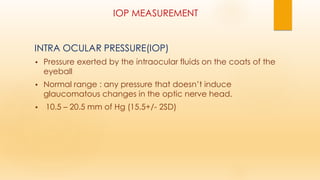
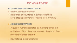
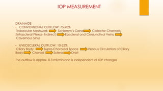
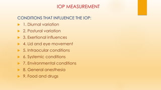
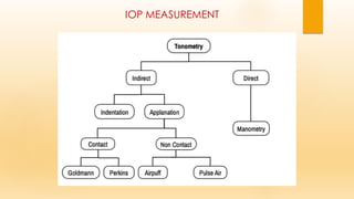
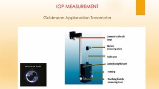
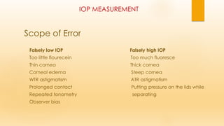
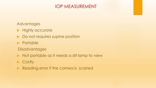
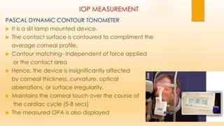
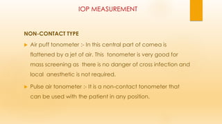
2. IOP MEASUREMENT
INTRA OCULAR PRESSURE(IOP)
• Pressure exerted by the intraocular fluids on the coats of the
eyeball
• Normal range : any pressure that doesn’t induce
glaucomatous changes in the optic nerve head.
• 10.5 – 20.5 mm of Hg (15.5+/- 2SD)
3. IOP MEASUREMENT
FACTORS AFFECTING LEVEL OF IOP:
• Rate of aqueous secretion
• Resistance encountered in outflow channels
• Level of Episcleral Venous Pressure (N 8-10 mmHG)
AQUEOUS FORMATION:
▪ Aqueous humor is secreted by the Nonpigmented
epithelium of the ciliary processes of ciliary body from a
substrate of blood plasma.
• Flow rate is 2-3 microlitre/ min.
4. IOP MEASUREMENT
Significance of Aqueous humor
1. Nutrition to lens, cornea and iris
2. Medium for removal of metabolic toxic products
3. Has an RI of 1.33
4. Inflates the globe and maintains IOP
5. Facilitates cellular and humoral response of eye
towards inflammation & infection.
5. IOP MEASUREMENT
The aqueous enters the posterior chamber by:
• ACTIVE : Energy dependent secretion of certain ions and
substrates
• PASSIVE: Diffusion & Ultrafiltration
6. IOP MEASUREMENT
DRAINAGE
• CONVENTIONAL OUTFLOW: 75-90%
Trabecular Meshwork Schlemm’s Canal Collector Channels
(Intrascleral Plexus- Indirect) Episcleral and Conjunctival Veins
Cavernous Sinus
• UVEOSCLERAL OUTFLOW: 10-25%
Ciliary Body Supra-Choroidal Space Venous Circulation of Ciliary
Body Choroid Sclera Orbit
The outflow is approx. 0.3 ml/min and is independent of IOP changes
7. IOP MEASUREMENT
Why IOP measurement?
Because ….
- IOP is the only modifiable risk factor in Glaucoma
- most frequently examined parameter in the follow up
of a glaucoma patient.
8. IOP MEASUREMENT
CONDITIONS THAT INFLUENCE THE IOP:
1. Diurnal variation
2. Postural variation
3. Exertional influences
4. Lid and eye movement
5. Intraocular conditions
6. Systemic conditions
7. Environmental conditions
8. General anesthesia
9. Food and drugs
9. IOP MEASUREMENT
DIURNAL VARIATION:
• The most common pattern is that the IOP is maximum in the morning
and decreases as day progresses and becomes minimum in the
evening because of endogeneous catecholamines.
It once again starts to rise as the night progresses.
• The variation is about 3 to 6 mm Hg in normal individuals and about 10
mm Hg or more in a glaucomatous eye.
• It is due to cyclic fluctuation of blood levels of adrenocortical
steroids. Maximum IOP is reached 3 to 4 hours after the peak of plasma
cortisol.
10. IOP MEASUREMENT
• The night time elevated IOP is due to the supine position along with
the fluctuating cortisol levels.
There are four types of diurnal variation curves.
Falling type: maximal at 6–8 am followed by a continuous decline
Rising type: maximal at 4–6 pm
Double variation type: with 2 peaks 9–11 am and 6 pm
Flat type of curve
11. IOP MEASUREMENT
• POSTURE: The IOP rises (0.3–6 mm Hg) when a person is lying down.
This may be because of increase in the episcleral venous pressure in
the supine posture.
• EXERTION: Valsalva maneuvers increases IOP (by increasing
episcleral venous pressure) while prolonged exercise decreases IOP
(by metabolic acidosis and increased colloid osmotic pressure)
• HARD LID SQUEEZING: Increases IOP because of increased
orbicularis tone
12. IOP MEASUREMENT
INTRAOCULAR CONDITIONS:
• Acute anterior uveitis causes a slight reduction in IOP because of
decreased aqueous humour production.
• Rhegmatogenous retinal detachment also causes a reduction
because of reduced aqueous humor production as well as shunting
of aqueous humour from the posterior chamber through the vitreous
and retinal hole into the subretinal space.
14. IOP MEASUREMENT
• ENVIRONMENTAL CONDITIONS:
Exposure to cold decreases IOP (because of lowered episcleral
venous pressure)
Reduced gravity increases IOP
• ANESTHETIC AGENTS: In general,
general anesthetic agents reduce IOP.
However, tricholoroethylene, ketamine, succinylcholine and
suxamethonium increase IOP
15. IOP MEASUREMENT
• FOOD AND DRUGS:
Factors increasing IOP: Factors decreasing IOP:
i. Caffeine i. Alcohol
ii. Tobacco smoking ii. Heroin and marijuana
• HEREDITY: IOP tends to be higher in individuals with enlarged cup-
disk ratio and in relatives of open-angle glaucoma
16. IOP MEASUREMENT
Also known as tonometry
Device used to measure intra ocular pressure is tonometer.
In 1865, Donders designed the first tonometer intended for
use against the sclera and
Priestley Smith in 1884, came up with something similar.
17. IOP MEASUREMENT
FACTORS INFLUENCING TONOMETRY:
CENTRAL CORNEAL THICKNESS:
Increase in corneal rigidity increased IOP reading
ASTIGMATISM
CORNEAL CURVATURE
OCCULAR RIGIDITY:
• resistance to deformation of the ocular coats.
• It is decreased in Acutely elevated IOP, Osteogenesis imperfecta,
Miotic therapy, Vasodilator therapy, Myopic eyes.
• The increase in ocular rigidity is seen in ARMD, Hypermetropic eyes
and long standing glaucoma
20. IOP MEASUREMENT
Digital assessment
It is the response of the intact eyeball to the pressure exerted
on it by the pulp of finger.
Alternately just enough pressure is applied on the eyeball
to indent it with the index finger of one hand while feeling
the compliance with the other.
21. IOP MEASUREMENT
Advantages:
It is easy to perform
no equipment required
No anesthesia is required
Helps to estimate the IOP in irregular corneas,
where applanation tonometry isn’t possible.
Disadvantages :
It is inaccurate and often subjective;
leads to over or under estimation of the IOP.
22. IOP MEASUREMENT
IDEAL TONOMETER:
• Must be accurate in its measurement
• Should be convenient to use
• Calibration should be simple
• Easy Standardization
• Maintenance should be hassle-free
23. IOP MEASUREMENT
MANOMETRY:
▪ The only direct measurement of IOP.
▪ A needle is introduced into the AC or the vitreous.
▪ And then connected to a mercury or
water manometer
24. IOP MEASUREMENT
Uses:
Continuous measurements of IOP;
Used in animal eyes for research and experimental purposes
Disadvantages:
An impractical method for use on humans;
Needs general anesthesia;
Introduction of needle produces breakdown of blood aqueous
barrier and release of prostaglandins which alter the IOP
25. IOP MEASUREMENT
Indentation Tonometry
•The shape of deformation is a truncated cone.
•It is based on the principle that the plunger would indent a soft eye
more than the hard one
•SCHIOTZ TONOMETER
It was devised in 1905
The most popular,
26. IOP MEASUREMENT
Indentation Tonometry
•When the tonometer is placed on the cornea, the following
forces become relevant :
W- Weight of the tonometer
A- Area of indentation
Vc- Volume of fluid displaced after indentation
T- Tensile forces of the outer coats of the eyeball
27. IOP MEASUREMENT
The resting intraocular raises to a new artificial value.
The scale reading of tonometer actually measures the
artificially raised Intraocular pressure.
The conversion ofread pressure to resting pressure is elicited
from the conversion tables developed by Friedenwald.
The calibration was carried by experiments in cadaveric
eyes connected with manometer through cannula.
The observation were plotted on semi log scale, which
serve as Friedenwald nomogram where the indicated scale
reading and the plunger weight are converted to an IOP
measurement
More the plunger indents the cornea, higher the scale
reading and lower the IOP.
29. IOP MEASUREMENT
COMPONENTS OF SCHIOTZ
Handle to hold the instrument in vertically
onto the cornea
Footplate that rests on the corneal surface
A Plunger that moves freely within a shaft in
footplate
A Bent lever whose short arm rests on upper
end of plunger.
A long arm which acts as pointer needle
Weights of 5.5 gm that is permanently fixed to
the plunger and can be increased to 7.5 and
10gm.
30. IOP MEASUREMENT
Schiotz Tonometry
Patient should be anesthetised with 4% lignocaine or 0.5%
proparacaine.
With the patient in supine position, looking up at a fixation target,
while the examiner would separate the lids and lower the tonometer
plate to rest on the cornea so that plunger becomes free to move.
5.5 gm is the initial weight that is used.
If the scale reading is 4 or less, additional weight is added.
IOP measurement is repeated until 3 consecutive readings agree
within 0.5 scale units.
Conversion table is used to derive IOP in mmHg. from scale reading
and plunger weight.
31. IOP MEASUREMENT
Scope of Error
In the instrument,
Might be due to the difference in shape, weight, size and
curvature of footplate
Contraction of extra ocular muscles tend to increase IOP
Due to accommodation,
With the patient looking at the tonometer resulting in
accommodation causes lowering of IOP because of
contraction of ciliary muscle
32. IOP MEASUREMENT
Scope of Error
Moses effect:
In low scale reading, the cornea might mold into the space
between the Plunger and hole, pushing the plunger up and giving
false high IOP reading.
► Due to ocular rigidity
► Due to variation in corneal curvature:
Steep and thick cornea will cause greater displacement of fluid
causing high IOP readings
33. IOP MEASUREMENT
ADVANTAGES
Simple Technique
Elegant design
Portable
No need for slit lamp or power supply
Economical
Widely used tonometer
DISADVANTAGES
Falsely high/low IOP in Ocular rigidity
Cannot be used in traumatic cases and corneal
pathologies
34. IOP MEASUREMENT
APPLANATION TONOMETRY
It was introduced by Goldmann in 1954.
The shape of deformation is flattening
It is based on IMBERT FICK LAW.
The prism applanates the cornea in an area of 3.06 mm diameter.
Variable force, Fixed area of applanation
For a very thin walled sphere, perfectly elastic and dry,
the internal pressure is equivalent to the force per unit
area of applanation.
35. IOP MEASUREMENT
APPLANATION TONOMETRY
It states that the pressure inside an ideal sphere (P) is equal to
force (W) required to flatten area(A)
P=W/A
GAT is influenced by corneal thickness,
corneal curvature,
structure of the cornea
36. IOP MEASUREMENT
Modified Imbert-Fick’s Law
W + S = P A1 + B
W = Tonometer force
S = Surface tension of pre-corneal tear film
P = Intra-ocular pressure
A1 = Inner corneal area of applanation
B = Corneal rigidity
37. IOP MEASUREMENT
Goldmann Applanation Tonometer
Most popular and accurate(Gold Standard)
Double prism mounted on slit lamp
Measures the force required to applanate the cornea over
a circular area of 3.06mm².
Significance of 3.06 mm² area is
* amount of fluid displacement is negligible(0.5μl)
* surface tension of precorneal film and force required to
counteract corneal rigidity act opposite to each other
* tonometer force becomes equal to the force in mmHg
❑ Area applanated on the cornea is 7.35mm²
39. IOP MEASUREMENT
Technique
Topical anesthesia is given, correct eye height is set via
the chin rest.
The tear film is stained with fluorescein.
The patient should blink the eyes once or twice to
spread the fluorescein stained tear film over the cornea.
Keep eyes open wide
40. IOP MEASUREMENT
Technique
The cornea and biprisms are illuminated with cobalt blue light.
Meticulously the apex of cornea is just touched by the biprism.
Applanation force against cornea is adjusted until inner edges of
two semicircles just touches.
Applanation tonometry rings viewed through Goldmann biprism
41. IOP MEASUREMENT
Scope of Error
Falsely low IOP Falsely high IOP
Too little flourecein Too much fluoresce
Thin cornea Thick cornea
Corneal edema Steep cornea
WTR astigmatism ATR astigmatism
Prolonged contact Putting pressure on the lids while
Repeated tonometry separating
Observer bias
42. IOP MEASUREMENT
Advantages
Highly accurate
Do not requires supine position
Portable
Disadvantages
Not portable as it needs a slit lamp to view
Costly
Reading error if the cornea is scarred
43. IOP MEASUREMENT
Perkins Tonometer
It uses the same biprism as the Goldmann applanation.
The light source is powered by battery.
The readings are consistent and compared quite well with
the Goldmann applanation.
ADVANTAGES OVER GAT:
Portable & counter-balanced, so it can be used
in any position.
Useful in examining Infants and children
For bedside examination for non-ambulatory
patients who cannot be examined at the slit lamp.
44. IOP MEASUREMENT
Pneumatic tonometer
Cornea is applanated by touching apex by silastic
diaphragm covering sensing nozzle.
It is connected to central chamber containing pressurized
air.
There is pneumatic to electronic transducer.
It converts the air pressure to recording on
paper strip and IOP is read.
45. IOP MEASUREMENT
Pneumatic tonometer
High displacement tonometer
Can also be used to measure outflow facility
Printouts of IOP over time
measuring IOP fluctutations possible.
Measurements independent of CCT
Post LASIK
46. IOP MEASUREMENT
Tonopen
This is small, handheld Mackay Marg type
computerised pocket tonometer
The instrument is 18 cm in length and weighs 60 g.
It converts IOP into electric waves
Wave form is internally analyzed by a
microprocessor
Average of 3 to 6 readings of IOP
47. IOP MEASUREMENT
Tonopen
As the area of applanation of the Tonopen is smaller
than GAT therefore, theoretically the difference
between applanating pressure & IOP is reduced
due to reduced corneal resistance of a smaller
contact area
It is particularly useful in community eye camps, on
ward rounds, children, irregular surfaces, measuring
through an amniotic membrane patch graft, to
read from the sclera
A disposable latex cover which is discarded after
each use provides infection control
48. IOP MEASUREMENT
PASCAL DYNAMIC CONTOUR TONOMETER
It is a slit lamp mounted device.
The contact surface is contoured to compliment the
average corneal profile.
Contour matching- Independent of force applied
or the contact area
Hence, the device is insignificantly affected
by corneal thickness, curvature, optical
aberrations, or surface irregularity.
Maintains the corneal touch over the course of
the cardiac cycle (5-8 secs)
The measured OPA is also displayed
49. IOP MEASUREMENT
NON-CONTACT TYPE
Air puff tonometer :- In this central part of cornea is
flattened by a jet of air. This tonometer is very good for
mass screening as there is no danger of cross infection and
local anesthetic is not required.
Pulse air tonometer :- It is a non-contact tonometer that
can be used with the patient in any position.
50. IOP MEASUREMENT
GROLMAN NON-CONTACT TONOMETER
Introduced by Grolman in 1972 and works
on the principle of a time interval.
Measuring the time from initial generation
of the puff of air where cornea gets flattened
(in milliseconds) to the point where the
timing device stops
51. IOP MEASUREMENT
The NCT has 3 sub-systems:
1. Alignment system:
It aligns patient’s eye in 3 dimensions (axial/vertical/lateral)
2. Optoelectronic applanation monitoring system:
Comprises a transmitter, a receiver&detector and a timer
• Transmitter directs a collimated beam of light at corneal apex
• Receiver & detector accept only parallel coaxial rays of light
reflected from cornea
• Timer measures from an internal reference to the point of
peak light intensity.
3. Pneumatic system:
It generates a puff of room air directed against cornea
52. IOP MEASUREMENT
A puff of air jet creates a constant force that
momentarily
flattens the cornea.
The force of air jet generated by a solenoid-activated
piston increases linearly over time, when the reflected
light is at peak intensity, the cornea is presumed to be
flattened.
The time elapsed is directly related to the force of jet
necessary to flatten the cornea & correspondingly to
IOP
The time from an internal reference point to the
moment of flattening is measured & converted to IOP.
53. IOP MEASUREMENT
Non contact tonometer
A fast and simple way to screen for high IOP
Modern non contact tonometers have been shown to
correlate well with Goldmann applanation tonometery
measurements.
Particularly useful
in children and other non compliant patient groups
Reduces the potential for disease transmission.
54. IOP MEASUREMENT
REBOUND TONOMETER
It determines IOP by bouncing a small plastic tipped
metal probe against the cornea.
The device uses an induction coil to magnetize the
probe and fire it against the cornea.
The rebound tonometer is a handheld portable
instrument with distinct advantages for the pediatric
population because the light-weight probe makes
only momentary contact with the cornea
(i.e. 0.25 to 0.30 meters/seconds)
55. IOP MEASUREMENT
REBOUND TONOMETER
Its main advantage
▪ Topical anesthesia is not required.
▪ It is particularly useful in children who do not
tolerate Goldmann applanation tonometry.
▪ It has also been used in home settings
▪ In situations when patients are unable to
follow-up for frequent IOP assessments
▪ To monitor diurnal variations
(Icare HOME; Icare USA)
56. IOP MEASUREMENT
OCULAR RESPONSE ANALYSER
It measures the corneal response to indentation by a rapid
air pulse.
A fully automated alignment system positions an air tube to a
precise position relative to the apex of the cornea.
Once aligned, a 25 millisecond air pulse applies pressure
to the cornea. The air pulse causes the cornea to move
inward, past applanation and into a slight concavity before
returning to normal curvature.
Corneal deformation is recorded via an electrooptical
infrared (IR) detection system
(similar to the classical air-puff tonometer).
57. IOP MEASUREMENT
It directs the air jet against the cornea and measures
not one but two pressures at which applanation occurs
1) when the air jet flattens the cornea as the cornea is bent
inward, and
2) as the air jet lessens in force and the cornea recovers.
58. IOP MEASUREMENT
The first is the resting intraocular pressure. The difference between
the first and the second applanation pressure is called corneal
hysteresis.It is the "energy absorption capability" of the cornea
This is because of the speed at which the cornea is deformed
during the dynamic bi-directional applanation process in ORA
59. IOP MEASUREMENT
TRANSPALPEBRAL TONOMETER
Diaton tonometer (BICOM Inc )
Measuring intraocular pressure through the eyelid.
Transpalpebral tonometry does not involve contact with the
cornea and does not require sterilization of the device or topical
anesthetic during routine use
Moderate correlation with those provided by applanation
tonometer.
60. IOP MEASUREMENT
TRANSPALPEBRAL TONOMETER
Diaton tonometer calculates pressure by
measuring the response of a free falllling rod.
The principle is based on Newton’s
second law, as it rebounds against the tarsal
plate of the eyelid.
The patient is positioned so that the tip of the device and lid are
overlying sclera.
61. IOP MEASUREMENT
SPECIAL SITUATIONS
In eyes with corneal edema, significant surface irregularity,
scarring and recent corneal grafts:
• PNEUMOTONOMETRY
• TONOPEN,
• DCT (It gives more accurate measurement than the GAT)
In children:
• PERKIN’S
• I-CARE (Rebound tonometry)
• TONOPEN
62. IOP MEASUREMENT
Post keratorefractive surgery:
• DCT
• Ocular Response Analyser
In Supine position:
• PERKIN’s
• PNEUMOTONOMETER
• TONOPEN: Less accurate but more practical and commonly used
Tonometry over Soft Contact Lenses
• PNEUMOTONOMETER
• TONOPEN can measure with reasonable accuracy the IOP through bandage
contact lenses.
63. IOP MEASUREMENT
Tonometry in Gas-Filled Eyes
• Intraocular gas significantly affects scleral rigidity.
In a study with irregular corneas after vitrectomy and air-gas
fluid exchange, readings with the TONOPEN and
PNEUMOTONOMETER were highly correlated.
Tonometry in Flat Anterior Chamber
In human autopsy eyes with flat anterior chambers,
IOP readings from the GAT, PNEUMOTONOMETER and TONOPEN did
not correlate well with manometrically determined pressures