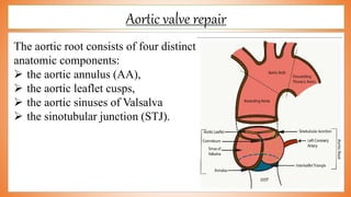
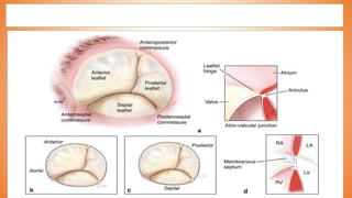
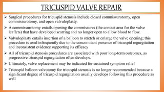
Best Rate (Hyderabad) Call Girls Jahanuma ⟟ 8250192130 ⟟ High Class Call Girl...
valve replacement and reconstruction.pptx
1. college of nursing
madras medical college
Chennai-03
valve reconstruction
and
valve replacement
By
Edwin jose.l
Msc(n) ii year
College of nursing
Madras medical college
Chennai-03
2. introduction
Cardiac surgery is usually performed in one of the largest suites in the OR as these
procedures have the most personnel and equipment needed for the operation.
The central role of the mitral and aortic valves is to prevent the regurgitation of blood flow
during the cardiac cycle.
When either valve fails through stenosis, regurgitation, or the combination of both, the
complexity of these structures become fully apparent.
the conical nature of the left ventricle, intact and functioning aortic and mitral valves are
essential to overall cardiac function.
However, because these valves are intimately associated with each other, pathology is
often coexistent. .
3. history
Theodore Tuffier is recognized to have performed the first aortic valve repair in a patient
with aortic stenosis in 1913.
The operation involved digital invagination of the anterior ascending aorta wall and
“tearing” of the stenotic valve.
Prior to the popularization of Gibbon’s and Lillehei’s methods for extracorporeal circulation
in the mid 1950s, efforts to correct aortic regurgitation surgically were limited to closed
correction.
Since then, a variety of reports have been published on repair of aortic insufficiency (AI) by
suturing two adjacent cusps together to correct prolapse or by excising the non-coronary
cusp and its aortic sinus and narrowing the aortic root and proximal ascending aorta,
thereby converting the aortic valve into a bicuspid valve
4. Relavent anatomy
Valve anatomy is complex structure
The mitral and tricuspid atrioventricular (AV) valves separate the atria from
the ventricles, while the aortic and pulmonary semilunar (SL) valves separate
the ventricles from the great arteries.
The aortic root is made up of the fibrous valve annulus region and the arterial
tissue within the sinuses of Valsalva.
The AV valves are characterized by large asymmetric leaflets hinged to ring
shaped annuli on the secured end and tethered to the ventricles by an elaborate
apparatus made up of the chordae tendineae and papillary muscles on the
mobile end.
5. Cont….
The fibrous skeleton of the heart is continuous with the annulus fibrosa that
constitutes the interconnected fibrous cartilage-like support apparatus of the
tricuspid, mitral, and aortic valves.
The annulus fibrosa is connected to the muscle of the heart in a manner that is
analogous to the attachment of tendon to skeletal muscle
The pulmonary valve is separated from the other valves by a muscular sleeve
and has a poorly defined, less substantial annulus structure.
The annuli of the AV valves are ring-shaped; however, the annulus of the
aortic valve is crown-shaped resulting in the “semilunar” shape of the
individual cusps
11. Valve reconstruction- meaning
Valve reconstruction is a surgical procedures in which the valves of the
heart is repaired.
This is done by either of the following
Opening up the stenotic valve (commissurotomy)
Tightening the opening of the leaking valve (annuloplasty)
Chordoplasty
12. commissurotomy
Repair may be made to the commissures between the leaflets in a procedure
known as commissurotomy
The most common valvuloplasty procedure is commissurotomy.
The leaflets may adhere to one another and close the commissure (ie,
stenosis).
Less commonly, the leaflets fuse in such a way that, in addition to stenosis,
the leaflets are also prevented from closing completely, resulting in a
backward flow of blood (ie, regurgitation).
A commissurotomy is the procedure performed to separate the fused leaflets.
13.
14. CLOSED COMMISSUROTOMY
Closed commissurotomies do not require cardiopulmonary bypass.
The valve is not directly visualized.
The patient receives a general anesthetic, a midsternal incision is made, a
small hole is cut into the heart, and the surgeon’s finger or a dilator is used to
break open the commissure.
This type of commissurotomy has been performed for mitral, aortic, tricuspid,
and pulmonary valve disease.
16. OPEN COMMISSUROTOMY
Open commissurotomies are performed with direct visualization of the
valve.
The patient is under general anesthesia, and a median sternotomy or left
thoracic incision is made.
Cardiopulmonary bypass is initiated, and an incision is made into the
heart.
A finger, scalpel, balloon, or dilator may be used to open the
commissures.
advantage of direct visualization of the valve is that thrombus may be
identified and removed, calcifications can be seen, and if the valve has
chordae or papillary muscles, they may be surgically repaired
https://mmcts.org/tutorial/623
17. Balloon valvuloplasty
Balloon valvuloplasty is another type of closed commissurotomy beneficial
for mitral valve stenosis in younger patients, for aortic valve stenosis in
elderly patients, and for patients with complex medical conditions that place
them at high risk for the complications of more extensive surgical procedures.
Most commonly used for mitral and aortic valve stenosis, balloon
valvuloplasty also has been used for tricuspid and pulmonic valve stenosis.
The procedure is performed in the cardiac catheterization laboratory, and the
patient may receive a local anesthetic.
Patients remain in the hospital 24 to 48 hours after the procedure
19. annuloplasty
Annuloplasty is the repair of the annulus.
General anesthesia and cardiopulmonary bypass are required for all annuloplasties.
The procedure narrows the diameter of the valve’s orifice and is useful for the treatment of
valvular regurgitation.
There are two annuloplasty techniques.
One technique uses an annuloplasty ring
The leaflets of the valve are sutured to a ring, creating an annulus of the desired size. When
the ring is in place, the tension created by the moving blood and contracting heart is borne
by the ring rather than by the valve or a suture line, and progressive regurgitation is
prevented by the repair.
20. The other technique involves tacking the valve leaflets to the atrium with
sutures or taking tucks to tighten the annulus.
Because the valve’s leaflets and the suture lines are subjected to the direct
forces of the blood and heart muscle movement, the repair may degenerate
more quickly than with the annuloplasty ring technique..
21.
22. Chordoplasty
Chordoplasty is the repair of the chordae tendineae.
The mitral valve is involved with chordoplasty (because it has the chordae
tendineae); seldom is chordoplasty required for the tricuspid valve.
Regurgitation may be caused by stretched, torn, or shortened chordae
tendineae.
Stretched chordae tendineae can be shortened, torn ones can be reattached to
the leaflet, and shortened ones can be elongated.
Regurgitation may also be caused by stretched papillary muscles, which can be
shortened.
23. Non-surgical valve repair
Percutaneous or balloon valvuloplasty/valvotomy is used for stiffened or
narrowed (stenosed) pulmonary, mitral or aortic valves. A balloon tip on the
end of the catheter is positioned in the narrowed valve and inflated to enlarge
the opening
Percutaneous mitral valve repair methods - such as edge-to-edge repair -
can fix a leaky mitral valve in a patient who is considered high risk for
surgery. A catheter holding a clip is inserted into the groin and up into the left
side of the heart. The open clip is positioned beyond the leaky valve and then
pulled back so it catches the flaps (leaflets) of the mitral valve. Once closed,
the clip holds the leaflets together and stops the valve from leaking
24. Aortic valve repair
The aortic root consists of four distinct
anatomic components:
the aortic annulus (AA),
the aortic leaflet cusps,
the aortic sinuses of Valsalva
the sinotubular junction (STJ).
25. indications
Aortic stenosis
Calcified tricuspid aortic valves
The number of leaflets, leaflet thickness, free margin appearance,
and leaflet excursion during the cardiac cycle represent the most
important information to determine reparability of the aortic
valve.
Information regarding the morphology of the aortic sinuses, STJ,
and ascending aorta is also important.
26. patient selection
Current recommendations include symptomatic patients with severe aortic
regurgitation or asymptomatic patients with severe AI and left ventricular
dysfunction (ejection fraction less than 50%) or progressive left ventricular
dilation.
aortic insufficiency due to prolapse of a BAV
children with subaortic ventricular septal defect and AI
Aortic valve repair is a valuable operative procedure for patients with AI due
to prolapse of an aortic cusp or due to dilation of the aortic root with normal
aortic cusps.
Individuals with dilated STJ and aortic sinuses but with normal aortic cusps
by echocardiography may also be candidates for aortic valve repair, although
a more complex reconstruction of the aortic root is needed.
27. Pre operative assessment
Patients younger than 40 years of age and without coronary artery risk factors
do not routinely require coronary angiography before surgery, but it should be
performed in older patients.
It is important that patients undergoing attempted aortic valve repair
understand in advance of surgery that repair may not be feasible. .
For this reason, the possibility of valve replacement, including prosthesis
options, must be discussed with the patient before surgery.
As with all valve operations, poor dental hygiene and other potential sources
of postoperative bacteremia must be addressed prior to elective operations
28. Surgical procedure
Performed via median sternotomy
CBP is established
Electro mechanical arrest by providing initial dose of cardioplegic solution
The component of aortic root is carefully assessed
4.0 polypropylene horizontal matress sutures are placed on the top of each
commissure.
Gentle traction is applied
Finally the valves sutures are examined for geometry, coaptation,
fenestrations, and mobility.
29. Mitral valve anatomy
The bicuspid mitral valve is one of the most complex structures of the human heart, its
complexity lies in its multifaceted anatomy.
The concept of “form follows function” is particularly applicable to the mitral valve.
Because each part of the valve’s anatomy is intimately related to function, there are a
variety of pathways whereby regurgitation may be created.
If one part of the valvular apparatus fails, regurgitation can result.
There are five discrete components to the mitral valve complex:
the annulus,
the two leaflets (anterior and posterior),
the chordae,
the papillary muscles, and
the LV
30.
31. Indications for mitral valve surgery
Surgery is indicated for severe symptoms (NYHA class III or IV), moderate
or severe mitral stenosis (mitral valve area ≤ 1.5 cm2 ) or systemic emboli.
Prognosis is determined by the severity of the stenosis, the size of the atrium,
the onset of AF, rising pulmonary artery pressure and the unpredictable risk
of embolism from a large, fibrillating atrium
Surgical options include commissurotomy or valvotomy, which may be
closed or open, mitral valve repair or mitral valve replacement.
Since the 1980s, percutaneous mitral balloon valvotomy (PMBV), a catheter-
based approach, has become an accepted alternative to surgical approaches in
selected patients
32.
33. Mitral valve repair
Closed mitral valvotomy
The heart is approached through a left thoracotomy and purse-string sutures are placed at
the apex of the left ventricle and in the left atrial appendage.
A finger is introduced into the left atrial appendage and the mitral valve is assessed by
direct palpation.
A special dilator (‘the Tubbs dilator’) is inserted through the left ventricular apex and
across the mitral valve.
The dilator is opened and the fused commissures are split. Some regurgitation may occur
and the process may have to be repeated after 10–15 years.
In experienced hands, the mortality rate is < 1%. It is now uncommonly performed in
developed countries as similar results can be achieved with PMBV
34. Open mitral valvotomy
With the development of CPB in the 1960s, open mitral valvotomy (or commissurotomy)
became accepted practice.
It allows direct inspection of the mitral valve apparatus and, under direct vision, division of
the commissures, splitting of fused chordae tendinae and papillary muscles, and
debridement of calcium deposits.
Amputation of the left atrial appendage may reduce the chance of subsequent
thromboembolic events.
It has became clear that restoration of normal valve function is preferable to replacement
and, as experience in open valvotomy has increased, there have been improvements in the
complexity and variety of reconstructive mitral repair techniques available, particularly
those to deal with mitral regurgitation.
35.
36. Tricuspid anatomy
The tricuspid valve apparatus comprises the leaflets, annulus, chordae, and papillary
muscles.
There are three main leaflets (anterior, posterior, and septal) separated by clefts or
commissures.
Smaller commissural leaflets are found in between the three main leaflets.
The leaflets attach at their base to the annulus and at their free edge and body to chordae,
which in turn attach to papillary muscles.
Chordae from the septal leaflet, as well as the septal half of the anterior leaflet, attach
directly to the septum.
The papillary muscles are attached to the free wall of the right ventricle and septum.
Changes in the size and geometry of the right ventricle, particularly with increased
eccentricity, can therefore cause leaflet tethering with reduced coaptation resulting in TR .
39. TRICUSPID VALVE REPAIR
Surgical procedures for tricuspid stenosis include closed commissurotomy, open
commissurotomy, and open valvuloplasty.
A commissurotomy entails opening the commissures (the contact area for the valve
leaflets) that have developed scarring and no longer open to allow blood to flow.
Valvuloplasty entails insertion of a balloon to stretch or enlarge the valve opening; this
procedure is used infrequently due to the concomitant presence of tricuspid regurgitation
and inconsistent evidence supporting its efficacy
All of tricuspid stenosis procedures are associated with poor long-term outcomes, as
progressive tricuspid regurgitation often develops.
Ultimately, valve replacement may be indicated for sustained symptom relief
Tricuspid balloon valvotomy for tricuspid stenosis is no longer recommended because a
significant degree of tricuspid regurgitation usually develops following this procedure as
well
41. Valve replacement- meaning
Valve replacement is a surgical procedure in which the heart valves were
replaced with mechanical or biological prosthesis valves made from cow, pig, or
human tissue.
This procedure is done if the valves are not able to repaired.
42. VALVE SUBSTITUTES
Mechanical valves
Biological valves.
Stented prostheses (either porcine or bovine),
Stentless prostheses,
Homografts, and
Pulmonary autografts (ross procedure).
Stentless bioprostheses are especially useful in patients requiring aortic root
replacement with a biological valve or in patients with small aortic annuli, in
order to avoid PPM.
These valves are not widely used because implantation is more complex than
stented AVR
43. Mechanical valves
Mechanical valves can be used in any age group to replace any valve
They are extremely durable but the components of the valve are thrombogenic
and therefore the patient requires systemic anticoagulation, usually with
warfarin.
This subjects the patients to a lifetime of blood tests, medication and the
constant threat of haemorrhagic complications (intracerebral, epistaxis,
gastrointestinal bleed)
44.
45.
46. Advantages:
Durability of these vales are long lasting.
Disadvantages
Need for anticoagulant therapy is life long.
Risk of thrombo-embolism is high.
Other associated problems included are para-valvular leak and endocarditis
47. Biological valves
Biological valves include homograft (or allograft) valves, removed from
cadavers; autografts, a patient’s own valve; and, most commonly, heterografts
(or xenografts) prepared from animal tissues.
All share the basic design of three semilunar leaflets with central flow, so
decreasing pressure gradients and minimizing turbulence
Heterograft ‘tissue’ valves are the most commonly used valves and can be
stented with a limited durability of 10–15 years, whereas stentless (or
frameless) valves are expected to have less late calcific degeneration but are
more technically difficult to insert.
48.
49. Advantages of Tissue Valves
Need for anticoagulation therapy is only for a short duration in
xenograft and only there is rare need for anticoagulation therapy in
autograft and allograft tissues.
Thrombogenicity is low in biologic tissue valves.
The cost of the valve is low.
50. Disadvantages
Tendency for early calcification and tissue degeneration is high and durability
is limited.
Biological valves may be preferred in patients who cannot take anticoagulant
therapy e.g. women of child-bearing age, Age below 18 years and very old
person (after 70 years).
51. Aortic valve replacement
An aortic valve replacement involves removing a faulty or damaged
valve and replacing it with a new valve made from synthetic
materials or animal tissue.
It's a major operation that isn't suitable for everyone and can take a
long time to recover from.
56. mitral valve replacement
Mitral valve replacement is an operation to replace the mitral valve with a
mechanical valve or a valve made from animal tissue (a bioprosthetic valve).
This is usually only done if you have mitral stenosis or mitral prolapse or
regurgitation and are unable to have a valve repair.
62. Pre operative nursing care
Prior to cardiac surgery, the patient should have a complete physical examination with
special attention given to the cardiovascular examination.
A new history and physical examination, chest radiograph, electrocardiogram (ECG),
complete blood count, serum electrolytes, coagulation screen, and typing and
crossmatching of blood are performed. Preoperative anemia increases the risk of
postoperative adverse events.
These data provide information about other disease conditions and cardiac problems. .
Patients with symptomatic carotid bruits should undergo carotid duplex to assess for
carotid stenosis.
Patients with pre-existing cerebrovascular disease are at increased risk for neurological
complications postoperative.
Patients with chronic lung disease should undergo pulmonary function testing and arterial
blood gas testing because they may have difficulty weaning from the ventilator
63. Cont…..
Patients undergoing valve surgery should complete a dental evaluation and work before
valve repair or replacement to reduce the chance of dental disease being a source of
bacteremia and possible prosthetic valve endocarditis.
Patients are maintained on antianginal, antihypertensives, and heart failure medications
until surgery.
Antiplatelet medications are usually discontinued before surgery: aspirin, clopidogrel, and
nonsteroidal anti-inflammatory agents should be stopped before surgery to prevent
perioperative bleeding.
The Society of Thoracic Surgeon’s workforce recommends that for elective patients and for
high-risk aspirin sensitive patients that aspirin should be stopped 3 to 5 days before
surgery.
Patients on warfarin usually have their dose withheld 3 to 5 days preoperatively. Patients
on warfarin for previous mechanical valve replacements may be admitted 1 to 2 days
before surgery for intravenous heparin.
Heparin is withheld 1 to 2 hours before surgery, whereas enoxaparin is usually stopped 12
hours beforehand.
64. Pre operative nursing diagnosis
1.Ineffective Breathing Pattern related valvular dysfunction
2.Fear/Anxiety related to Change in health status; threat of death
3.Risk for Injury related to complications of drug therapy
4.Risk for Infection related to Presence of pathogens/contaminants,
environmental exposure, invasive procedures
5.Risk for Imbalanced Body Temperature related to Exposure to cool
environment
6.Deficient Knowledge about surgical procedures related to lack of exposures
7.Altered Sensory/Thought Perception related to Therapeutically restricted
environments; excessive sensory stimuli
8.Risk for Deficient Fluid Volume related to Restriction of oral intake
65. Post operative Nursing assessment
Patient background (age, sex)
Type of operation (CABG, valve, elective vs. urgent etc)
Indications for operation
Pre-operative cath report (vessels involved, LVEF)
Success of operation (completely or incompletely revascularized, difficulties,
complications)
CPB time and aortic cross-clamp time
Ease of separation from CPB ( dysrhythmias, need for inotropes, pacing, etc). Difficulty
coming off pump may imply problems with myocardial preservation or with the
revascularization.
66. Cont….
Current inotropes, vasopressors, or anti-hypertensives (if any)
Need for cardiac pacing
Use of Intra-aortic balloon pump (IABP), ventricular assist devices (VAD), or
nitric oxide (NO).
Significant bleeding
Other significant co morbidity, with emphasis on those conditions that may
alter the post-operative management or course (carotid artery disease, COPD,
asthma, diabetes, renal failure, hepatic failure, etc.)
Pre-operative medications
Allergies
67. Physical exam and assessment
Assure that the endotracheal tube is in proper position and the patient has
equal air entry bilaterally. Remember that tube displacement or
pneumothoraxes can occur or become apparent at any moment.
Verify that the patient's oxygen saturation is adequate. Check the ABG results
as soon as they are available.
Verify correct ventilator settings.
Check the initial hemodynamic readings (HR, BP, cardiac output and index,
CVP, PCWP) and determine what vasoactive infusions the patient is on and at
what rates.
Check the patient's heart rhythm. Verify pacemaker settings if the patient is
connected to one.
68. Cont….
Check the chest and mediastinal drainage sumps to ensure they are patent and
that the patient is not bleeding excessively.
Examine heart sounds. Listen for murmurs particularly if the patient has had
valve surgery.
Check all peripheral pulses. Do repeated assessments if there is concern for
acute limb ischemia or if the patient has a femoral arterial line or IABP in
place.
Examine the abdomen.
Check pupillary reflexes. Do a more complete neurologic exam when the
patient begins to awaken from GA.
69. Warming
CPB is usually accompanied by hypothermia to < 32 C. Patients are usually warmed to at
least 34 C before transfer to the ICU.
Effects of hypothermia
Predisposes to ventricular dysrhythmias and lowers VF threshold
Increases SVR; increases afterload and myocardial workload
Patient shivering causes increased peripheral O2 consumption
Decreases CO2 production; a patient who has a respiratory alkalosis (low PCO2) on initial
ABG usually will increase their PCO2 with rewarming
Coagulopathy; impairs platelet function and the coagulation cascade. Rewarming is an
important part of the treatment of a bleeding patient.
Patients are rewarmed using the "Bear Hugger". This blows warm air over the body surface
to warm by convection.
70. Nursing diagnosis
Decreased cardiac output related to blood loss and compromised valvular
function
Impaired gas exchange related to trauma of extensive chest surgery
Risk for deficient fluid volume and electrolyte imbalance related to alterations
in blood volume
High risk for impaired tissue integrity related to bypass pump, Hypothermia.
Risk of infection related to invasive procedure and exposure to pathogens.
Risk for injury related to chemical, physical, and electrical hazards.
Risk for alteration in fluid and electrolyte balance related to abnormal blood
loss and NPO status.
71. Nursing management
Patients who have had valvuloplasty or valve replacements are admitted to the
intensive care unit; care focuses on recovery from anesthesia and
hemodynamic stability.
Vital signs are assessed every 5 to 15 minutes and as needed until the patient
recovers from anesthesia or sedation and then assessed every 2 to 4 hours and
as needed.
Intravenous medications to increase or decrease blood pressure and to treat
dysrhythmias or altered heart rates are administered and their effects
monitored.
The intravenous medications are gradually decreased until they are no longer
required or the patient takes needed medication by another route (eg, oral,
topical)
72. Patient assessments are conducted every 1 to 4 hours and as needed, with
particular attention to neurologic, respiratory, and cardiovascular systems .
After the patient has recovered from anesthesia and sedation, is
hemodynamically stable without intravenous medications, and assessment
values are stable, the patient is usually transferred to a telemetry unit,
typically within 24 to 72 hours after surgery.
Nursing care continues as for most postoperative patients, including wound
care and patient teaching regarding diet, activity, medications, and self-care..
73. The nurse educates the patient about long-term anticoagulant therapy,
explaining the need for frequent follow-up appointments and blood laboratory
studies, and provides teaching about any prescribed medication: the name of
the medication, dosage, its actions, prescribed schedule, potential side effects,
and any drug-drug or drug-food interactions.
Patients with a mechanical valve prosthesis require education to prevent
bacterial endocarditis with antibiotic prophylaxis, which is prescribed before
all dental and surgical interventions.
Patients are discharged from the hospital in 3 to 7 days.
Home care and office or clinic nurses reinforce all new information and self-
care instructions with the patient and family for 4 to 8 weeks after the
procedure.
Extracorporeal -situated or occurring outside the body
The sinuses of Valsalva, also known as aortic sinuses, are the anatomic spaces at the aortic root bounded internally by the aortic valve leaflets and externally by outward bulges of the aortic wall.
The aortic annulus is a fibrous ring at the aortic orifice to the front and right of the atrioventricular aortic valve and is considered the transition point between the left ventricle and aortic root.
Each valve has leaflets; the site where the leaflets meet is called the commissure.
valve annulus (ie, junction of the valve leaflets and the muscular heart wall)
Tacking is a sailing maneuver by which a sailing vessel, whose desired course is into the wind
Percutaneous or catheter-based procedures are done without any incisions in the chest or stopping the heart. Instead, a thin flexible tube called a catheter is inserted into a blood vessel in your groin or arm and then threaded through the blood vessels into your heart.
The entire circumference of the ventriculoaortic junction (VAJ) is defined by a band of connective tissue, which is referred to as the aortic annulus
The aortic leaflets, or cusps, possess a semilunar shape, with the base of each leaflet attached to the AA in a scalloped fashion. The center of each leaflet inserts on the AA at the lowest point of the scallop, or nadir.
The segments of arterial wall that are delineated by the AA proximally and by the STJ distally are called the sinuses of Valsalva
Bicuspid aortic valve (BAV)
BAV is the most common congenital heart defect, affecting 1.3% of the general population and evidence suggests an underlying genetic basis.
form follows function- This means that the purpose of a building should be the starting point for its design.
Pannus is a type of extra growth
Pannus is an abnormal layer of fibrovascular tissue or granulation tissue.
Leonardo de Freitas , 2018,retroseptive, multicentre study, on Long-Term Outcomes After Transcatheter Aortic Valve-in-Valve Replacement among 116 consecutive patients had ViV-TAVR between 2009 and 2015 in 9 center.
Eligibility for ViV-TAVR, valve type, and access route was determined at each center by the local heart team. Clinical, procedural, and echocardiographic data were prospectively collected in a database at each participating center.
showed that about one-fourth of patients had died after a median follow-up of 3 years. Valve hemodynamics remained overall stable over time (up to 5-year follow-up), but some degree of SVD was detected in 18.2% of patients(structural valve degeneration)