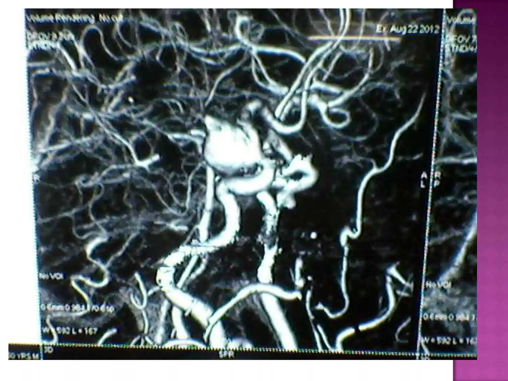
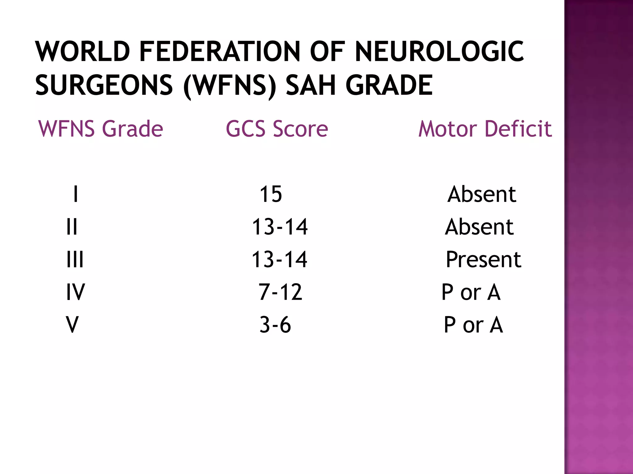
Dr. Manoj Tripathi
 75% of subarachnoid hemorrhages
 27,000 American/year
 6-49 per 100,00 year depending on location
 Female predominance
 Age 40-60
 Ruptured intracranial aneurysm (IA)
 20% morbidity
 20% mortality
 Unruptured IA
 4% morbidity
 0-2% mortality
 More than 90% of cerebral aneurysm occur at the
following locations-
1-The origin of the posterior communicating artery
2- The region of the anterior communicating artery
3- Middle cerebral artery bifurcation
4- apex of basilar artery
5- internal carotid artery bifurcation.
 Acquired vascular lesions secondary to
degenerative changes in the muscular and elastic
components of the vessel wall.
 Usually occuring at the branching points of the
major cerebral vessels.
 A deficiency of type III collagen in arteries is
assosiated with SAH.
 Congenital influences may play a role.
 Disease processes associated with an increased
risk of IA
 Polycystic kidney
 Hypertension
 Coarctation of the aorta
 Ehler- Danlos syndrome
 Fibromuscular disease
 smoking
 Small – less than 12 mm 78%
 Large – 12-24 mm 20%
 Giant - 24mm 2%
 Majority of aneurysms that bleed are less than 1
cm of diameter.
 Aneurysms that are less than .5 cm diameter
have less risk of bleeding.
 Hypertension
 Pregnancy
 Smoking
 Heavy drinking
 Strenuous activity
 Causes increase ICP
 Increased ICP causes decrease CBF
 Bleeding stops with decreased CBF
 Decreased consciousness
 2 clinical scenarios are seen typically
 Return to normal ICP and CBF with return of function
 High ICP continues with low CBF
 Grade 0 - Aneurysm is not ruptured
 Grade 1 - Asymptomatic, min. headache and sl. nuchal
rigidity
 Grade 2 - Moderate to severe headache, nuchal rigidity, but
no neurologic deficit other than cranial nerve palsy
 Grade 3 - Drowsiness, confusion, mild focal deficits
 Grade 4 - Stupor, mild or severe hemiparesis, possible early
decerebrate rigidity, vegetative disturbances
 Grade 5 - Deep coma, decerebrate rigidity, moribund
appearance
WFNS Grade GCS Score Motor Deficit
I 15 Absent
II 13-14 Absent
III 13-14 Present
IV 7-12 P or A
V 3-6 P or A
 It is very important to assess the degree of SAH.
There are different grading scales for this
purpose.
 Modified Hunt and Hess grading scale is most
commonly used because of ease of application.
 Extent of vasospasm is related to the amount of
subarachnoid blood present.
 CT scan is graded according to the Fisher grade
 Grade 1 – No blood detected
 Grade 2- Diffuse thin layer of subarachnoid
blood ( vertical layers less than 1 mm thick)
 Grade 3 – Localised clot or thick layer of
suarachnoid blood( vertical layer = 1 mm thick)
 Grade 4 – Intracerebral or intraventricular blood
with diffuse or no subarachnoid blood
 The clinical management of cerebral aneurysms
centers on the reduction of risk of hemorrhage in
uruptured cases and of repeat hemarrhage in
SAH.
 The major complications of SAH are –
1- Aneurysmal rebleeding
2- delayed cerebral ischemia secondary to
vasospasm
 Incidence of rebleeding is 14-30 % .
 Peak incidence at the end of the first week of
SAH.
 High risk of rebleed during angiography
 Assosiated with high rate of mortality and
morbidity.
 Blood pressure control is of critical importance
in reduction of risk of rebleeding.
 Antifibrinolytic agents have been used
successfully to control rebleeding
 Vasospasm is the leading cause of morbidity and
mortality in patients who initially survive SAH
 Radiological evidence of vasospasm is noted in upto
70% of patients .
 Clinical vasospasm occur in almost 30% of patients
 Clinical vasospasm occur after 4-9 days of SAH
 It typically does not occur after 2 weeks of
aneurysmal rupture.
 Pathological changes occur are contraction of
vascular smooth muscles and thickening of the
vessel wall
 Prostaglandins , biological amines , peptides , cyclic
neucleotides , calcium , lipid peroxidation and free
redicals are implicated .
 Conventional cerebral angiography , xenon-
enhanced CT and transcranial doppler is used to
confirm the presence of vasospasm
 There is a correlation between the amount of
subarachnoid blood after aneurysmal rupture and
the occurrence and severity of vasospasm
 Because of this , extensive removal of subarachnoid
blood by early surgery is attempted to decrease the
incidence of vasospasm.
 Nimodipine , a calcium channel blocker is
successfully used .
 Triple H therapy – hypertension , hypervolumia
and hemodilution is used in treatment of
vasospasm.
 A new method for symptomatic vasospasm
includes use of cerebral angioplasty to dilate
constricted major cerebral vessels.

PREOPERATIVE ASSESSMENT
DETERMINATION OF
ANAESTHETIC STRATEGY
PREOPERATIVE
PREPARATION
INDUCTION
MAINTENANCE
EMERGENCE
 Assesment of patients neuroloical condition and
clinical grading of SAH
 A review of patient,s intracranial pathological
conditions including CT scan and angiograms.
 Monitoring of ICP and transcranial doppler
ultrasonography.
 Evaluation of patients other systemic functions ,
premorbid as well as present
 Systems known to affected by SAH
 Communication with the neurosurgeon regarding
positioning and special monitoring
 Optimisation of patient,s condition by correcting
any biochemical and physiological condition
 To assess the CNS , as we have discussed before
there are grading scales-
1. Modified Hunt and Hess grading
2. WFNS grade scale
3. Fisher grading of CT scan
 The greater the clinical grade , more likely
vasospasm , elevated ICP , impaired autoregulation
and disordered response to hypocapnia will occur
 Worse clinical grade is also assosited with cardiac
arrythmia , myocardial dysfunction , hypovolumia
and hyponatremia.
 ECG abnormalities
 Very common
 Many changes seen
 cannon t wave, Q-T prolongation, ST changes
 Autonomic surge may in fact cause some
subendocardial injury from increase myocardial wall
tension
 Cardiac dysfunction does not appear to affect
morbidity or mortality (studies from Zaroff and
Browers)
 Prolonged Q-T with increased incidence of
ventricular arrhythmias
 PVC’s are seen in 80%
 ECG changes occur during the first 48 hrs of SAH and
correlate with amount of intracranial bleed.
 ECG changes reflect the severity of neurogenic
damage and have not shown to contribute
perioperative mortality and morbidity
 The decision to operate should not be influenced
by these ECG changes.
 Hydrocephalous
 Seizures
 13%
 Vasospasm may be cause
 Increased risk of rebleed
 Treat and prophylaxis
 Headache, visual field changes, motor
deficits
 SIADH
 Cerebral salt wasting syndrome
 release of naturetic peptide
 hypovolemia, increased urine NA and volume
contraction
 Distinguish between the two and treat
accordingly
 Neurogenic pulmonary edema
 1-2% with SAH
 Hyperactivity of the sympathetic nervous system
 Pneumonia in 7-12% of hospitalized patients with
SAH
 0-3 days post bleed appears to be optimal
 Improved outcome within 6 hours of rupture
despite high H/H grade
 If delayed, should be done after 10 days post
bleed after fibrinolytic phase
 The results are worst with surgery performed
between 7 to 10 days.
 Avoid abrupt changes in BP
 Maintain CBF with normal to high blood pressure
 Avoid increase of ICP
 Assess immobility & vital signs control
 Achieve brain relaxation
 Allow for swift emergence & neurologic assessment
 Be prepared for disaster
 Arterial blood pressure- beat to beat monitoring
of MAP
 ECG- myocardial ischemia/ arrhythmia
 Pulse oximetry- systemic hypoxia
 EtCO2- trend monitor for Paco2/ detection of VAE
 Temperature- via oesophageal lead; to allow
modest, passive hypothermia(~35o C)
 Urine output- adequacy of renal function &
hydration
 Blood glucose/ serum electrolytes/ osmolality
-particularly if mannitol is used
 Hemoglobin & hematocrit- to estimate extent
of bleeding/ permissible blood loss
 Jugular venous bulb monitoring- adequacy of
cerebral perfusion & oxygenation
 EEG- CMR/ cerebral ischemia/ depth of
anaesthesia
 Evoked potentials- intactness of specific CNS
pathways
 Transcranial oximetry- noninvasive information
on regional cerebral oxygenation
 TCD ultrasonorgaphy
 TCD is a indirect measure CBF
 It is unreliable as a measure of CBF in patients of
SAH because of changes in vessel diameter
 But it has become valuable for diagnosing
vasospasm noninvasively before the onset of
clinical symptoms
 TCD has been successfully used in the
perioperative management of patients with
cerebral aneurysm.
 Continuous TCD monitoring may improve the safety
of induced hypotension by correlating the blood
velocity change to the decline in the blood
pressure.
 It has been used perioperatively to confirm the
diagnosis of aneurysmal rupture.
 Patients should receive their regular dose of
nimodipine and dexamethasone
 Tab Loarazepam 1-2 mg and tab rantac 150
should be given in night before surgery
 To relieve anxiety inj midazolam in incremental
dose of 1 mg is given in the morning of surgery.
 There is risk of rupture of aneurysm at the time
of induction due to high blood pressure during
tracheal intubation
 As a general principle , the patients blood
pressure should be reduced by 20-25% below the
baseline value and hypertensive response to the
tracheal intubation should be alleviated.
 Another useful approach is to balance the risk of
ischemia from a decrease in CPP against the
benefit of a reduced chance of aneurysmal
rupture from a decrease TMP.
 Conceptually induction phase is consisting of 2
parts
Induction to achieve loss of consciousness
 Thiopental ( 3- 5 mg/kg ) or propofol (1-2.5
mg/kg ) in combination with fentanyl (3-7 ug/kg)
or sufentanil(.3-.7 ug/kg) is suitable
 Other alternatives include etomidate (.3-.4 mg/kg)
and midazolam ( .1-.2mg/kg)
Prophylaxis against rise in BP during laryngoscopy
 Many agents have been used successfully to
alleviate hypertensive response of intubation.
Fentanyl ( 5-10 ug/kg)
Sufentanil ( .5-1 ug/kg)
Esmolol (.5 mg/kg)
Labetolol (10-20 mg)
Intraveous or topical lidocaine (1.5-2 mg/kg)
Second dose of thiopental ( 1-2 mg/kg)
 Intravenous adjuncts are preferred in patients
with poor SAH grades whereas deep inhalational
anesthetics are preferred in patients with good
SAH grades.
Choice of muscle relaxant
 Vecuronium is most hemodyanamically stable
and suitable muscle relaxant.
 Succinylcholine causes incease in ICP.
 Atracurium may cause hypotension.
 Pancuronium causes tachycardia and
hypertension
 The location and size of aneurysm generally
determine the position of patient.
 Anterior circulation aneurysm are usually
approached using fronto-temporal incision with the
patient in supine position
 Basilar tip aneurysms are approached using
subtemporal incision with the patient in lareral
position
 Vertebral and basilar trunk aneurysms approached using
suboccipital incision with the patient in sitting or park
bench position
 Avoid extreme positioning (extreme rotation or flexion
of neck to avoid IJV compression)
 Padding/ fixing of regions susceptible to injury by
pressure/ abrasion/ movement -groin, breasts, axillary
region
-falling extremities
-knees kept in mild flexion to prevent
backache postoperatively
 Mild head-up position (to aid venous cerebral drainage)
 Elevation of contralateral shoulder by wedge/ roll
(to prevent brachial plexus stretch injury if head is
turned laterally)
 Meticulous attention to specific problems in prone/
lateral/ parkbench/ sitting positions
 Care of ETT –easy intraoperative accessibility
-fixed & packed securely to
prevent accidental extubation, or abrasions
resulting from movement
 Care of eyes- taped occlusively to prevent corneal
damage (from exposure/ irrigation with antiseptic
solutions)
APPLICATION OF SKULL PIN HOLDER FRAME
 Pain- provides maximal nociceptive stimulus
- must be blocked adequately by
i. deepening of anaesthesia (i.v. bolus of
thiopentone 1mg/kg or propofol 0.5 mg/kg)
ii. analgesia (i.v. bolus of fentanyl 1-3
mcg/kg or alfentanil 10-20 mcg/kg or remifentanil 0.25-1
mcg/kg)
iii. local anaesthetic infiltration at pin site
iv. antihypertensive β-blockers e.g.
Esmolol 1 mg/kg or Labetalol 0.5-1 mg/kg
 VAE- may occur with pin insertion
 Positioning of Anaesthetist
-optimal patient monitoring
-access to airway/ intravenous & intraarterial
lines
The goals during maintainance of anesthesia are --
 To provide a relaxed or ‘slack’ brain that will allow
minimum retraction pressure
 To maintain perfusion to the brain
 To reduce TMP if necessary during dissection of the
aneurysm and final clipping
 Allow prompt awakening and assessment of
patients with good SAH grades
 Maintenance
CHOICE OF TECHNIQUE
Volatile agents Intravenous agents
Advantages Controlability/ predictability/ early
awakening
Good control of CBF, ICP, & brain
bulk
-cerebrovasoconstriction
↓ in ICP
Disadvantages Poor control of CBF, ICP, & brain
bulk
-cerebrovasodilation
↑ in ICP
Prolonged/ unpredictable
awakening
May interfere with D/D of delayed
awakening
May require emergent CT scan
to rule out surgical complications
Type of
surgery
Simple, low risk of ↑ed ICP Complex, high risk of ↑ed ICP
 Maintenance
CHOICE OF TECHNIQUE
Volatile agents Intravenous agents
Early institution of
moderate
hyperventilation
Mandatory Optional
Concurrent use with
N2O
Ideal agent
Usually avoided
-synergistic effects in ↑ing CBF &
CMR
-if used, ensure ↓in ICP by
i. hyperventilation
Ii. osmotic diuretics
Iii. BP control
Iv. adequate positioning/ cerebral
venous drainage
v. lumbar drainage
Vi. Use of < 1 MAC (e.g. < 1.15% of
isoflurane)
No
Can be used without
significant problems
Yes
Fluid Therapy
 Fluid therapy should be guided by intraoperative blood
loss, urine output and CVP/PAWP
 The aim is to maintain normovolumia before
aneurysmal clipping and slight hypervolumia and
hypertension after clipping.
 Avoidance of hyperglycemia (worsens consequences of
cerebral ischemia)
 Avoidance of hypoosmolality – can cause brain
oedema
i. Target osmolality: 290-320 mOsm/kg)
ii. Colloid oncotic pressure plays no significant role
in brain oedema
iii. Avoidance of glucose-containing & hypoosmolar
solutions (e.g. Ringer’s lactate, 254 mOsm/kg)
 Preferred solutions – crystalloids: 0.9% NaCl
colloids: 6% HES (304 mOsm/kg)
 Hematocrit- Target for >28%
 Warming of I.V. solutions– may be avoided to
permit establishment of mild hypothermia (~350 C)
for neuroprotection
-must be essentially warmed at the end of
procedure to ensure normothermia for emergence
from anaesthesia
 Hemodynamic control
-Undesirable CNS arousal & hemodynamic activation may
occur despite adequate depth of anaesthesia &
analgesia
-Consider use of i. Esmolol (1mg/kg: initial dose)
ii. Labetalol (0.5-1mg/kg: initial
dose)
iii. Clonidine (0.5-1mcg/kg: initial
dose)
 Moderate hypothermia (~350C)
-may confer a degree of brain protection if ischemic
event occurs
 Prevention
1. No over hydration
2. Sedation/ analgesia/ anxiolysis
3. Avoidance of application of any noxious stimulus with
sedation/ local anaesthesia
4. Head-up position
5. Osmotic agents (mannitol/ hypertonic saline)
6. β-blockers/ clonidine/ lignocaine
7. Adequate hemodynamics: MAP, CVP, PCWP, HR
8. Adequate ventilation: PaO2>100mmHg;
PaCO2~35mmHg
9. Minimal possible intrathoracic pressure
10. Hyperventilation on demand (before induction)
11. Use of total I.V. anaesthestic agents for induction
& maintenance
12. Avoidance of cerebral vasodilators (e.g.
nitroglycerine)
 Treatment
1.Hyperventilation
2.Osmotic agents
3.CSF drainage (if ventricular/ lumbar catheter in situ)
4.Augmentation of anaesthesia with I.V. anaesthetic
agents (e.g. propofol, thiopentone, etomidate)
5.Adequate muscle relaxation
6. Venous drainage (head-up/ avoidance of PEEP/
reduction of inspiratory time)
7.Mild controlled hypertension (if autoregulation is
present)
 5-7 minutes of occlusion with prompt reperfusion
are usually well tolerated but this duration is
insufficient for clipping difficult or giant aneurysms
 A number of regimens have been used to extend
the occlusion duration
 High dose Mannitol 2g/kg
 SENDAI COCKTAIL - mannitol (500 ml of 20%
solution) + vitamin E (500 mg) + dexamethasone
(50 mg)
 Pharmacological metabolic suppression by
thiopentone ( 5-6 mg/kg) or etomidate (.4-.5
mg/kg)
 Etomidate is preferred over thiopental due to
greater hemodyanmic stability
 Moderate hypothermia has also been to extend the
duration of tolerable occlusion
 If the surgical procedure is uneventful , SAH grade I
and II patients should be extubated.
 Because hypertensive therapy is useful in reversing
delated cerebral ischemia from vasospasm , modest
level of postoperative hypertension (<180mm hg )
should not be aggressively treated.
 Depending on preoperative ventilatory status and
duration and difficulty of surgical procedure
 SAH grade III patients may or may not be extubated.
 Patients with preoperative SAH grade IV and V
usually require postoperative ventilatory support and
neurointensive care.
 In the postoperative period blood pressure should be
maintained above 140-150 mm hg and less than 180
mm hg.
 To distinguish residual anesthesia from surgical
cause following general guidelines are useful
1- Anesthesia causes global depression and any new
focal neurological deficit should alert to a surgical
cause
2-The effect of potent inhaled anesthetics should
have larly dissipated after 30-60 minutes
3- patients whose pupils are midsized and having no
respiratoty depression are unlikly to experience a
narcotic overdose.
4- unequal pupils not present before surgery always
suggest a surgical cause.
 Neurological assessment should be done every 15
minutes in the recovery room.
Thank you

Intracranial aneurysm surgery and anesthesia

  • 1.
  • 2.
     75% ofsubarachnoid hemorrhages  27,000 American/year  6-49 per 100,00 year depending on location  Female predominance  Age 40-60
  • 3.
     Ruptured intracranialaneurysm (IA)  20% morbidity  20% mortality  Unruptured IA  4% morbidity  0-2% mortality
  • 6.
     More than90% of cerebral aneurysm occur at the following locations- 1-The origin of the posterior communicating artery 2- The region of the anterior communicating artery 3- Middle cerebral artery bifurcation 4- apex of basilar artery 5- internal carotid artery bifurcation.
  • 8.
     Acquired vascularlesions secondary to degenerative changes in the muscular and elastic components of the vessel wall.  Usually occuring at the branching points of the major cerebral vessels.  A deficiency of type III collagen in arteries is assosiated with SAH.
  • 9.
     Congenital influencesmay play a role.  Disease processes associated with an increased risk of IA  Polycystic kidney  Hypertension  Coarctation of the aorta  Ehler- Danlos syndrome  Fibromuscular disease  smoking
  • 10.
     Small –less than 12 mm 78%  Large – 12-24 mm 20%  Giant - 24mm 2%  Majority of aneurysms that bleed are less than 1 cm of diameter.  Aneurysms that are less than .5 cm diameter have less risk of bleeding.
  • 11.
     Hypertension  Pregnancy Smoking  Heavy drinking  Strenuous activity
  • 12.
     Causes increaseICP  Increased ICP causes decrease CBF  Bleeding stops with decreased CBF  Decreased consciousness  2 clinical scenarios are seen typically  Return to normal ICP and CBF with return of function  High ICP continues with low CBF
  • 13.
     Grade 0- Aneurysm is not ruptured  Grade 1 - Asymptomatic, min. headache and sl. nuchal rigidity  Grade 2 - Moderate to severe headache, nuchal rigidity, but no neurologic deficit other than cranial nerve palsy  Grade 3 - Drowsiness, confusion, mild focal deficits  Grade 4 - Stupor, mild or severe hemiparesis, possible early decerebrate rigidity, vegetative disturbances  Grade 5 - Deep coma, decerebrate rigidity, moribund appearance
  • 14.
    WFNS Grade GCSScore Motor Deficit I 15 Absent II 13-14 Absent III 13-14 Present IV 7-12 P or A V 3-6 P or A
  • 15.
     It isvery important to assess the degree of SAH. There are different grading scales for this purpose.  Modified Hunt and Hess grading scale is most commonly used because of ease of application.  Extent of vasospasm is related to the amount of subarachnoid blood present.  CT scan is graded according to the Fisher grade
  • 16.
     Grade 1– No blood detected  Grade 2- Diffuse thin layer of subarachnoid blood ( vertical layers less than 1 mm thick)  Grade 3 – Localised clot or thick layer of suarachnoid blood( vertical layer = 1 mm thick)  Grade 4 – Intracerebral or intraventricular blood with diffuse or no subarachnoid blood
  • 17.
     The clinicalmanagement of cerebral aneurysms centers on the reduction of risk of hemorrhage in uruptured cases and of repeat hemarrhage in SAH.  The major complications of SAH are – 1- Aneurysmal rebleeding 2- delayed cerebral ischemia secondary to vasospasm
  • 18.
     Incidence ofrebleeding is 14-30 % .  Peak incidence at the end of the first week of SAH.  High risk of rebleed during angiography  Assosiated with high rate of mortality and morbidity.
  • 19.
     Blood pressurecontrol is of critical importance in reduction of risk of rebleeding.  Antifibrinolytic agents have been used successfully to control rebleeding
  • 20.
     Vasospasm isthe leading cause of morbidity and mortality in patients who initially survive SAH  Radiological evidence of vasospasm is noted in upto 70% of patients .  Clinical vasospasm occur in almost 30% of patients  Clinical vasospasm occur after 4-9 days of SAH  It typically does not occur after 2 weeks of aneurysmal rupture.
  • 21.
     Pathological changesoccur are contraction of vascular smooth muscles and thickening of the vessel wall  Prostaglandins , biological amines , peptides , cyclic neucleotides , calcium , lipid peroxidation and free redicals are implicated .  Conventional cerebral angiography , xenon- enhanced CT and transcranial doppler is used to confirm the presence of vasospasm
  • 22.
     There isa correlation between the amount of subarachnoid blood after aneurysmal rupture and the occurrence and severity of vasospasm  Because of this , extensive removal of subarachnoid blood by early surgery is attempted to decrease the incidence of vasospasm.  Nimodipine , a calcium channel blocker is successfully used .
  • 23.
     Triple Htherapy – hypertension , hypervolumia and hemodilution is used in treatment of vasospasm.  A new method for symptomatic vasospasm includes use of cerebral angioplasty to dilate constricted major cerebral vessels.
  • 24.
  • 25.
    PREOPERATIVE ASSESSMENT DETERMINATION OF ANAESTHETICSTRATEGY PREOPERATIVE PREPARATION INDUCTION MAINTENANCE EMERGENCE
  • 26.
     Assesment ofpatients neuroloical condition and clinical grading of SAH  A review of patient,s intracranial pathological conditions including CT scan and angiograms.  Monitoring of ICP and transcranial doppler ultrasonography.
  • 27.
     Evaluation ofpatients other systemic functions , premorbid as well as present  Systems known to affected by SAH  Communication with the neurosurgeon regarding positioning and special monitoring  Optimisation of patient,s condition by correcting any biochemical and physiological condition
  • 28.
     To assessthe CNS , as we have discussed before there are grading scales- 1. Modified Hunt and Hess grading 2. WFNS grade scale 3. Fisher grading of CT scan
  • 29.
     The greaterthe clinical grade , more likely vasospasm , elevated ICP , impaired autoregulation and disordered response to hypocapnia will occur  Worse clinical grade is also assosited with cardiac arrythmia , myocardial dysfunction , hypovolumia and hyponatremia.
  • 30.
     ECG abnormalities Very common  Many changes seen  cannon t wave, Q-T prolongation, ST changes  Autonomic surge may in fact cause some subendocardial injury from increase myocardial wall tension
  • 31.
     Cardiac dysfunctiondoes not appear to affect morbidity or mortality (studies from Zaroff and Browers)  Prolonged Q-T with increased incidence of ventricular arrhythmias  PVC’s are seen in 80%  ECG changes occur during the first 48 hrs of SAH and correlate with amount of intracranial bleed.
  • 32.
     ECG changesreflect the severity of neurogenic damage and have not shown to contribute perioperative mortality and morbidity  The decision to operate should not be influenced by these ECG changes.
  • 33.
     Hydrocephalous  Seizures 13%  Vasospasm may be cause  Increased risk of rebleed  Treat and prophylaxis  Headache, visual field changes, motor deficits
  • 34.
     SIADH  Cerebralsalt wasting syndrome  release of naturetic peptide  hypovolemia, increased urine NA and volume contraction  Distinguish between the two and treat accordingly
  • 35.
     Neurogenic pulmonaryedema  1-2% with SAH  Hyperactivity of the sympathetic nervous system  Pneumonia in 7-12% of hospitalized patients with SAH
  • 36.
     0-3 dayspost bleed appears to be optimal  Improved outcome within 6 hours of rupture despite high H/H grade  If delayed, should be done after 10 days post bleed after fibrinolytic phase  The results are worst with surgery performed between 7 to 10 days.
  • 37.
     Avoid abruptchanges in BP  Maintain CBF with normal to high blood pressure  Avoid increase of ICP  Assess immobility & vital signs control  Achieve brain relaxation  Allow for swift emergence & neurologic assessment  Be prepared for disaster
  • 38.
     Arterial bloodpressure- beat to beat monitoring of MAP  ECG- myocardial ischemia/ arrhythmia  Pulse oximetry- systemic hypoxia  EtCO2- trend monitor for Paco2/ detection of VAE  Temperature- via oesophageal lead; to allow modest, passive hypothermia(~35o C)  Urine output- adequacy of renal function & hydration
  • 39.
     Blood glucose/serum electrolytes/ osmolality -particularly if mannitol is used  Hemoglobin & hematocrit- to estimate extent of bleeding/ permissible blood loss  Jugular venous bulb monitoring- adequacy of cerebral perfusion & oxygenation  EEG- CMR/ cerebral ischemia/ depth of anaesthesia
  • 40.
     Evoked potentials-intactness of specific CNS pathways  Transcranial oximetry- noninvasive information on regional cerebral oxygenation  TCD ultrasonorgaphy
  • 41.
     TCD isa indirect measure CBF  It is unreliable as a measure of CBF in patients of SAH because of changes in vessel diameter  But it has become valuable for diagnosing vasospasm noninvasively before the onset of clinical symptoms  TCD has been successfully used in the perioperative management of patients with cerebral aneurysm.
  • 42.
     Continuous TCDmonitoring may improve the safety of induced hypotension by correlating the blood velocity change to the decline in the blood pressure.  It has been used perioperatively to confirm the diagnosis of aneurysmal rupture.
  • 43.
     Patients shouldreceive their regular dose of nimodipine and dexamethasone  Tab Loarazepam 1-2 mg and tab rantac 150 should be given in night before surgery  To relieve anxiety inj midazolam in incremental dose of 1 mg is given in the morning of surgery.
  • 44.
     There isrisk of rupture of aneurysm at the time of induction due to high blood pressure during tracheal intubation  As a general principle , the patients blood pressure should be reduced by 20-25% below the baseline value and hypertensive response to the tracheal intubation should be alleviated.
  • 45.
     Another usefulapproach is to balance the risk of ischemia from a decrease in CPP against the benefit of a reduced chance of aneurysmal rupture from a decrease TMP.  Conceptually induction phase is consisting of 2 parts Induction to achieve loss of consciousness  Thiopental ( 3- 5 mg/kg ) or propofol (1-2.5 mg/kg ) in combination with fentanyl (3-7 ug/kg) or sufentanil(.3-.7 ug/kg) is suitable
  • 46.
     Other alternativesinclude etomidate (.3-.4 mg/kg) and midazolam ( .1-.2mg/kg) Prophylaxis against rise in BP during laryngoscopy  Many agents have been used successfully to alleviate hypertensive response of intubation. Fentanyl ( 5-10 ug/kg) Sufentanil ( .5-1 ug/kg) Esmolol (.5 mg/kg) Labetolol (10-20 mg) Intraveous or topical lidocaine (1.5-2 mg/kg) Second dose of thiopental ( 1-2 mg/kg)
  • 47.
     Intravenous adjunctsare preferred in patients with poor SAH grades whereas deep inhalational anesthetics are preferred in patients with good SAH grades.
  • 48.
    Choice of musclerelaxant  Vecuronium is most hemodyanamically stable and suitable muscle relaxant.  Succinylcholine causes incease in ICP.  Atracurium may cause hypotension.  Pancuronium causes tachycardia and hypertension
  • 49.
     The locationand size of aneurysm generally determine the position of patient.  Anterior circulation aneurysm are usually approached using fronto-temporal incision with the patient in supine position  Basilar tip aneurysms are approached using subtemporal incision with the patient in lareral position
  • 50.
     Vertebral andbasilar trunk aneurysms approached using suboccipital incision with the patient in sitting or park bench position  Avoid extreme positioning (extreme rotation or flexion of neck to avoid IJV compression)  Padding/ fixing of regions susceptible to injury by pressure/ abrasion/ movement -groin, breasts, axillary region -falling extremities -knees kept in mild flexion to prevent backache postoperatively  Mild head-up position (to aid venous cerebral drainage)
  • 51.
     Elevation ofcontralateral shoulder by wedge/ roll (to prevent brachial plexus stretch injury if head is turned laterally)  Meticulous attention to specific problems in prone/ lateral/ parkbench/ sitting positions  Care of ETT –easy intraoperative accessibility -fixed & packed securely to prevent accidental extubation, or abrasions resulting from movement
  • 52.
     Care ofeyes- taped occlusively to prevent corneal damage (from exposure/ irrigation with antiseptic solutions) APPLICATION OF SKULL PIN HOLDER FRAME  Pain- provides maximal nociceptive stimulus - must be blocked adequately by i. deepening of anaesthesia (i.v. bolus of thiopentone 1mg/kg or propofol 0.5 mg/kg) ii. analgesia (i.v. bolus of fentanyl 1-3 mcg/kg or alfentanil 10-20 mcg/kg or remifentanil 0.25-1 mcg/kg)
  • 53.
    iii. local anaestheticinfiltration at pin site iv. antihypertensive β-blockers e.g. Esmolol 1 mg/kg or Labetalol 0.5-1 mg/kg  VAE- may occur with pin insertion
  • 54.
     Positioning ofAnaesthetist -optimal patient monitoring -access to airway/ intravenous & intraarterial lines
  • 55.
    The goals duringmaintainance of anesthesia are --  To provide a relaxed or ‘slack’ brain that will allow minimum retraction pressure  To maintain perfusion to the brain  To reduce TMP if necessary during dissection of the aneurysm and final clipping  Allow prompt awakening and assessment of patients with good SAH grades
  • 56.
     Maintenance CHOICE OFTECHNIQUE Volatile agents Intravenous agents Advantages Controlability/ predictability/ early awakening Good control of CBF, ICP, & brain bulk -cerebrovasoconstriction ↓ in ICP Disadvantages Poor control of CBF, ICP, & brain bulk -cerebrovasodilation ↑ in ICP Prolonged/ unpredictable awakening May interfere with D/D of delayed awakening May require emergent CT scan to rule out surgical complications Type of surgery Simple, low risk of ↑ed ICP Complex, high risk of ↑ed ICP
  • 57.
     Maintenance CHOICE OFTECHNIQUE Volatile agents Intravenous agents Early institution of moderate hyperventilation Mandatory Optional Concurrent use with N2O Ideal agent Usually avoided -synergistic effects in ↑ing CBF & CMR -if used, ensure ↓in ICP by i. hyperventilation Ii. osmotic diuretics Iii. BP control Iv. adequate positioning/ cerebral venous drainage v. lumbar drainage Vi. Use of < 1 MAC (e.g. < 1.15% of isoflurane) No Can be used without significant problems Yes
  • 58.
    Fluid Therapy  Fluidtherapy should be guided by intraoperative blood loss, urine output and CVP/PAWP  The aim is to maintain normovolumia before aneurysmal clipping and slight hypervolumia and hypertension after clipping.  Avoidance of hyperglycemia (worsens consequences of cerebral ischemia)
  • 59.
     Avoidance ofhypoosmolality – can cause brain oedema i. Target osmolality: 290-320 mOsm/kg) ii. Colloid oncotic pressure plays no significant role in brain oedema iii. Avoidance of glucose-containing & hypoosmolar solutions (e.g. Ringer’s lactate, 254 mOsm/kg)  Preferred solutions – crystalloids: 0.9% NaCl colloids: 6% HES (304 mOsm/kg)
  • 60.
     Hematocrit- Targetfor >28%  Warming of I.V. solutions– may be avoided to permit establishment of mild hypothermia (~350 C) for neuroprotection -must be essentially warmed at the end of procedure to ensure normothermia for emergence from anaesthesia
  • 61.
     Hemodynamic control -UndesirableCNS arousal & hemodynamic activation may occur despite adequate depth of anaesthesia & analgesia -Consider use of i. Esmolol (1mg/kg: initial dose) ii. Labetalol (0.5-1mg/kg: initial dose) iii. Clonidine (0.5-1mcg/kg: initial dose)  Moderate hypothermia (~350C) -may confer a degree of brain protection if ischemic event occurs
  • 62.
     Prevention 1. Noover hydration 2. Sedation/ analgesia/ anxiolysis 3. Avoidance of application of any noxious stimulus with sedation/ local anaesthesia 4. Head-up position 5. Osmotic agents (mannitol/ hypertonic saline) 6. β-blockers/ clonidine/ lignocaine
  • 63.
    7. Adequate hemodynamics:MAP, CVP, PCWP, HR 8. Adequate ventilation: PaO2>100mmHg; PaCO2~35mmHg 9. Minimal possible intrathoracic pressure 10. Hyperventilation on demand (before induction) 11. Use of total I.V. anaesthestic agents for induction & maintenance 12. Avoidance of cerebral vasodilators (e.g. nitroglycerine)
  • 64.
     Treatment 1.Hyperventilation 2.Osmotic agents 3.CSFdrainage (if ventricular/ lumbar catheter in situ) 4.Augmentation of anaesthesia with I.V. anaesthetic agents (e.g. propofol, thiopentone, etomidate) 5.Adequate muscle relaxation 6. Venous drainage (head-up/ avoidance of PEEP/ reduction of inspiratory time) 7.Mild controlled hypertension (if autoregulation is present)
  • 65.
     5-7 minutesof occlusion with prompt reperfusion are usually well tolerated but this duration is insufficient for clipping difficult or giant aneurysms  A number of regimens have been used to extend the occlusion duration  High dose Mannitol 2g/kg  SENDAI COCKTAIL - mannitol (500 ml of 20% solution) + vitamin E (500 mg) + dexamethasone (50 mg)
  • 66.
     Pharmacological metabolicsuppression by thiopentone ( 5-6 mg/kg) or etomidate (.4-.5 mg/kg)  Etomidate is preferred over thiopental due to greater hemodyanmic stability  Moderate hypothermia has also been to extend the duration of tolerable occlusion
  • 67.
     If thesurgical procedure is uneventful , SAH grade I and II patients should be extubated.  Because hypertensive therapy is useful in reversing delated cerebral ischemia from vasospasm , modest level of postoperative hypertension (<180mm hg ) should not be aggressively treated.  Depending on preoperative ventilatory status and duration and difficulty of surgical procedure
  • 68.
     SAH gradeIII patients may or may not be extubated.  Patients with preoperative SAH grade IV and V usually require postoperative ventilatory support and neurointensive care.
  • 69.
     In thepostoperative period blood pressure should be maintained above 140-150 mm hg and less than 180 mm hg.  To distinguish residual anesthesia from surgical cause following general guidelines are useful 1- Anesthesia causes global depression and any new focal neurological deficit should alert to a surgical cause
  • 70.
    2-The effect ofpotent inhaled anesthetics should have larly dissipated after 30-60 minutes 3- patients whose pupils are midsized and having no respiratoty depression are unlikly to experience a narcotic overdose. 4- unequal pupils not present before surgery always suggest a surgical cause.  Neurological assessment should be done every 15 minutes in the recovery room.
  • 71.