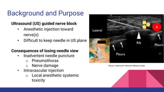
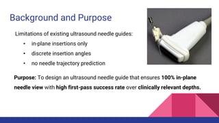
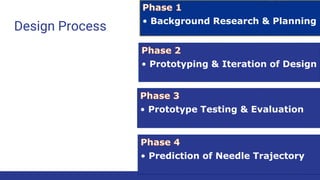
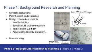
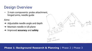
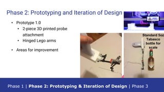
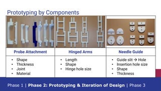
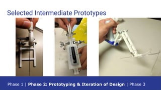
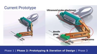
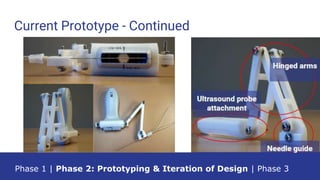
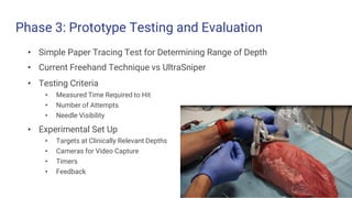
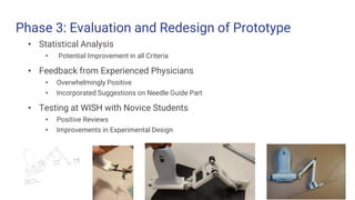
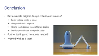
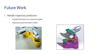
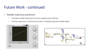
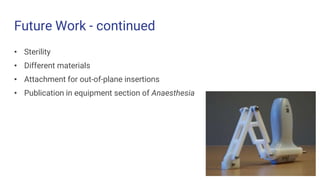
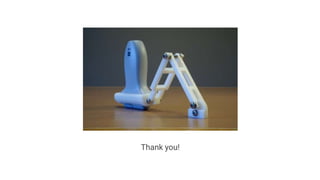
UltraSniper:
An Ultrasound Needle Guide to
Aid Nerve Block Procedures
Kate Fox, Binhan Pham, Minghui Shi, Jonathan Yu
UW Bioengineering Capstone Symposium
May 23, 2016
Kate Fox, Binhan Pham, Minghui Shi, Jonathan Yu
Dr. Christopher Neils, Dr. Alyssa Taylor
Dr. Daniel Low Anesthesiologist
Dr. Timothy Casias Anesthesiologist
Keith Williams Director of Hardware Engineering
Paul Dunham Principal Transducer Engineer
Saeed Aliakbari Mechanical Engineer
Angel Brown System Engineer
Our Team
Overview
• Background and Purpose
• Current Solutions
• Design Overview
• Evolution of Design
• Prototype Testing and Results
• Conclusions
• Future Directions
G.E. Healthcare
Background and Purpose
Ultrasound (US) guided nerve block
• Anesthetic injection toward
nerve(s)
• Difficult to keep needle in US plane
G.E. Healthcare
Source: Dartmouth-Hitchcock Medical Center
Consequences of losing needle view
• Inadvertent needle puncture
o Pneumothorax
o Nerve damage
• Intravascular injection
o Local anesthetic systemic
toxicity
Current Solutions
Brattain et. al. IEEE EMBS Conference 2011
Fixed Angle Clear Guide One CIVCO Infinity Harvard Design
Limited needle path Expensive,
No physical
guidance
Does not support distal
injection sites
Short arms, sterility not
addressed,
limited clinical relevance
Background and Purpose
Limitations of existing ultrasound needle guides:
• in-plane insertions only
• discrete insertion angles
• no needle trajectory prediction
Purpose: To design an ultrasound needle guide that ensures 100% in-plane
needle view with high first-pass success rate over clinically relevant depths.
Design Process • Background Research & Planning
• Prototyping & Iteration of Design
• Prototype Testing & Evaluation
• Prediction of Needle Trajectory
Phase 1: Background Research and Planning
Phase 1: Background Research & Planning | Phase 2 | Phase 3
• Clinical observations
• Patent search and evaluation
➢ Design criteria & constraints
○ Needle visibility
○ SonoSite L38 probe compatible
○ Target depth: 0.5-6 cm
○ Adjustability, Sterility, Durability...
• Brainstorming
Design Overview
Phase 1: Background Research & Planning | Phase 2 | Phase 3
• 3 main components: probe attachment,
hinged arms, needle guide
Aims:
➔ Adjustable needle angle and depth
➔ Maintain needle in US plane
➔ Improved accuracy and safety
Phase 2: Prototyping and Iteration of Design
Standard 5oz
Tabasco
bottle for
scale
Phase 1 | Phase 2: Prototyping & Iteration of Design | Phase 3
• Prototype 1.0
• 2-piece 3D printed probe
attachment
• Hinged Lego arms
• Areas for improvement
Prototyping by Components
Phase 1 | Phase 2: Prototyping & Iteration of Design | Phase 3
Probe Attachment Hinged Arms Needle Guide
• Shape
• Thickness
• Joint
• Material
• Length
• Shape
• Hinge hole size
• Guide slit  Hole
• Insertion hole size
• Shape
• Thickness
Selected Intermediate Prototypes
Phase 1 | Phase 2: Prototyping & Iteration of Design | Phase 3
Current Prototype
Phase 1 | Phase 2: Prototyping & Iteration of Design | Phase 3
Current Prototype - Continued
Phase 1 | Phase 2: Prototyping & Iteration of Design | Phase 3
Phase 3: Prototype Testing and Evaluation
• Simple Paper Tracing Test for Determining Range of Depth
• Current Freehand Technique vs UltraSniper
• Testing Criteria
• Measured Time Required to Hit
• Number of Attempts
• Needle Visibility
• Experimental Set Up
• Targets at Clinically Relevant Depths
• Cameras for Video Capture
• Timers
• Feedback
Phase 3: Evaluation and Redesign of Prototype
• Statistical Analysis
• Potential Improvement in all Criteria
• Feedback from Experienced Physicians
• Overwhelmingly Positive
• Incorporated Suggestions on Needle Guide Part
• Testing at WISH with Novice Students
• Positive Reviews
• Improvements in Experimental Design
Conclusion
• Device meets original design criteria/constraints?
• Easier to keep needle in plane
• Compatible with L38 probe
• Able to reach desired range of target depths
• Sterility: possibly use extra probe cover
• Further testing and iterations needed
• Worked well as a team
Future Work
• Needle trajectory prediction
• 3 potentiometers to measure angles
• Separate potentiometer holder
Future Work - continued
• Needle trajectory prediction
• Calculate needle trajectory from arm angles using LabView
• Overlay trajectory on ultrasound screen or display projected needle depth
Future Work - continued
• Sterility
• Different materials
• Attachment for out-of-plane insertions
• Publication in equipment section of Anaesthesia
Thank you!

UltraSniper Capstone Symposium Presentation

  • 1.
    UltraSniper: An Ultrasound NeedleGuide to Aid Nerve Block Procedures Kate Fox, Binhan Pham, Minghui Shi, Jonathan Yu UW Bioengineering Capstone Symposium May 23, 2016
  • 2.
    Kate Fox, BinhanPham, Minghui Shi, Jonathan Yu Dr. Christopher Neils, Dr. Alyssa Taylor Dr. Daniel Low Anesthesiologist Dr. Timothy Casias Anesthesiologist Keith Williams Director of Hardware Engineering Paul Dunham Principal Transducer Engineer Saeed Aliakbari Mechanical Engineer Angel Brown System Engineer Our Team
  • 3.
    Overview • Background andPurpose • Current Solutions • Design Overview • Evolution of Design • Prototype Testing and Results • Conclusions • Future Directions G.E. Healthcare
  • 4.
    Background and Purpose Ultrasound(US) guided nerve block • Anesthetic injection toward nerve(s) • Difficult to keep needle in US plane G.E. Healthcare Source: Dartmouth-Hitchcock Medical Center Consequences of losing needle view • Inadvertent needle puncture o Pneumothorax o Nerve damage • Intravascular injection o Local anesthetic systemic toxicity
  • 5.
    Current Solutions Brattain et.al. IEEE EMBS Conference 2011 Fixed Angle Clear Guide One CIVCO Infinity Harvard Design Limited needle path Expensive, No physical guidance Does not support distal injection sites Short arms, sterility not addressed, limited clinical relevance
  • 6.
    Background and Purpose Limitationsof existing ultrasound needle guides: • in-plane insertions only • discrete insertion angles • no needle trajectory prediction Purpose: To design an ultrasound needle guide that ensures 100% in-plane needle view with high first-pass success rate over clinically relevant depths.
  • 7.
    Design Process •Background Research & Planning • Prototyping & Iteration of Design • Prototype Testing & Evaluation • Prediction of Needle Trajectory
  • 8.
    Phase 1: BackgroundResearch and Planning Phase 1: Background Research & Planning | Phase 2 | Phase 3 • Clinical observations • Patent search and evaluation ➢ Design criteria & constraints ○ Needle visibility ○ SonoSite L38 probe compatible ○ Target depth: 0.5-6 cm ○ Adjustability, Sterility, Durability... • Brainstorming
  • 9.
    Design Overview Phase 1:Background Research & Planning | Phase 2 | Phase 3 • 3 main components: probe attachment, hinged arms, needle guide Aims: ➔ Adjustable needle angle and depth ➔ Maintain needle in US plane ➔ Improved accuracy and safety
  • 10.
    Phase 2: Prototypingand Iteration of Design Standard 5oz Tabasco bottle for scale Phase 1 | Phase 2: Prototyping & Iteration of Design | Phase 3 • Prototype 1.0 • 2-piece 3D printed probe attachment • Hinged Lego arms • Areas for improvement
  • 11.
    Prototyping by Components Phase1 | Phase 2: Prototyping & Iteration of Design | Phase 3 Probe Attachment Hinged Arms Needle Guide • Shape • Thickness • Joint • Material • Length • Shape • Hinge hole size • Guide slit  Hole • Insertion hole size • Shape • Thickness
  • 12.
    Selected Intermediate Prototypes Phase1 | Phase 2: Prototyping & Iteration of Design | Phase 3
  • 13.
    Current Prototype Phase 1| Phase 2: Prototyping & Iteration of Design | Phase 3
  • 14.
    Current Prototype -Continued Phase 1 | Phase 2: Prototyping & Iteration of Design | Phase 3
  • 15.
    Phase 3: PrototypeTesting and Evaluation • Simple Paper Tracing Test for Determining Range of Depth • Current Freehand Technique vs UltraSniper • Testing Criteria • Measured Time Required to Hit • Number of Attempts • Needle Visibility • Experimental Set Up • Targets at Clinically Relevant Depths • Cameras for Video Capture • Timers • Feedback
  • 17.
    Phase 3: Evaluationand Redesign of Prototype • Statistical Analysis • Potential Improvement in all Criteria • Feedback from Experienced Physicians • Overwhelmingly Positive • Incorporated Suggestions on Needle Guide Part • Testing at WISH with Novice Students • Positive Reviews • Improvements in Experimental Design
  • 18.
    Conclusion • Device meetsoriginal design criteria/constraints? • Easier to keep needle in plane • Compatible with L38 probe • Able to reach desired range of target depths • Sterility: possibly use extra probe cover • Further testing and iterations needed • Worked well as a team
  • 19.
    Future Work • Needletrajectory prediction • 3 potentiometers to measure angles • Separate potentiometer holder
  • 20.
    Future Work -continued • Needle trajectory prediction • Calculate needle trajectory from arm angles using LabView • Overlay trajectory on ultrasound screen or display projected needle depth
  • 21.
    Future Work -continued • Sterility • Different materials • Attachment for out-of-plane insertions • Publication in equipment section of Anaesthesia
  • 22.

Editor's Notes

  • #2 Subheading
  • #5 Second major bullet animated in
  • #6 First 3 commercially available. Fixed angle: Several companies, including CIVCO, Protek, and GE, have several lines of these types of needle guides for various applications. Clear guide one optical tracking with real-time feedback of needle trajectory by tracking the needle’s current position. Custom and proprietary components are required while no physical guidance is given. Regular calibration and servicing is required. Still highly dependent on anesthesiologist's technique as no physical guidance is provided. CIVCO Infinity: Disposable guide slit. Allow more freedom than fixed angle but is still attached to the probe not allowing for distal injections and providing no depth prediction or guidance on target angle within slit area. Harvard design: 10 cm arms can be locked into place. Needle must be inserted through hole, but no discussion of sterility. Short arms do not allow to reach distant depths and based on our review, we found this device to not be clinically relevant
  • #8 Lower flow chart and include all 4 and pick up where we left off
  • #9 Bold top criteria ? Take out descrip. And make bigger over white background. Sp(?)
  • #15 LIVE DEMO. Figure larger, text on the figure