Downloaded 160 times


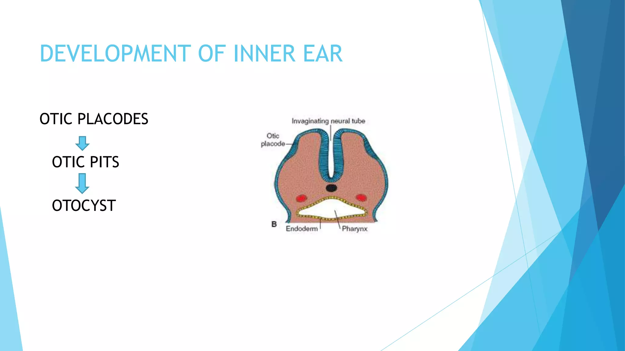


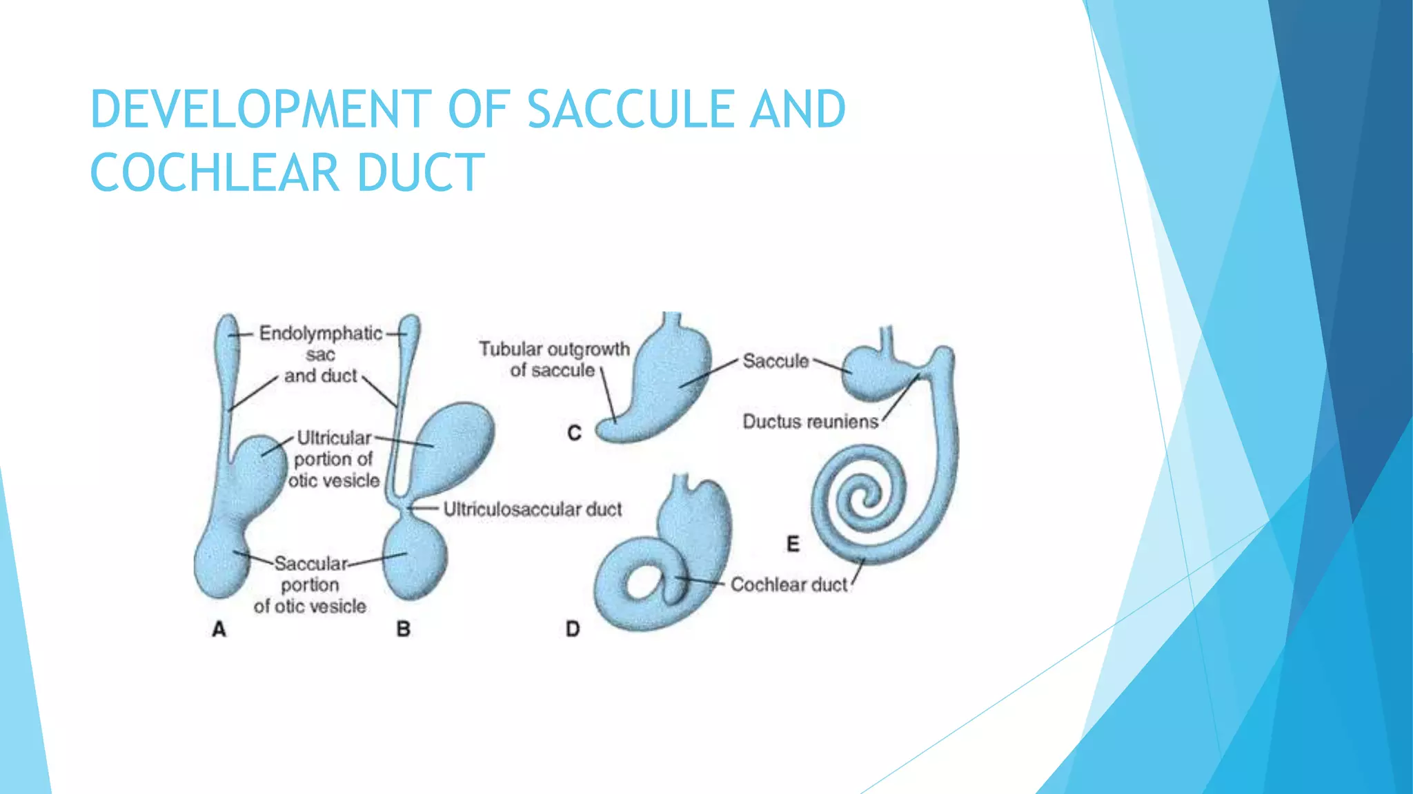


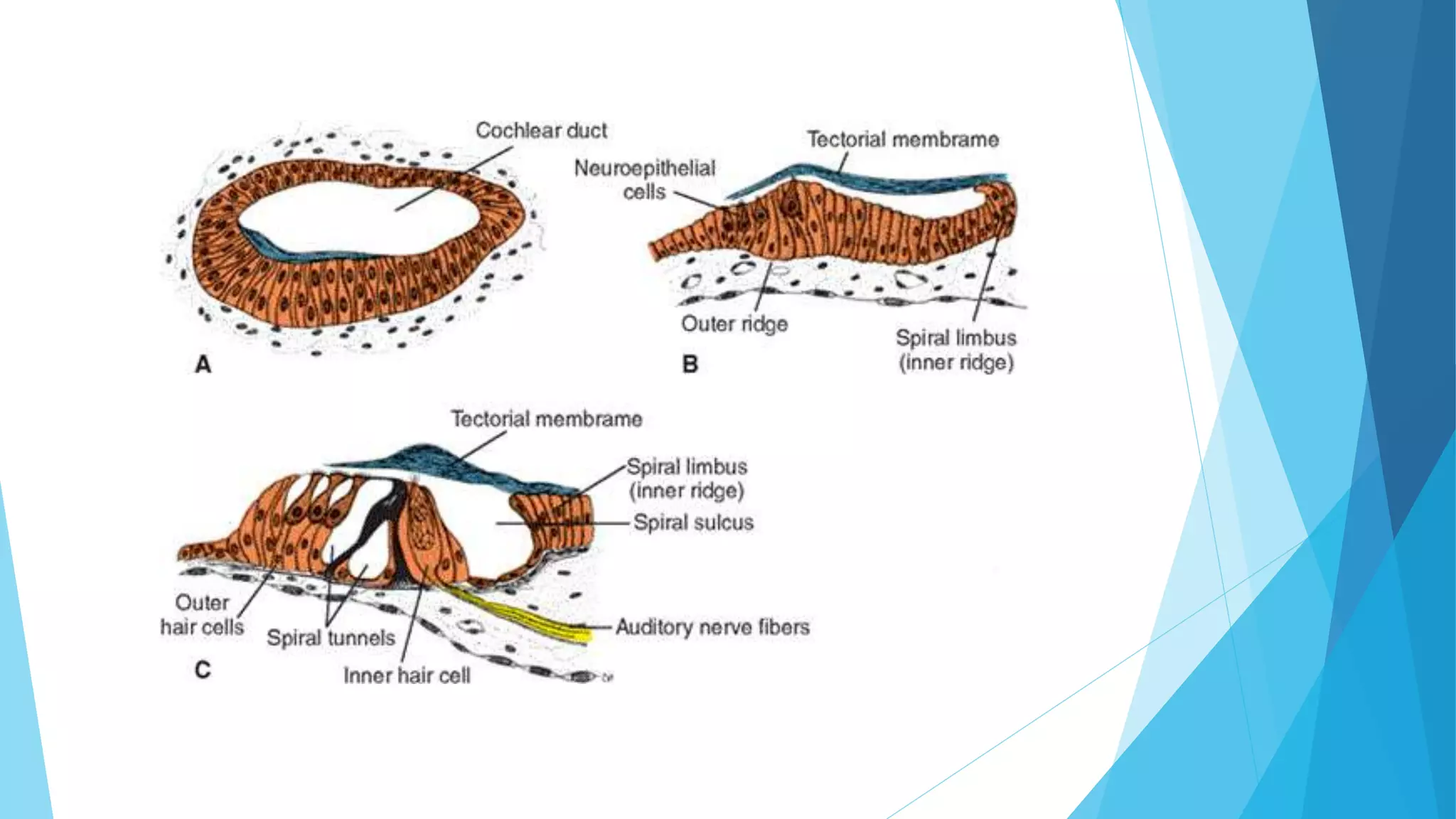

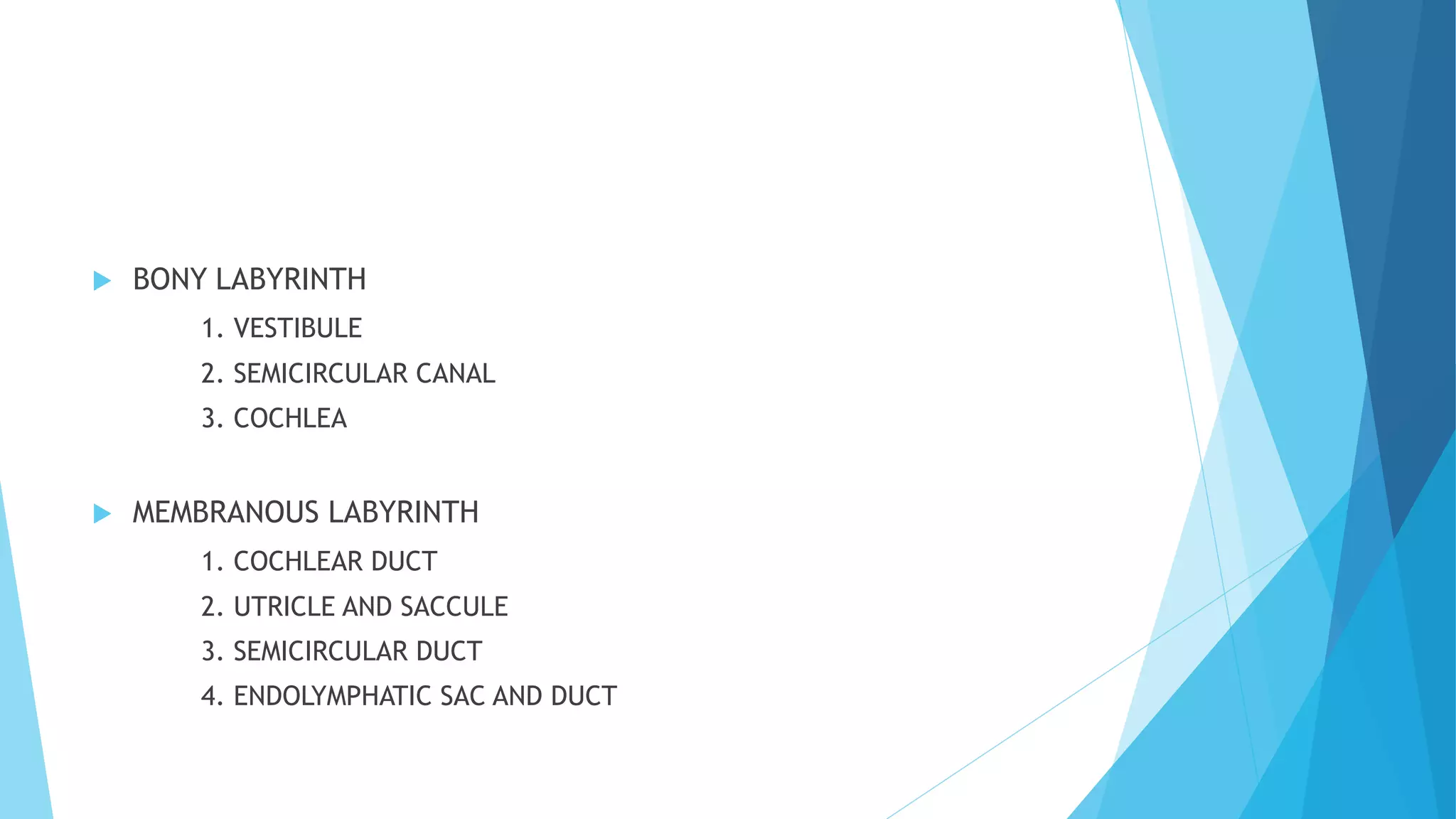


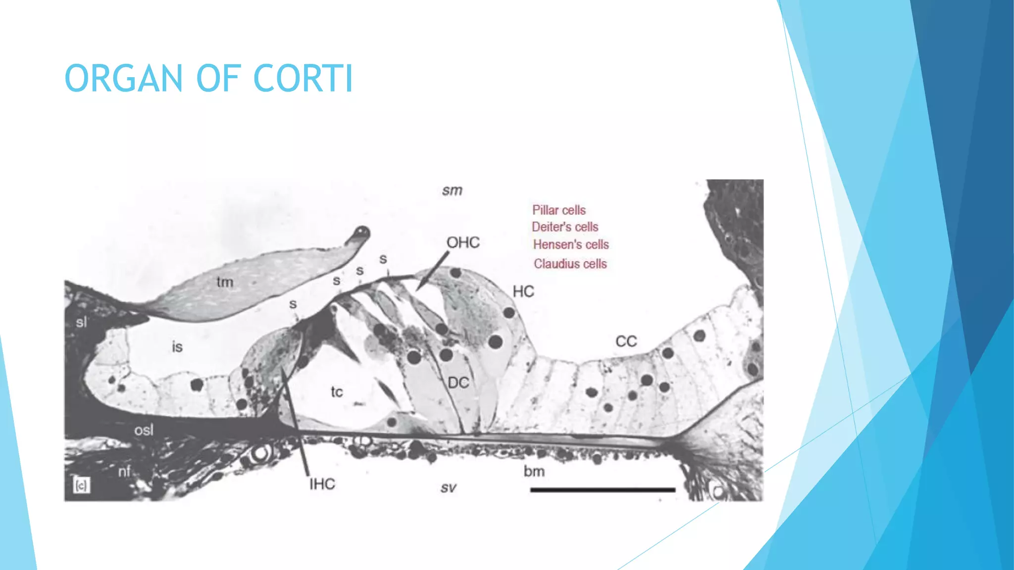


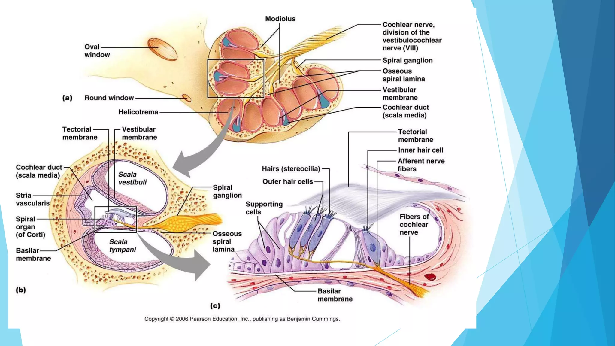


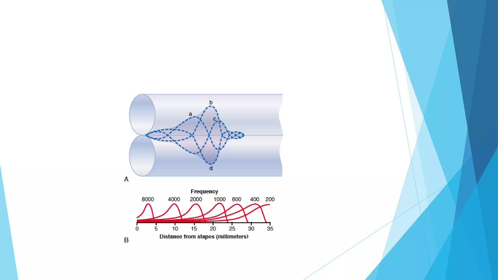


The document summarizes the anatomy and development of the inner ear. It discusses the embryological development from otic placodes to the formation of the membranous labyrinth. The inner ear anatomy includes the bony labyrinth containing the vestibule, semicircular canals and cochlea, as well as the membranous labyrinth containing the cochlear duct, utricle, saccule and endolymphatic structures. The organ of Corti is described as the sensory receptor organ of the cochlea containing inner and outer hair cells. The mechanism of hearing is also briefly outlined involving mechanical conduction of sound and the traveling wave theory of sound transmission in the cochle


