In order to obtain a diagnosis of cervical cancer information, the characteristics of each cell nucleus must be identified and evaluated properly through a Pap smear test. The presence of inflammatory cells in Pap smear images can complicate the process of identification of cell nuclei in the early detection of cervical cancer. Inflammatory cells need to be eliminated to assist pathologists in reading Pap smear slides. In this work, we developed a novel method to extract the inflammatory cells that allow detection of cell nuclei more accuracy. The proposed algorithm consists of two stages: extraction of inflammatory cells using the distance criterion and image transformation. This experiment applied to the 1358 cells comprising 378 nuclei cells and 980 inflammatory cells from 25 Pap smear images. The results showed that our method can significantly reduce the amount of inflammation that can disrupt the cell nuclei in the detection process. The proposed method has promising results with a sensitivity level of 97% and a specificity of 84.38%.
The document proposes a Modified Fuzzy C-Means (MFCM) clustering algorithm to segment chromosomal images. The MFCM includes preprocessing steps of median filtering and image enhancement to address noise sensitiveness and segmentation error problems in existing methods. It achieves improved segmentation accuracy of 61.6% compared to 56.4%, 55.47%, and 57.6% for Fuzzy C-Means, Adaptive Fuzzy C-Means, and Improved Adaptive Fuzzy C-Means respectively. The MFCM results in higher quality segmented images as indicated by its lower mean square error and higher peak signal-to-noise ratio values.
Segmentation of Tumor Region in MRI Images of Brain using Mathematical Morpho...CSCJournals
This paper introduces an efficient detection of brain tumor from cerebral MRI images. The methodology consists of two steps: enhancement and segmentation. To improve the quality of images and limit the risk of distinct regions fusion in the segmentation phase an enhancement process is applied. We applied mathematical morphology to increase the contrast in MRI images and to segment MRI images. Some of experimental results on brain images show the feasibility and the performance of the proposed approach.
MEDICAL IMAGE TEXTURE SEGMENTATION USINGRANGE FILTERcscpconf
Medical image segmentation is a frequent processing step in image understanding and computer
aided diagnosis. In this paper, we propose medical image texture segmentation using texture
filter. Three different image enhancement techniques are utilized to remove strong speckle noise as well enhance the weak boundaries of medical images. We propose to exploit the concept of range filtering to extract the texture content of medical image. Experiment is conducted on ImageCLEF2010 database. Results show the efficacy of our proposed medical image texture segmentation.
Defect Fruit Image Analysis using Advanced Bacterial Foraging Optimizing Algo...IOSR Journals
This document presents a method for segmenting defect areas on fruit images using an improved bacterial foraging optimization algorithm (ABFOA). The algorithm first decomposes the input fruit image into its red, green, and blue color components. It then applies the ABFOA to each color component separately to calculate individual thresholds. The final threshold is calculated as the average of the individual thresholds. This threshold is then applied to the original image to segment the defected areas. The method is tested on images of apples with defects like scab, rot, and blotch disease. Results show the ABFOA approach more accurately segments the defect areas compared to Otsu thresholding in terms of entropy, standard deviation, and peak signal-to
Face Recognition Using Neural Network Based Fourier Gabor Filters & Random Pr...CSCJournals
Face detection and recognition has many applications in a variety of fields such as authentication, security, video surveillance and human interaction systems. In this paper, we present a neural network system for face recognition. Feature vector based on Fourier Gabor filters is used as input of our classifier, which is a Back Propagation Neural Network (BPNN). The input vector of the network will have large dimension, to reduce its feature subspace we investigate the use of the Random Projection as method of dimensionality reduction. Theory and experiment indicates the robustness of our solution.
HYBRID APPROACH FOR NOISE REMOVAL AND IMAGE ENHANCEMENT OF BRAIN TUMORS IN MA...acijjournal
In medical image processing, Magnetic Resonance Imaging (MRI) is one of significant diagnostic
techniques. It provides high quality of important information about the analysis of human soft tissue when
measured with CT imaging modalities; hence it is suitable for diagnosis at best. However, if it gives quality
of information, image may distorted by noise because of image acquisition device and transmission. The
noises in MR image reduces the quality of image and also damages the segmentation task which can lead
to faulty diagnosis. Noises have to reduce at the same time there is no information loss. This paper propose
a hybrid approach to enhance the brain tumor MRI images using combined features of Anisotropic
Diffusion Filter (ADF) with Modified Decision Based Unsymmetric Trimmed Median Filter (MDBUTMF).
ADF scheme provides a superior performance by removing noise while preserving image details and
enhancing edges. MDBUTMF helps in image denoising as well as preserving edges satisfactorily when the
noise level is high. The performance of this filter is evaluated by carrying out a qualitative comparison of
this method with other filters namely, ADF filter, Modified Decision Algorithm, Median filter, MDBUTMF.
Feature Extraction of an Image by Using Adaptive Filtering and Morpological S...IOSR Journals
Abstract: For enhancing an image various enhancement schemes are used which includes gray scale manipulation, filtering and Histogram Equalization, Where Histogram equalization is one of the well known image enhancement technique. It became a popular technique for contrast enhancement because it is simple and effective. The basic idea of Histogram Equalization method is to remap the gray levels of an image. Here using morphological segmentation we can get the segmented image. Morphological reconstruction is used to segment the image. Comparative analysis of different enhancement and segmentation will be carried out. This comparison will be done on the basis of subjective and objective parameters. Subjective parameter is visual quality and objective parameters are Area, Perimeter, Min and Max intensity, Avg Voxel Intensity, Std Dev of Intensity, Eccentricity, Coefficient of skewness, Coefficient of Kurtosis, Median intensity, Mode intensity. Keywords: Histogram Equalization, Segmentation, Morphological Reconstruction .
International Journal of Engineering Research and Applications (IJERA) is an open access online peer reviewed international journal that publishes research and review articles in the fields of Computer Science, Neural Networks, Electrical Engineering, Software Engineering, Information Technology, Mechanical Engineering, Chemical Engineering, Plastic Engineering, Food Technology, Textile Engineering, Nano Technology & science, Power Electronics, Electronics & Communication Engineering, Computational mathematics, Image processing, Civil Engineering, Structural Engineering, Environmental Engineering, VLSI Testing & Low Power VLSI Design etc.
A New Approach for Segmentation of Fused Images using Cluster based ThresholdingIDES Editor
This paper proposes the new segmentation technique
with cluster based method. In this, the multi source medical
images like MRI (Magnetic Resonance Imaging), CT
(computed tomography) & PET (positron emission
tomography) are fused and then segmented using cluster based
thresholding approach. The edge details of an image have
become an essential technique in clinical and researchoriented
applications. The more edge details of the fused image
have obtainable with this method. The objective of the
clustering process is to partition a fused image coefficients
into a number of clusters having similar features. These
features are useful to generate the threshold value for further
segmentation of fused image. Finally the segmented output
is compared with standard FCM method and modified Otsu
method. Experimental results have shown that the proposed
cluster based thresholding method is able to effectively extract
important edge details of fused image.
This document summarizes a research paper that proposes a new method for detecting moving objects in videos using background subtraction and morphological techniques. The method establishes a reliable background updating model and uses dynamic thresholding to obtain a more complete segmentation of moving objects. The algorithm is implemented on a Microblaze soft processor in VHDL and tested on a Spartan-3 FPGA board. Experimental results show the area and speed of the algorithm. In conclusion, the proposed method allows inherently parallel processing of video frames and can improve detection accuracy by operating at the region level using morphological operations.
Automatic Determination Number of Cluster for NMKFC-Means Algorithms on Image...IOSR Journals
This document discusses image segmentation techniques using clustering algorithms. It introduces Fuzzy C-Means (FCM) clustering, which allows data points to belong to multiple clusters with varying degrees of membership. However, FCM does not work well on noisy or non-linearly separable data. The document proposes the Kernel Fuzzy C-Means (KFCM) algorithm, which uses a kernel function to map data to a higher dimensional space, making separation easier. While improving results for noisy images, KFCM does not consider neighboring pixels. Finally, the document introduces the Novel Modified Kernel Fuzzy C-Means (NMKFCM) algorithm, which incorporates neighborhood information into the objective function to further improve segmentation accuracy, especially for noisy images
An artificial neural network approach for detecting skin cancerTELKOMNIKA JOURNAL
This study aims to present diagnose of melanoma skin cancer at an early stage. It applies feature
extraction method of the first order for feature extraction based on texture in order to get high degree of
accuracy with method of classification using artificial neural network (ANN). The method used is training
and testing phases with classification of Multilayer Perceptron (MLP) neural network. The results showed
that the accuracy of test image with 4 sets of training for image not suspected of melanoma and melanoma
with the lowest accuracy of 80% and the highest accuracy of 88.88%, respectively. The 4 sets of training
used consisted of 23 images. Of the 23 images used as a training consisted of 6 as not suspected of
melanoma images and 17 as suspected melanoma images.
International Journal of Engineering Research and Applications (IJERA) is an open access online peer reviewed international journal that publishes research and review articles in the fields of Computer Science, Neural Networks, Electrical Engineering, Software Engineering, Information Technology, Mechanical Engineering, Chemical Engineering, Plastic Engineering, Food Technology, Textile Engineering, Nano Technology & science, Power Electronics, Electronics & Communication Engineering, Computational mathematics, Image processing, Civil Engineering, Structural Engineering, Environmental Engineering, VLSI Testing & Low Power VLSI Design etc.
This document discusses the challenges of applying compressed sensing (CS) to reconstruct phase images from under-sampled gradient echo MRI data needed for quantitative susceptibility mapping (QSM). Existing CS methods do not work well because they do not account for or preserve the important structural information contained in the phase images. A new CS method is needed that can separately enforce sparsity on the magnitude and phase to better reconstruct images from data with a strong phase component while preserving anatomical details, as current methods result in poor image quality and loss of details.
Detection of Malignancy in Digital Mammograms from Segmented Breast Region Us...IOSR Journals
This document presents a technique for detecting malignancy in digital mammograms using morphological operations. The proposed method involves noise removal using Gaussian filtering, image enhancement, removing background information through thresholding and morphological operations, performing image subtraction on the segmented image and converted RGB image to obtain tumors, and applying erosion to reduce small scale details and region sizes. The method was tested on images from a cancer hospital and implemented in Matlab. Experimental results show the technique can effectively preprocess images and segment regions to identify malignant data for assessment. Future work may focus on improving edge detection, segmentation algorithms, and producing more accurate cancer detection results.
Application of Digital Image Processing in Drug IndustryIOSRjournaljce
This document summarizes four digital image processing techniques used to detect defects in tablet strips: morphology operations, template matching, mathematical manipulation, and Euler's method. Morphology operations can detect broken tablets, template matching and mathematical manipulation can find broken and missing tablets, and Euler's method identifies holes in tablets. The techniques are applied to tablet strip images in MATLAB and effectively detect various defects. In summary, digital image processing provides a way to automatically inspect tablet strips for defects in pharmaceutical manufacturing.
This document provides a survey of various image segmentation techniques used in image processing. It begins with an introduction to image segmentation and its importance in fields like pattern recognition and medical imaging. It then categorizes and describes different segmentation approaches like edge-based, threshold-based, region-based, etc. The literature survey section summarizes several papers on specific segmentation algorithms or applications. It concludes with a table comparing the advantages and disadvantages of different segmentation techniques. The overall document aims to provide an overview of segmentation methods and their uses in computer vision.
This document summarizes various image segmentation methods that can be used for diagnosing dermatitis diseases. It discusses thresholding methods like global thresholding, Otsu's method, and Bayesian thresholding. It also covers region-based methods such as region growing, seeded region growing, and GMM-based segmentation. Additionally, it reviews shape-based/model-based approaches like deformable surfaces, level sets, and edge detection methods. The document provides an overview of the key concepts and applications of these segmentation techniques for skin disease diagnosis.
Spot Edge Detection in cDNA Microarray Images using Window based Bi-Dimension...idescitation
Ongoing Microarray is an increasingly playing a crucial role applied in the field
of medical and biological operations. The initiator of Microarray technology is M. Schena et
al. [1] and from past few years microarrays have begun to be used in many fields such as
biomedicine, mostly on cancer and Diabetic, and medical diagnoses. A Deoxyribonucleic
Acid (DNA) microarray is a collection of microscopic DNA spots attached to a solid surface,
such as glass, plastic or silicon chip forming an array. Processing of DNA microarray image
analysis includes three tasks: gridding, segmentation and intensity extraction and at the
stage of processing, the irregularities of shape and spot position which leads to generate
significant errors. This article presents a new spot edge detection method using Window
based Bi-dimensional Empirical Mode Decomposition. On separating spots form the
background area and to decreases the probability of errors and gives more accurate
information about the states of spots we are proposing a spot edge detection via WBEMD.
By using this method we can identify the spots with low density, which leads to increasing
the performance of cDNA microarray images.
Segmentation of medical images using metric topology – a region growing approachIjrdt Journal
A metric topological approach to the region growing based segmentation is presented in this article. Region based growing techniques has gained a significant importance in the medical image processing field for finest of segregation of tumor detected part in the image. Conventional algorithms were concentrated on segmentation at the coarser level which failed to produce enough evidence for the validity of the algorithm. In this article a novel technique is proposed based on metric topological neighbourhood also with the introduction of new objective measure entropy, apart from the traditional validity measures of Accuracy, PSNR and MSE. This measure is introduced to prove the amount of information lost after segmentation is reduced to greater extent which elucidates the effectiveness of the algorithm. This algorithm is tested on the well known benchmarking of testing in ground truth images in par with the proposed region based growing segmented images. The results validated show the validation of effectiveness of the algorithm.
Analysis of microscope images_FINAL PRESENTATIONGeorge Livanos
This document outlines the presentation scheme for a thesis on the analysis of microscope images. The thesis will analyze tissue samples using both polarimetric imaging at a macroscopic level and microscope imaging at a cellular level. For polarimetric imaging, the thesis will develop statistical models to characterize tissue properties based on how polarized light interacts with tissue elements. For microscope imaging, it will automatically segment cells from immunohistochemistry images and evaluate biomarkers like Her2 to characterize cancer impacts. Key techniques will include membrane boundary estimation, image clustering, and watershed transforms. The goal is both material characterization from polarimetric signatures and cancer analysis from cellular-level microscope images.
OBJECT SEGMENTATION USING MULTISCALE MORPHOLOGICAL OPERATIONSijcseit
Object segmentation plays an important role in human visual perception, medical image processing and content based image retrieval. It provides information for recognition and interpretation. This paper uses mathematical morphology for image segmentation. Object segmentation is difficult because one usually does not know a priori what type of object exists in an image, how many different shapes are there and what regions the image has. To carryout discrimination and segmentation several innovative segmentation methods, based on morphology are proposed. The present study proposes segmentation method based on multiscale morphological reconstructions. Various sizes of structuring elements have been used to segment simple and complex shapes. It enhances local boundaries that may lead to improve segmentation accuracy.The method is tested on various datasets and results shows that it can be used for both interactive and automatic segmentation.
A Review on Image Segmentation using Clustering and Swarm Optimization Techni...IJSRD
The process of dividing an image into multiple regions (set of pixels) is known as Image segmentation. It will make an image easy and smooth to evaluate. Image segmentation objective is to generate image more simple and meaningful. In this paper present a survey on image segmentation general segmentation techniques, clustering algorithms and optimization methods. Also a study of different research also been presented. The latest research in each of image segmentation methods is presented in this study. This paper presents the recent research in biologically inspired swarm optimization techniques, including ant colony optimization algorithm, particle swarm optimization algorithm, artificial bee colony algorithm and their hybridizations, which are applied in several fields.
The document outlines the methodology for brain tumor detection from MRI images. It involves four main stages: pre-processing, skull stripping, segmentation, and feature extraction. In pre-processing, MRI images are converted to grayscale and filters are applied to remove noise. Skull stripping removes non-brain tissues. Segmentation uses Otsu's thresholding and watershed methods to separate brain regions. Feature extraction uses morphological operators to extract the tumor region by subtracting it from the original grayscale image.
International Journal of Engineering Research and Applications (IJERA) is an open access online peer reviewed international journal that publishes research and review articles in the fields of Computer Science, Neural Networks, Electrical Engineering, Software Engineering, Information Technology, Mechanical Engineering, Chemical Engineering, Plastic Engineering, Food Technology, Textile Engineering, Nano Technology & science, Power Electronics, Electronics & Communication Engineering, Computational mathematics, Image processing, Civil Engineering, Structural Engineering, Environmental Engineering, VLSI Testing & Low Power VLSI Design etc.
SEGMENTATION OF MAGNETIC RESONANCE BRAIN TUMOR USING INTEGRATED FUZZY K-MEANS...ijcsit
Segmentation is a process of partitioning the image into several objects. It plays a vital role in many fields
such as satellite, remote sensing, object identification, face tracking and most importantly in medical field.
In radiology, magnetic resonance imaging (MRI) is used to investigate the human body processes and
functions of organisms. In hospitals, this technique has been using widely for medical diagnosis, to find the
disease stage and follow-up without exposure to ionizing radiation.Here in this paper, we proposed a novel
MR brain image segmentation method for detecting the tumor and finding the tumor area with improved
performance over conventional segmentation techniques such as fuzzy c means (FCM), K-means and even
that of manual segmentation in terms of precision time and accuracy. Simulation performance shows that
the proposed scheme has performed superior to the existing segmentation methods.
International Journal of Engineering Research and Applications (IJERA) is an open access online peer reviewed international journal that publishes research and review articles in the fields of Computer Science, Neural Networks, Electrical Engineering, Software Engineering, Information Technology, Mechanical Engineering, Chemical Engineering, Plastic Engineering, Food Technology, Textile Engineering, Nano Technology & science, Power Electronics, Electronics & Communication Engineering, Computational mathematics, Image processing, Civil Engineering, Structural Engineering, Environmental Engineering, VLSI Testing & Low Power VLSI Design etc.
Filter technique of medical image on multiple morphological gradient methodTELKOMNIKA JOURNAL
Filter technique is supportive for reducing image noise. This paper presents a study on filtering medical images, i.e., CT-Scan, Chest X-ray and Panoramic X-ray collected from two of the most prominent public hospitals in Padang City, Indonesia. The aim of this study preserved to facilitate in diagnosing objects in x-ray medical images. This study used filter technique, i.e. Blur, Emboss, Gaussian, Laplacian, Roberts, Sharpen, or Sobel techniques as pre-processing step. The filter process performed before edge detection and edge clarification. MMG method used in this study to clarify the edge detection. Thus, this research showed the hesitation decline (confidence increase) of the diagnosis of objects contained in medical images.
IRJET - Lung Cancer Detection using GLCM and Convolutional Neural NetworkIRJET Journal
This document summarizes a research paper that proposes a method for detecting lung cancer using CT scan images with convolutional neural networks. The method involves preprocessing images using median filtering to remove noise, segmenting images using k-means clustering, extracting features using gray-level co-occurrence matrix, and classifying images using convolutional neural networks. The researchers achieved 96% accuracy in classifying tumors as malignant or benign, which is more accurate than traditional neural network methods.
This document proposes a computer-aided lung cancer classification system using curvelet features and an ensemble classifier. It first pre-processes CT images using adaptive histogram equalization to improve contrast. Then it segments the images using kernelized fuzzy c-means clustering. Curvelet features are extracted from the segmented regions and an ensemble classifier is applied to classify regions as benign or malignant. The proposed approach achieves reliable and accurate classification results compared to existing methods, with better performance metrics like accuracy, sensitivity and specificity.
The document presents a new approach called Modified Fuzzy C-Means Clustering Algorithm (MFCM) for segmenting chromosome images. MFCM includes preprocessing steps of median filtering and image enhancement to address noise sensitiveness and segmentation error problems in existing methods. It was tested on M-FISH chromosome images and achieved a higher segmentation ratio of 61.6% compared to 56.4%, 55.47%, and 57.6% for FCM, AFCM, and IAFCM respectively. MFCM also had lower mean square error and higher peak signal-to-noise ratio, indicating better image quality. The preprocessing steps in MFCM improved segmentation accuracy by separating superimposed foreground and background data in the complex chromosome
SEGMENTATION OF LUNG GLANDULAR CELLS USING MULTIPLE COLOR SPACESIJCSEA Journal
Early detection of lung cancer is a challenging problem, the world faces today. Prior to classify glandular cells as malignant or benign a reliable segmentation technique is required. In this paper we present a novel lung glandular cell segmentation technique. The technique uses a combination of multiple color spaces and various clustering algorithms to automatically find the best possible segmentation result. Unsupervised clustering methods of K-means and Fuzzy C-means were used on multiple color spaces such as HSV, LAB, LUV, xyY. Experimental results of segmentation using various color spaces are provided to show the performance of the proposed system.
Live and Dead Cells Counting from Microscopic Trypan Blue Staining Images usi...IJECEIAES
Cell counting is a required procedure in biomedical experiments and drug testing. Manual cell counting performed with a hemocytometer is time consuming and individual dependence. This study reportedthe development of a computer-assisted program for trypan blue stained-cell counting using digital image analysis. Images of trypan blue-stained breast cancer cells line were obtained by a microscope with a digital camera. Undesired noise and debris were removed by applying a guided image filter. Color space HSV (Hue, Saturation and Value)conversion and grayscale conversion were performed for distinguishing between live and dead cells. Image thresholding and morphological operators were applied for image segmentation. Live and dead cells were counted after image segmentation and the results were compared with manual counting by three well-experienced counters. The computer-assisted cell counting from thirty-six trypan blue-stained microscopic images had a high correlation coefficient with the live cell results of the experts (r=0.99). The correlation coefficient of the number of dead cells comparing the computer-assisted count and the experts’ count was 0.74. Our approach offers high accuracy (>85%)on counting live cells compared with the experts’ counting. This automated cell counting approach can assist biomedical researchers for both live and dead cells counting.
The Effects of Segmentation Techniques in Digital Image Based Identification ...TELKOMNIKA JOURNAL
This paper presents the effects of segmentation techniques in the identification of Ethiopian
coffee variety. In Ethiopia, coffee varieties are classified based on their growing region. The most widely
coffee growing regions in Ethiopia are Bale, Harar, Jimma, Limu, Sidamo and Welega. Coffee beans of
these regions very in color shape and texture. We investigated various segmentation techniques for
efficient coffee beans variety identification system. Images of six different coffee beans varieties in Oromia
and Southern Ethiopia were acquired and analyzed. For this study Otsu, Fuzzy-C-Means (FCM) and Kmeans
segmentation techniques are considered. For classification of the varieties of Ethiopian coffee
beans back propagation neural network (BPNN) is used. From the experiment 94.54% accuracy is
achieved when BPNN is used on FCM segmentation technique.
brain tumor detection by thresholding approachSahil Prajapati
This technical paper proposes a method for detecting tumors in MRI brain images using thresholding and morphological operations. The methodology involves preprocessing images using sharpening filters, histogram equalization, and median filtering. Threshold segmentation is then used to create binary images, and morphological operations like erosion and dilation are applied. Finally, tumor regions are extracted using image subtraction, which removes closely packed pixels. The authors found that this approach, combining thresholding with morphological operations and subtraction, was effective at detecting and segmenting tumor regions in MRI brain images.
Chest radiograph image enhancement with wavelet decomposition and morphologic...TELKOMNIKA JOURNAL
Medical image processing algorithms significantly affect the precision of disease diagnostic
process. This makes it crucial to improve the quality of a medical image with the goal to enhance
perceivability of the points of interest in order to obtain accurate diagnosis of a patient. Despite the
reliance of various medical diagnostics on X-rays, they are usually plagued by dark and low contrast
properties. Sought-after details in X-rays can only be accessed by means of digital image processing
techniques, despite the fact that these techniques are far from being perfect. In this paper, we implement
a wavelet decomposition and reconstruction technique to enhance radiograph properties, using a series of
morphological erosion and dilation to improve the visual quality of the chest radiographs for the detection of
cancer nodules.
A statistical approach on pulmonary tuberculosis detection system based on X-...TELKOMNIKA JOURNAL
This paper presented the research result on the design of pulmonary TB (Tuberculosis) detection systems using a statistical approach. The study aimed to address two problems in detecting pulmonary TB by doctors, especially in remote areas of Indonesia, namely the long waiting time for patients to get the doctor's diagnosis and the doctor's subjectivity. We used hundreds of X-ray images from radiology department of Sardjito Hospital, Yogyakarta, as primary data and thirty data from various sources on the internet as secondary data. Using statistical approach, we exploited statistical image feature from image histogram, examined two statistical methods of PCA and LDA transformation for feature extraction, and two minimum distance classifier in image classification. We also used histogram equalization in the image enhancement process and bicubic interpolation in image segmentation and template making. Test results on primary and secondary data images show the identification accuracy of 94% and 83.3%, respectively.
This document presents a new approach for segmenting skin lesions in dermoscopic images using a fixed-grid wavelet network (FGWN). The FGWN takes R, G, and B color values as inputs and determines the network structure without training. The image is then segmented and the exact lesion boundary is extracted. Experimental results on 30 images showed the FGWN approach achieved better segmentation accuracy than other methods according to 11 evaluation metrics, extracting lesion boundaries more precisely. In conclusion, the FGWN provides an effective tool for automated skin lesion segmentation in dermoscopy images.
Blood image analysis to detect malaria using filtering image edges and classi...TELKOMNIKA JOURNAL
Malaria is a most dangerous mosquito borne disease and its infection spread through the infected mosquito. It especially affects the pregnant females and Children less than 5 years age. Malarial species commonly occur in five different shapes, Therefore, to avoid this crucial disease the contemporary researchers have proposed image analysis based solutions to mitigate this death causing disease. In this work, we propose diagnosis algorithm for malaria which is implemented for testing and evaluation in Matlab. We use Filtering and classification along with median filter and SVM classifier. Our proposed method identifies the infected cells from rest of blood images. The Median filtering smoothing technique is used to remove the noise. The feature vectors have been proposed to find out the abnormalities in blood cells. Feature vectors include (Form factor, measurement of roundness, shape, count total number of red cells and parasites). Primary aim of this research is to diagnose malaria by finding out infected cells. However, many techniques and algorithm have been implemented in this field using image processing but accuracy is not up to the point. Our proposed algorithm got more efficient results along with high accuracy as compared to NCC and Fuzzy classifier used by the researchers recently.
This document summarizes a research paper that proposes a novel approach for enhancing digital images using morphological operators. The approach aims to improve contrast in images with poor lighting conditions. It uses two morphological operators based on Weber's law - the first employs blocked analysis while the second uses opening by reconstruction to define a multi-background. The performance of the proposed operators is evaluated on images with various backgrounds and lighting conditions. Key steps include dividing images into blocks, estimating minimum/maximum intensities in each block to determine background criteria, and applying contrast enhancement transformations based on the criteria. Opening by reconstruction is also used to approximate image background without modifying structures. Experimental results demonstrate the approach enhances images with poor lighting.
Automatic Diagnosis of Abnormal Tumor Region from Brain Computed Tomography I...ijcseit
The research work presented in this paper is to achieve the tissue classification and automatically
diagnosis the abnormal tumor region present in Computed Tomography (CT) images using the wavelet
based statistical texture analysis method. Comparative studies of texture analysis method are performed
for the proposed wavelet based texture analysis method and Spatial Gray Level Dependence Method
(SGLDM). Our proposed system consists of four phases i) Discrete Wavelet Decomposition (ii)
Feature extraction (iii) Feature selection (iv) Analysis of extracted texture features by classifier. A
wavelet based statistical texture feature set is derived from normal and tumor regions. Genetic Algorithm
(GA) is used to select the optimal texture features from the set of extracted texture features. We construct
the Support Vector Machine (SVM) based classifier and evaluate the performance of classifier by
comparing the classification results of the SVM based classifier with the Back Propagation Neural network
classifier(BPN). The results of Support Vector Machine (SVM), BPN classifiers for the texture analysis
methods are evaluated using Receiver Operating Characteristic (ROC) analysis. Experimental results
show that the classification accuracy of SVM is 96% for 10 fold cross validation method. The system
has been tested with a number of real Computed Tomography brain images and has achieved satisfactory
results.
Fast Segmentation of Sub-cellular OrganellesCSCJournals
Segmentation and counting sub-cellular structure is a very challenging problem even for medical experts. A fast and efficient method for segmentation and counting of sub-cellular structure is proposed. The proposed method uses a hybrid combination of several image processing techniques and is effective in segmenting the sub-cellular structures in a fast and effective manner.
This paper discusses techniques for digital image processing, including noise reduction, edge detection, and histogram equalization. Noise reduction techniques discussed include mean, Gaussian, and median filters to remove salt and pepper noise and Gaussian noise. Edge detection algorithms like Sobel and Laplacian are introduced to reduce image data while preserving object boundaries. Histogram equalization is used for image enhancement by spreading pixel values across the full intensity range for increased contrast. The goal is recognizing objects in images through these preprocessing steps.
A SIMPLE IMAGE PROCESSING APPROACH TO ABNORMAL SLICES DETECTION FROM MRI TUMO...ijma
This paper proposed a method for brain tumor detection from the magnetic resonance imaging (MRI) of
human head scans. The proposed work explained the tumor detection process by means of image
processing transformations and thresholding technique. The MRI images are preprocessed by
transformation techniques and thus enhance the tumor region. Then the images are checked for
abnormality using fuzzy symmetric measure (FSM). If abnormal, then Otsu’s thresholding is used to extract
the tumor region. Experiments with the proposed method were done on 17 datasets. Various evaluation
parameters were used to validate the proposed method. The predictive accuracy (PA) and dice coefficient
(DC) values of proposed method reached maximum.
Similar to Inflammatory Cell Extraction in Pap smear Images: A Combination of Distance Criterion and Image Transformation Approach (20)
Amazon products reviews classification based on machine learning, deep learni...TELKOMNIKA JOURNAL
In recent times, the trend of online shopping through e-commerce stores and websites has grown to a huge extent. Whenever a product is purchased on an e-commerce platform, people leave their reviews about the product. These reviews are very helpful for the store owners and the product’s manufacturers for the betterment of their work process as well as product quality. An automated system is proposed in this work that operates on two datasets D1 and D2 obtained from Amazon. After certain preprocessing steps, N-gram and word embedding-based features are extracted using term frequency-inverse document frequency (TF-IDF), bag of words (BoW) and global vectors (GloVe), and Word2vec, respectively. Four machine learning (ML) models support vector machines (SVM), logistic regression (RF), logistic regression (LR), multinomial Naïve Bayes (MNB), two deep learning (DL) models convolutional neural network (CNN), long-short term memory (LSTM), and standalone bidirectional encoder representations (BERT) are used to classify reviews as either positive or negative. The results obtained by the standard ML, DL models and BERT are evaluated using certain performance evaluation measures. BERT turns out to be the best-performing model in the case of D1 with an accuracy of 90% on features derived by word embedding models while the CNN provides the best accuracy of 97% upon word embedding features in the case of D2. The proposed model shows better overall performance on D2 as compared to D1.
Design, simulation, and analysis of microstrip patch antenna for wireless app...TELKOMNIKA JOURNAL
In this study, a microstrip patch antenna that works at 3.6 GHz was built and tested to see how well it works. In this work, Rogers RT/Duroid 5880 has been used as the substrate material, with a dielectric permittivity of 2.2 and a thickness of 0.3451 mm; it serves as the base for the examined antenna. The computer simulation technology (CST) studio suite is utilized to show the recommended antenna design. The goal of this study was to get a more extensive transmission capacity, a lower voltage standing wave ratio (VSWR), and a lower return loss, but the main goal was to get a higher gain, directivity, and efficiency. After simulation, the return loss, gain, directivity, bandwidth, and efficiency of the supplied antenna are found to be -17.626 dB, 9.671 dBi, 9.924 dBi, 0.2 GHz, and 97.45%, respectively. Besides, the recreation uncovered that the transfer speed side-lobe level at phi was much better than those of the earlier works, at -28.8 dB, respectively. Thus, it makes a solid contender for remote innovation and more robust communication.
Design and simulation an optimal enhanced PI controller for congestion avoida...TELKOMNIKA JOURNAL
This document describes using a snake optimization algorithm to tune the gains of an enhanced proportional-integral controller for congestion avoidance in a TCP/AQM system. The controller aims to maintain a stable and desired queue size without noise or transmission problems. A linearized model of the TCP/AQM system is presented. An enhanced PI controller combining nonlinear gain and original PI gains is proposed. The snake optimization algorithm is then used to tune the parameters of the enhanced PI controller to achieve optimal system performance and response. Simulation results are discussed showing the proposed controller provides a stable and robust behavior for congestion control.
Improving the detection of intrusion in vehicular ad-hoc networks with modifi...TELKOMNIKA JOURNAL
Vehicular ad-hoc networks (VANETs) are wireless-equipped vehicles that form networks along the road. The security of this network has been a major challenge. The identity-based cryptosystem (IBC) previously used to secure the networks suffers from membership authentication security features. This paper focuses on improving the detection of intruders in VANETs with a modified identity-based cryptosystem (MIBC). The MIBC is developed using a non-singular elliptic curve with Lagrange interpolation. The public key of vehicles and roadside units on the network are derived from number plates and location identification numbers, respectively. Pseudo-identities are used to mask the real identity of users to preserve their privacy. The membership authentication mechanism ensures that only valid and authenticated members of the network are allowed to join the network. The performance of the MIBC is evaluated using intrusion detection ratio (IDR) and computation time (CT) and then validated with the existing IBC. The result obtained shows that the MIBC recorded an IDR of 99.3% against 94.3% obtained for the existing identity-based cryptosystem (EIBC) for 140 unregistered vehicles attempting to intrude on the network. The MIBC shows lower CT values of 1.17 ms against 1.70 ms for EIBC. The MIBC can be used to improve the security of VANETs.
Conceptual model of internet banking adoption with perceived risk and trust f...TELKOMNIKA JOURNAL
Understanding the primary factors of internet banking (IB) acceptance is critical for both banks and users; nevertheless, our knowledge of the role of users’ perceived risk and trust in IB adoption is limited. As a result, we develop a conceptual model by incorporating perceived risk and trust into the technology acceptance model (TAM) theory toward the IB. The proper research emphasized that the most essential component in explaining IB adoption behavior is behavioral intention to use IB adoption. TAM is helpful for figuring out how elements that affect IB adoption are connected to one another. According to previous literature on IB and the use of such technology in Iraq, one has to choose a theoretical foundation that may justify the acceptance of IB from the customer’s perspective. The conceptual model was therefore constructed using the TAM as a foundation. Furthermore, perceived risk and trust were added to the TAM dimensions as external factors. The key objective of this work was to extend the TAM to construct a conceptual model for IB adoption and to get sufficient theoretical support from the existing literature for the essential elements and their relationships in order to unearth new insights about factors responsible for IB adoption.
Efficient combined fuzzy logic and LMS algorithm for smart antennaTELKOMNIKA JOURNAL
The smart antennas are broadly used in wireless communication. The least mean square (LMS) algorithm is a procedure that is concerned in controlling the smart antenna pattern to accommodate specified requirements such as steering the beam toward the desired signal, in addition to placing the deep nulls in the direction of unwanted signals. The conventional LMS (C-LMS) has some drawbacks like slow convergence speed besides high steady state fluctuation error. To overcome these shortcomings, the present paper adopts an adaptive fuzzy control step size least mean square (FC-LMS) algorithm to adjust its step size. Computer simulation outcomes illustrate that the given model has fast convergence rate as well as low mean square error steady state.
Design and implementation of a LoRa-based system for warning of forest fireTELKOMNIKA JOURNAL
This paper presents the design and implementation of a forest fire monitoring and warning system based on long range (LoRa) technology, a novel ultra-low power consumption and long-range wireless communication technology for remote sensing applications. The proposed system includes a wireless sensor network that records environmental parameters such as temperature, humidity, wind speed, and carbon dioxide (CO2) concentration in the air, as well as taking infrared photos.The data collected at each sensor node will be transmitted to the gateway via LoRa wireless transmission. Data will be collected, processed, and uploaded to a cloud database at the gateway. An Android smartphone application that allows anyone to easily view the recorded data has been developed. When a fire is detected, the system will sound a siren and send a warning message to the responsible personnel, instructing them to take appropriate action. Experiments in Tram Chim Park, Vietnam, have been conducted to verify and evaluate the operation of the system.
Wavelet-based sensing technique in cognitive radio networkTELKOMNIKA JOURNAL
Cognitive radio is a smart radio that can change its transmitter parameter based on interaction with the environment in which it operates. The demand for frequency spectrum is growing due to a big data issue as many Internet of Things (IoT) devices are in the network. Based on previous research, most frequency spectrum was used, but some spectrums were not used, called spectrum hole. Energy detection is one of the spectrum sensing methods that has been frequently used since it is easy to use and does not require license users to have any prior signal understanding. But this technique is incapable of detecting at low signal-to-noise ratio (SNR) levels. Therefore, the wavelet-based sensing is proposed to overcome this issue and detect spectrum holes. The main objective of this work is to evaluate the performance of wavelet-based sensing and compare it with the energy detection technique. The findings show that the percentage of detection in wavelet-based sensing is 83% higher than energy detection performance. This result indicates that the wavelet-based sensing has higher precision in detection and the interference towards primary user can be decreased.
A novel compact dual-band bandstop filter with enhanced rejection bandsTELKOMNIKA JOURNAL
In this paper, we present the design of a new wide dual-band bandstop filter (DBBSF) using nonuniform transmission lines. The method used to design this filter is to replace conventional uniform transmission lines with nonuniform lines governed by a truncated Fourier series. Based on how impedances are profiled in the proposed DBBSF structure, the fractional bandwidths of the two 10 dB-down rejection bands are widened to 39.72% and 52.63%, respectively, and the physical size has been reduced compared to that of the filter with the uniform transmission lines. The results of the electromagnetic (EM) simulation support the obtained analytical response and show an improved frequency behavior.
Deep learning approach to DDoS attack with imbalanced data at the application...TELKOMNIKA JOURNAL
A distributed denial of service (DDoS) attack is where one or more computers attack or target a server computer, by flooding internet traffic to the server. As a result, the server cannot be accessed by legitimate users. A result of this attack causes enormous losses for a company because it can reduce the level of user trust, and reduce the company’s reputation to lose customers due to downtime. One of the services at the application layer that can be accessed by users is a web-based lightweight directory access protocol (LDAP) service that can provide safe and easy services to access directory applications. We used a deep learning approach to detect DDoS attacks on the CICDDoS 2019 dataset on a complex computer network at the application layer to get fast and accurate results for dealing with unbalanced data. Based on the results obtained, it is observed that DDoS attack detection using a deep learning approach on imbalanced data performs better when implemented using synthetic minority oversampling technique (SMOTE) method for binary classes. On the other hand, the proposed deep learning approach performs better for detecting DDoS attacks in multiclass when implemented using the adaptive synthetic (ADASYN) method.
The appearance of uncertainties and disturbances often effects the characteristics of either linear or nonlinear systems. Plus, the stabilization process may be deteriorated thus incurring a catastrophic effect to the system performance. As such, this manuscript addresses the concept of matching condition for the systems that are suffering from miss-match uncertainties and exogeneous disturbances. The perturbation towards the system at hand is assumed to be known and unbounded. To reach this outcome, uncertainties and their classifications are reviewed thoroughly. The structural matching condition is proposed and tabulated in the proposition 1. Two types of mathematical expressions are presented to distinguish the system with matched uncertainty and the system with miss-matched uncertainty. Lastly, two-dimensional numerical expressions are provided to practice the proposed proposition. The outcome shows that matching condition has the ability to change the system to a design-friendly model for asymptotic stabilization.
Implementation of FinFET technology based low power 4×4 Wallace tree multipli...TELKOMNIKA JOURNAL
Many systems, including digital signal processors, finite impulse response (FIR) filters, application-specific integrated circuits, and microprocessors, use multipliers. The demand for low power multipliers is gradually rising day by day in the current technological trend. In this study, we describe a 4×4 Wallace multiplier based on a carry select adder (CSA) that uses less power and has a better power delay product than existing multipliers. HSPICE tool at 16 nm technology is used to simulate the results. In comparison to the traditional CSA-based multiplier, which has a power consumption of 1.7 µW and power delay product (PDP) of 57.3 fJ, the results demonstrate that the Wallace multiplier design employing CSA with first zero finding logic (FZF) logic has the lowest power consumption of 1.4 µW and PDP of 27.5 fJ.
Evaluation of the weighted-overlap add model with massive MIMO in a 5G systemTELKOMNIKA JOURNAL
The flaw in 5G orthogonal frequency division multiplexing (OFDM) becomes apparent in high-speed situations. Because the doppler effect causes frequency shifts, the orthogonality of OFDM subcarriers is broken, lowering both their bit error rate (BER) and throughput output. As part of this research, we use a novel design that combines massive multiple input multiple output (MIMO) and weighted overlap and add (WOLA) to improve the performance of 5G systems. To determine which design is superior, throughput and BER are calculated for both the proposed design and OFDM. The results of the improved system show a massive improvement in performance ver the conventional system and significant improvements with massive MIMO, including the best throughput and BER. When compared to conventional systems, the improved system has a throughput that is around 22% higher and the best performance in terms of BER, but it still has around 25% less error than OFDM.
Reflector antenna design in different frequencies using frequency selective s...TELKOMNIKA JOURNAL
In this study, it is aimed to obtain two different asymmetric radiation patterns obtained from antennas in the shape of the cross-section of a parabolic reflector (fan blade type antennas) and antennas with cosecant-square radiation characteristics at two different frequencies from a single antenna. For this purpose, firstly, a fan blade type antenna design will be made, and then the reflective surface of this antenna will be completed to the shape of the reflective surface of the antenna with the cosecant-square radiation characteristic with the frequency selective surface designed to provide the characteristics suitable for the purpose. The frequency selective surface designed and it provides the perfect transmission as possible at 4 GHz operating frequency, while it will act as a band-quenching filter for electromagnetic waves at 5 GHz operating frequency and will be a reflective surface. Thanks to this frequency selective surface to be used as a reflective surface in the antenna, a fan blade type radiation characteristic at 4 GHz operating frequency will be obtained, while a cosecant-square radiation characteristic at 5 GHz operating frequency will be obtained.
Reagentless iron detection in water based on unclad fiber optical sensorTELKOMNIKA JOURNAL
A simple and low-cost fiber based optical sensor for iron detection is demonstrated in this paper. The sensor head consist of an unclad optical fiber with the unclad length of 1 cm and it has a straight structure. Results obtained shows a linear relationship between the output light intensity and iron concentration, illustrating the functionality of this iron optical sensor. Based on the experimental results, the sensitivity and linearity are achieved at 0.0328/ppm and 0.9824 respectively at the wavelength of 690 nm. With the same wavelength, other performance parameters are also studied. Resolution and limit of detection (LOD) are found to be 0.3049 ppm and 0.0755 ppm correspondingly. This iron sensor is advantageous in that it does not require any reagent for detection, enabling it to be simpler and cost-effective in the implementation of the iron sensing.
Impact of CuS counter electrode calcination temperature on quantum dot sensit...TELKOMNIKA JOURNAL
In place of the commercial Pt electrode used in quantum sensitized solar cells, the low-cost CuS cathode is created using electrophoresis. High resolution scanning electron microscopy and X-ray diffraction were used to analyze the structure and morphology of structural cubic samples with diameters ranging from 40 nm to 200 nm. The conversion efficiency of solar cells is significantly impacted by the calcination temperatures of cathodes at 100 °C, 120 °C, 150 °C, and 180 °C under vacuum. The fluorine doped tin oxide (FTO)/CuS cathode electrode reached a maximum efficiency of 3.89% when it was calcined at 120 °C. Compared to other temperature combinations, CuS nanoparticles crystallize at 120 °C, which lowers resistance while increasing electron lifetime.
A progressive learning for structural tolerance online sequential extreme lea...TELKOMNIKA JOURNAL
This article discusses the progressive learning for structural tolerance online sequential extreme learning machine (PSTOS-ELM). PSTOS-ELM can save robust accuracy while updating the new data and the new class data on the online training situation. The robustness accuracy arises from using the householder block exact QR decomposition recursive least squares (HBQRD-RLS) of the PSTOS-ELM. This method is suitable for applications that have data streaming and often have new class data. Our experiment compares the PSTOS-ELM accuracy and accuracy robustness while data is updating with the batch-extreme learning machine (ELM) and structural tolerance online sequential extreme learning machine (STOS-ELM) that both must retrain the data in a new class data case. The experimental results show that PSTOS-ELM has accuracy and robustness comparable to ELM and STOS-ELM while also can update new class data immediately.
Electroencephalography-based brain-computer interface using neural networksTELKOMNIKA JOURNAL
This study aimed to develop a brain-computer interface that can control an electric wheelchair using electroencephalography (EEG) signals. First, we used the Mind Wave Mobile 2 device to capture raw EEG signals from the surface of the scalp. The signals were transformed into the frequency domain using fast Fourier transform (FFT) and filtered to monitor changes in attention and relaxation. Next, we performed time and frequency domain analyses to identify features for five eye gestures: opened, closed, blink per second, double blink, and lookup. The base state was the opened-eyes gesture, and we compared the features of the remaining four action gestures to the base state to identify potential gestures. We then built a multilayer neural network to classify these features into five signals that control the wheelchair’s movement. Finally, we designed an experimental wheelchair system to test the effectiveness of the proposed approach. The results demonstrate that the EEG classification was highly accurate and computationally efficient. Moreover, the average performance of the brain-controlled wheelchair system was over 75% across different individuals, which suggests the feasibility of this approach.
Adaptive segmentation algorithm based on level set model in medical imagingTELKOMNIKA JOURNAL
For image segmentation, level set models are frequently employed. It offer best solution to overcome the main limitations of deformable parametric models. However, the challenge when applying those models in medical images stills deal with removing blurs in image edges which directly affects the edge indicator function, leads to not adaptively segmenting images and causes a wrong analysis of pathologies wich prevents to conclude a correct diagnosis. To overcome such issues, an effective process is suggested by simultaneously modelling and solving systems’ two-dimensional partial differential equations (PDE). The first PDE equation allows restoration using Euler’s equation similar to an anisotropic smoothing based on a regularized Perona and Malik filter that eliminates noise while preserving edge information in accordance with detected contours in the second equation that segments the image based on the first equation solutions. This approach allows developing a new algorithm which overcome the studied model drawbacks. Results of the proposed method give clear segments that can be applied to any application. Experiments on many medical images in particular blurry images with high information losses, demonstrate that the developed approach produces superior segmentation results in terms of quantity and quality compared to other models already presented in previeous works.
Automatic channel selection using shuffled frog leaping algorithm for EEG bas...TELKOMNIKA JOURNAL
Drug addiction is a complex neurobiological disorder that necessitates comprehensive treatment of both the body and mind. It is categorized as a brain disorder due to its impact on the brain. Various methods such as electroencephalography (EEG), functional magnetic resonance imaging (FMRI), and magnetoencephalography (MEG) can capture brain activities and structures. EEG signals provide valuable insights into neurological disorders, including drug addiction. Accurate classification of drug addiction from EEG signals relies on appropriate features and channel selection. Choosing the right EEG channels is essential to reduce computational costs and mitigate the risk of overfitting associated with using all available channels. To address the challenge of optimal channel selection in addiction detection from EEG signals, this work employs the shuffled frog leaping algorithm (SFLA). SFLA facilitates the selection of appropriate channels, leading to improved accuracy. Wavelet features extracted from the selected input channel signals are then analyzed using various machine learning classifiers to detect addiction. Experimental results indicate that after selecting features from the appropriate channels, classification accuracy significantly increased across all classifiers. Particularly, the multi-layer perceptron (MLP) classifier combined with SFLA demonstrated a remarkable accuracy improvement of 15.78% while reducing time complexity.
Redefining brain tumor segmentation: a cutting-edge convolutional neural netw...IJECEIAES
Medical image analysis has witnessed significant advancements with deep learning techniques. In the domain of brain tumor segmentation, the ability to
precisely delineate tumor boundaries from magnetic resonance imaging (MRI)
scans holds profound implications for diagnosis. This study presents an ensemble convolutional neural network (CNN) with transfer learning, integrating
the state-of-the-art Deeplabv3+ architecture with the ResNet18 backbone. The
model is rigorously trained and evaluated, exhibiting remarkable performance
metrics, including an impressive global accuracy of 99.286%, a high-class accuracy of 82.191%, a mean intersection over union (IoU) of 79.900%, a weighted
IoU of 98.620%, and a Boundary F1 (BF) score of 83.303%. Notably, a detailed comparative analysis with existing methods showcases the superiority of
our proposed model. These findings underscore the model’s competence in precise brain tumor localization, underscoring its potential to revolutionize medical
image analysis and enhance healthcare outcomes. This research paves the way
for future exploration and optimization of advanced CNN models in medical
imaging, emphasizing addressing false positives and resource efficiency.
Discover the latest insights on Data Driven Maintenance with our comprehensive webinar presentation. Learn about traditional maintenance challenges, the right approach to utilizing data, and the benefits of adopting a Data Driven Maintenance strategy. Explore real-world examples, industry best practices, and innovative solutions like FMECA and the D3M model. This presentation, led by expert Jules Oudmans, is essential for asset owners looking to optimize their maintenance processes and leverage digital technologies for improved efficiency and performance. Download now to stay ahead in the evolving maintenance landscape.
Rainfall intensity duration frequency curve statistical analysis and modeling...bijceesjournal
Using data from 41 years in Patna’ India’ the study’s goal is to analyze the trends of how often it rains on a weekly, seasonal, and annual basis (1981−2020). First, utilizing the intensity-duration-frequency (IDF) curve and the relationship by statistically analyzing rainfall’ the historical rainfall data set for Patna’ India’ during a 41 year period (1981−2020), was evaluated for its quality. Changes in the hydrologic cycle as a result of increased greenhouse gas emissions are expected to induce variations in the intensity, length, and frequency of precipitation events. One strategy to lessen vulnerability is to quantify probable changes and adapt to them. Techniques such as log-normal, normal, and Gumbel are used (EV-I). Distributions were created with durations of 1, 2, 3, 6, and 24 h and return times of 2, 5, 10, 25, and 100 years. There were also mathematical correlations discovered between rainfall and recurrence interval.
Findings: Based on findings, the Gumbel approach produced the highest intensity values, whereas the other approaches produced values that were close to each other. The data indicates that 461.9 mm of rain fell during the monsoon season’s 301st week. However, it was found that the 29th week had the greatest average rainfall, 92.6 mm. With 952.6 mm on average, the monsoon season saw the highest rainfall. Calculations revealed that the yearly rainfall averaged 1171.1 mm. Using Weibull’s method, the study was subsequently expanded to examine rainfall distribution at different recurrence intervals of 2, 5, 10, and 25 years. Rainfall and recurrence interval mathematical correlations were also developed. Further regression analysis revealed that short wave irrigation, wind direction, wind speed, pressure, relative humidity, and temperature all had a substantial influence on rainfall.
Originality and value: The results of the rainfall IDF curves can provide useful information to policymakers in making appropriate decisions in managing and minimizing floods in the study area.
Introduction- e - waste – definition - sources of e-waste– hazardous substances in e-waste - effects of e-waste on environment and human health- need for e-waste management– e-waste handling rules - waste minimization techniques for managing e-waste – recycling of e-waste - disposal treatment methods of e- waste – mechanism of extraction of precious metal from leaching solution-global Scenario of E-waste – E-waste in India- case studies.
Design and optimization of ion propulsion dronebjmsejournal
Electric propulsion technology is widely used in many kinds of vehicles in recent years, and aircrafts are no exception. Technically, UAVs are electrically propelled but tend to produce a significant amount of noise and vibrations. Ion propulsion technology for drones is a potential solution to this problem. Ion propulsion technology is proven to be feasible in the earth’s atmosphere. The study presented in this article shows the design of EHD thrusters and power supply for ion propulsion drones along with performance optimization of high-voltage power supply for endurance in earth’s atmosphere.
artificial intelligence and data science contents.pptxGauravCar
What is artificial intelligence? Artificial intelligence is the ability of a computer or computer-controlled robot to perform tasks that are commonly associated with the intellectual processes characteristic of humans, such as the ability to reason.
› ...
Artificial intelligence (AI) | Definitio
2. TELKOMNIKA ISSN: 1693-6930
Inflammatory Cell Extraction in Pap smear Images:.... (Rahadian Kurniawan)
2049
Figure. 1. A part of Pap smear image, nuclei (red box) and inflammatory (white box)
Even though those researchers could extract cell nuclei and inflammatory cells pretty
well using feature extraction method, there are some inappropriate features involved to identify
cell nuclei and inflammatory cells. Another challenge to identify cell nuclei based on feature
extraction is Pap smear images taken by the low resolution camera. Furthermore, a feature
extraction method takes longer execution time. Therefore, this paper proposes a novel method
to extract and eliminate inflammatory cells using distance criteria and image transformation.
In this paper, there are three main phases to extract the inflammatory cell, among others: 1)
Segmentation of cytoplasm; 2) Segmentation of nuclei candidate, and 3) inflammatory cell
extraction. Further explanation will be described in subsequent subsections.
2. Method
2.1. Segmentation of Cytoplasm
The cytoplasm segmentation phase is needed to reduce the search area in the image.
In the first step, the h-minima transform [15] is applied individually for each color component of
initial image I (called Hm). The h value of image Hm at each layer of color (r, g, b) is given by,
( ) ( ) ( )
(1)
where is value of average intensities and is intensity value in the original image. Next, from
each filtered image, a grayscale image is produced through grayscale transformation as follows:
( ) ( ) ( ) (2)
A morphological open is then performed in order to flatten the intensity of the region of interest
using flat disk-shaped structuring element with radius of 5. Next, we perform sum of the image
of the morphological open processes (called M) and the image of the grayscale transformation
process (G) define as:
( ) ( ) ( ) (3)
After this operation, contrast enhancement filter is applied to complement image of J.
Furthermore, in order to expand the region of interest, we perform a morphological dilation using
flat disk-shaped structuring element with radius of the derived image (called D). In order to
obtain the boundaries of the cytoplasm, we perform the subtraction of two images. The first
image A is constructed through grayscale transformation of original images, image B is the
outcome of the application subtraction between image D and G. The result of the subtraction of
these two images is an image with all cytoplasm region sharp. Finally, the binary mask BW, with
the regions of cytoplasm included, is obtained by finding the pixel intensity that less than t value.
The value of t is defined as follows:
3. ISSN: 1693-6930
TELKOMNIKA Vol. 16, No. 5, October 2018: 2048-2056
2050
( )
(4)
where is value of average intensities. Furthermore, we do some noise reduction in BS image
by performing the morphological open using flat disk-shaped structuring element with radius
of 15. The resulted binary image is used as a mask to indicate the cytoplasm regions. The final
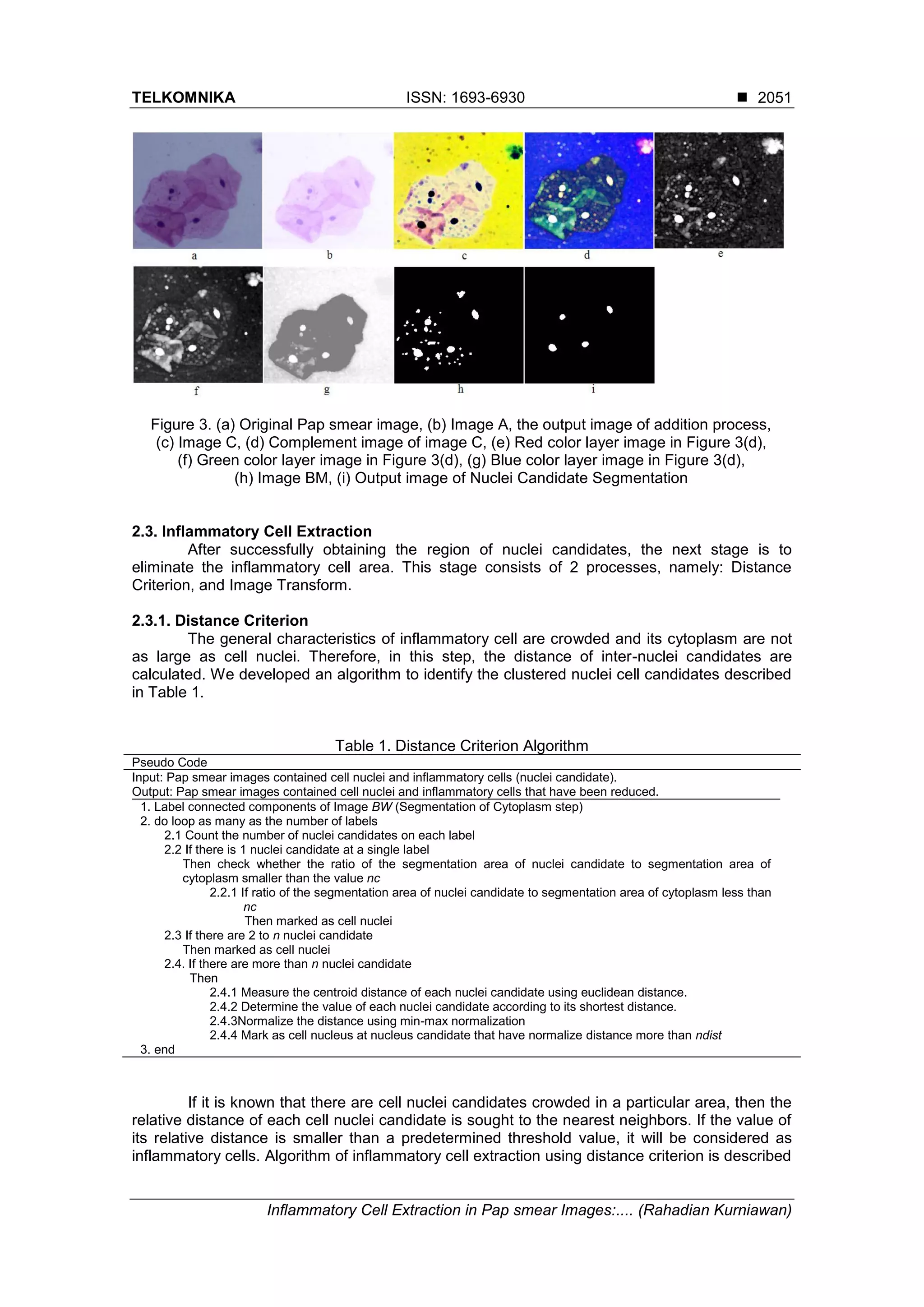
result of this part is shown in Figure 2.
Figure 2. (a) Original Pap smear image, and (b) binary mask, which is obtained after the
segmentation of cytoplasm process
2.2. Segmentation of Nuclei Candidate
This stage is done to find the area of candidate nuclei that is region consisting of nuclei
cell and inflammatory cell. First, Binary mask as a result from the previous phase uses to
remove the background image. This step is performed to remove noise occurred in the
background, while narrowing the search space nuclei candidates. This phase starts with each
color layer of original imageIis transformed to RGB image using h-minima transformation with a
value of h=115, called MT image. MT image is added toIimage to generate image A, as defined
in equation (5). Figure 3(b) shows the result of the adding process, A image.
( ) ( ) ( ) (5)
Furthermore, image A is subtracted with Hm image obtained from the cytoplasm
segmentation phase. Image resulting from the subtraction process is called image S. Erosion,
one of morphology process, is applied to image S using flat disk-shaped structuring element
with radius of 5, in order to enlarge the regions of candidate nuclei. Image resulting from the
erosion process called image C and shown in Figure 3(c), is enhanced using contrast
enhancement filter.
The average value of the grayscale intensity of each color layer in image C is
calculated. The color layer with the lowest average value, called image K, is chosen to be
subjected of global thresholding processes using the Otsu Thresholding method [16].
Figure 3(e, f, g) shows the grayscale image of each color layer in image C. As shown in those
Figure 3(e), red layer of image C, has the lowest average value of grayscale intensity among
another layer. Finally, the binary mask BM, with the regions of interest of the image included
given by,
(6)
where the image BS is an image obtained from the final result of cytoplasm segmentation
process and image TH is an image obtained from a global thresholding process using the Otsu
Thresholding method, in the previous step. Figure 3 (h) shows image BM. The last step of the
nuclei candidate segmentation process is cell separation process using the modified watershed
method [17] to handle the overlapping cell nuclei.
4. TELKOMNIKA ISSN: 1693-6930
Inflammatory Cell Extraction in Pap smear Images:.... (Rahadian Kurniawan)
2051
Figure 3. (a) Original Pap smear image, (b) Image A, the output image of addition process,
(c) Image C, (d) Complement image of image C, (e) Red color layer image in Figure 3(d),
(f) Green color layer image in Figure 3(d), (g) Blue color layer image in Figure 3(d),
(h) Image BM, (i) Output image of Nuclei Candidate Segmentation
2.3. Inflammatory Cell Extraction
After successfully obtaining the region of nuclei candidates, the next stage is to
eliminate the inflammatory cell area. This stage consists of 2 processes, namely: Distance
Criterion, and Image Transform.
2.3.1. Distance Criterion
The general characteristics of inflammatory cell are crowded and its cytoplasm are not
as large as cell nuclei. Therefore, in this step, the distance of inter-nuclei candidates are
calculated. We developed an algorithm to identify the clustered nuclei cell candidates described
in Table 1.
Table 1. Distance Criterion Algorithm
Pseudo Code
Input: Pap smear images contained cell nuclei and inflammatory cells (nuclei candidate).
Output: Pap smear images contained cell nuclei and inflammatory cells that have been reduced.
1. Label connected components of Image BW (Segmentation of Cytoplasm step)
2. do loop as many as the number of labels
2.1 Count the number of nuclei candidates on each label
2.2 If there is 1 nuclei candidate at a single label
Then check whether the ratio of the segmentation area of nuclei candidate to segmentation area of
cytoplasm smaller than the value nc
2.2.1 If ratio of the segmentation area of nuclei candidate to segmentation area of cytoplasm less than
nc
Then marked as cell nuclei
2.3 If there are 2 to n nuclei candidate
Then marked as cell nuclei
2.4. If there are more than n nuclei candidate
Then
2.4.1 Measure the centroid distance of each nuclei candidate using euclidean distance.
2.4.2 Determine the value of each nuclei candidate according to its shortest distance.
2.4.3Normalize the distance using min-max normalization
2.4.4 Mark as cell nucleus at nucleus candidate that have normalize distance more than ndist
3. end
If it is known that there are cell nuclei candidates crowded in a particular area, then the
relative distance of each cell nuclei candidate is sought to the nearest neighbors. If the value of
its relative distance is smaller than a predetermined threshold value, it will be considered as
inflammatory cells. Algorithm of inflammatory cell extraction using distance criterion is described
5. ISSN: 1693-6930
TELKOMNIKA Vol. 16, No. 5, October 2018: 2048-2056
2052
in Table 1. In Table 1, there are three parameters, namely nc, n and ndist. The value of each
parameter can be seen in Table 3. The value of nc used in this study is determined based on
average of nc value from a series of studies conducted previously to 521 normal nuclei different
types (superficial, intermediate, parabasal, and endocervix cells). Value of nc is obtained as
follows:
(7)
The value of n is the relative value of the number of nuclei permitted either adjacent or
overlap in a Pap smear image. Value of n used in this study is determined based on observation
of 421 Pap smear image which there are not overlapping nuclei more than four cells. It means
the value of n used for our dataset is 4. Next, the Normalization process is done using min-max
normalization method defined as,
(8)
where MDn is the shortest distance of each nuclei candidate, MDa is the shortest distance of
nuclei candidates in a cytoplasm, and MXa is the longest distance of nuclei candidates in a
cytoplasm. Figure 4 illustrates the proposed distance criterion method.
Box A in Figure 4(a) signifies the existence of crowded nuclei candidate while Box B
signifies nuclei candidate which do not have cytoplasm. It is indicated that in Box A,
inflammatory cell successfully identified and reduced based on a concept of the adjacent nuclei
candidate. In Box B, inflammatory cell successfully identified and reduced because the ratio of
nuclei candidate area to cytoplasm area is less than value of nc.
Figure 4. (a) The output of nuclei candidate segmentation (red), (b) The output of inflammatory
extraction using distance criterion
2.3.2. Image Transform
In this step, the remaining inflammatory cells from the previous step are reduced
utilizing image transformation. We propose a process for reducing the number of inflammatory
cells in the image based on image transformation. Figure 5 shows the sequence of the
proposed image transformation process.
Firstly, Grayscale transformation is performed in each of nuclei candidates, then median
filter is performed to attenuate noise in that area. Gaussian filter is performed in an image
obtained from median filter process to take the background area in the bounding box of nuclei
candidate. Subtraction process is performed between images from median filter and images
from Gaussian filter multiplied by a scaling factor for sharpening the contrast in nuclei area in,
this study the scaling factor is 0.9. Later, the thresholding process using the method proposed
by Ridler [18] is performed to segment image based in nuclei candidate texture. Synthesis
process for the texture pattern (geometrical structure) of nuclei candidate in the output image
from segmentation process is performed using Fast Fourier transformation [19]. Log
transformation is performed for compressing bright intensity of the pixel and enhancing the dark
6. TELKOMNIKA ISSN: 1693-6930
Inflammatory Cell Extraction in Pap smear Images:.... (Rahadian Kurniawan)
2053
intensity of pixels in an image. The last step, min-max normalization process is performed to
Log transformation output using equation defined as,
(9)
Each Nuclei
Candidate (in
Grayscale)
Median Filter Subtract Mask
Fourier TransformLog Transformnormalization
Figure 5. Flowchart for image transformation algorithm
From a series of experiments, mx value for nuclei area is around 0.13-0.29. Therefore,
nuclei candidate areas that have mx value around 0.13-0.29 is considered as nuclei area,
except that is considered as inflammatory cell. Figure 6 shows mx value of each nuclei
candidate.
Figure 6. Nuclei boundary (red), inflammatory boundary (blue)
3. Experiment
3.1. Data
Pap smear images used in this study are captured from the Laboratory CITO collection
of 25 Pap smear slides using NIKON D100 Microscopy. In those images, there are 1358 cell
nuclei candidate composed of 378 cell nuclei and 980 inflammatory cells. Those numbers have
been confirmed by our experts. Our experts interpreted those images twice at eight-day interval
randomly. That protocol needs to be done to prevent bias which is causing the experts still
remember the previous interpretation when the data are neither randomized nor given interval
7. ISSN: 1693-6930
TELKOMNIKA Vol. 16, No. 5, October 2018: 2048-2056
2054
time to another interpretation. The images are saved in JPEG format, 1280x960 pixel
dimensions.
3.2. Evaluation
3.2.1. Execution Time Testing
Table 2 shows time required to execute each step in order to test the efficiency of
proposed method using MATLAB software, 2.66 GHz Intel Pentium, and 4 GB RAM. Execution
time of distance criterion and the image transformation stage is diverse depending on the
number of nuclei candidates in a single image. Beside that the proportion of noise in the
background and the bad staining result are other factors that can affect the execution time. At
distance criterion stage, the number of crowded cells in a single image gives greatly effect at
execution time. The more cells crowded, the longer execution time needed.
Table 2. Execution Time
Step Time (sec)(mean std)
A. Distance Criterion 4.46 2.10
B. Image Transformation 2.75 1.12
3.2.2. Numerical Evaluation
This step compares the results between the proposed method of nuclei detection and
manual interpretation of the nuclei area by the experts (expert truth). System validity testing
used to calculate sensitivity and specificity level using Single Decision Threshold composed of:
a. TP (True Positive) when the system and the experts state “Cell Nuclei”.
b. TN (True Negative) when the system and the experts state “Inflammatory cell”.
c. FP (False Positive) when the experts state “Inflammatory cell” whereas system state “cell
nuclei”.
d. FN (False Negative) when the experts state “cell nuclei” whereas system state “Inflammatory
cell”.
e. Sensitivity (Se)
Formula for sensitivity is defined as,
Sensitivity (%) = x 100% (10)
f. Specificity (Sp)
Formula for specificity is defined as,
Specificity (%) = x 100% (11)
The average sensitivity rate of our proposed method was 97% and specificity rate was 84.38%
which are quite promising.
4. Discussion
Some parameter values used in proposed method can be shown in Table 3. In our
experiment, each of those parameters are acquired empirically by trial and error to 17 Pap
Smear images that are different images used for our dataset and 25 Pap Smear images used
as our dataset.
Table 3. Values of The Parameters
Step of Method Parameter Value
Distance Criterion the number of nuclei which may overlap (n) 4
a minimum distance between the candidates nuclei (dist) 0.1
the ratio between the nuclei and cytoplasm (nc) 0.37
Image Transformation value of Log Transform(mx) between 0.13 and 0.29
8. TELKOMNIKA ISSN: 1693-6930
Inflammatory Cell Extraction in Pap smear Images:.... (Rahadian Kurniawan)
2055
Table 4 shows the comparison between proposed method and the other proposed
method previously. It must be noted that it is difficult to compare the methods directly due to
differences of datasets and the unavailability of several parameters of data in studies, such as
in [20]. Moreover, the image size used in each study is different. This can affect the execution
time required for each method. Therefore, execution time testing cannot be done.
Table 4. Comparison of the Proposed Method and Other Methods
Method Slides Images Image Size Cells
Results
Sens Spec
[3] 15 38 1536x2048 5617 90.57% 75.28%
[14] 11 11 2000x1600 179 95% 98%
[20] * 50 * 122 49.82% 75.04%
This Work 25 25 1280x960 378 97% 84.38%
*
)
unknown
According to Table 4, in general, it can be concluded that the sensitivity and specificity
of our proposed method are better than those in [20] and [3], but the specificity of our proposed
method is worse than it in [14]. Even though having less specificity than it in [14], our proposed
method has more numerous and various cells. This can affect the robustness of the model
when is applied to a number of new data.
Figure 7 shows inflammatory cell extraction results contained FN and FP. According to
Figure 7, FN occurs because the cell nuclei are considered to have a transformed image which
tends to lower than the threshold value. On the contrary FP occurs because the inflammatory
cells are considered to have a transformed image which tends to higher than the threshold
value. A relative far distance between the centroids indicate the existence of true cell nuclei.
The remaining nuclei candidates that have relatively shorter distance are eliminated and marked
as inflammatory cells. Inflammatory cells are extracted and eliminated by changing their color
become the same color as the cytoplasm. Cell nuclei detected are remained with their original
color. Figure 8(b) shows the results of this procedure in the original image. According to
Figure 8(b), the color of inflammatory cells is changed in accordance with the color of cytoplasm
area.
Figure 7. The result of detection of nuclei cells
containing FP and FN
Figure 8. Application of the proposed method
(a) Original images, (b) resulted images
5. Conclusion
In this study, we proposed a method to extract inflammatory cells in the Pap smear
image. The main advantage of the proposed method is the extraction process is automated and
accurate (Se. 97% and Sp. 84.38%). The high specificity percentage indicates that proposed
9. ISSN: 1693-6930
TELKOMNIKA Vol. 16, No. 5, October 2018: 2048-2056
2056
method is very suitable used for extracting inflammatory cell since the proposed method can
distinguish cell nuclei and inflammatory correctly. The high sensitivity percentage indicates that
proposed method can be used for further research to classify the type of epithelial cell since
very few cell nuclei are not recognized. It means the possibility of loss of certain types of cell
nuclei in a single image can be reduced.
Acknowledgment
The research grant from Direktorat Penelitian dan Pengabdian Masyarakat (DPPM),
Universitas Islam Indonesia is gratefully acknowledged.
References
[1] J Na’am, J Harlan, S Madenda, EP Wibowo. Image Processing of Panoramic Dental X-Ray for
Identifying Proximal Caries. TELKOMNIKA (Telecommunication Comput. Electron. Control). 2017;
15(2): 702–708.
[2] S Wardoyo, AS Pramudyo, ED Rizanti, I Muttakin. Exudate and Blood Vessel Feature Extraction in
Diabetic Retinopathy Patients using Morphology Operation. TELKOMNIKA (Telecommunication
Comput. Electron. Control). 2016; 14(4): 1493–1501.
[3] ME Plissiti, C Nikou, A Charchanti. Automated Detection of Cell Nuclei in Pap Smear Images Using
Morphological Reconstruction and Clustering. 2011; 15(2): 233–241.
[4] I Muhimmah, R Kurniawan, Indrayanti. Automatic Epithelial Cells Detection of Pap smears images
using Fuzzy C-Means Clustering. 4th International Conference on Bioinformatics and Biomedical
Technology. 2012: 122–127.
[5] CH Lin, YK Chan, CC Chen. Detection and segmentation of cervical cell cytoplast and nucleus. Int. J.
Imaging Syst. Technol. 2009; 19(3): 260–270.
[6] SF Yang-Mao, YK Chan, YP Chu. Edge enhancement nucleus and cytoplast contour detector of
cervical smear images. IEEE Trans. Syst. Man Cybern. B, Cybern. 2008; 38(2): 353–366.
[7] A Garrido, NP de la Blanca. Applying deformable templates for cell image segmentation. Pattern
Recognit. 2000; 33: 821–832.
[8] E Bak, K Najarian, JP Brockway, POB Davidson. Efficient segmentation framework of cell images in
noise environments. Engineering in Medicine and Biology Society, 2004. IEMBS ’04. 26th Annual
International Conference of the IEEE. 2004; 1: 1802–1805.
[9] I Muhimmah, R Kurniawan. Shape-based nuclei area of digitized pap smear images. Fourth
International Conference on Digital Image Processing (ICDIP 2012). 2012; 8334(Icdip): 83344J–
83344J–5.
[10] I Muhimmah, R Kurniawan, Indrayanti. Automated Cervical Cell Nuclei Segmentation using
Morphological Operation and Watershed Transformation. IEEE International Conference on
Computational Intelligence and Cybernetics. 2012: 163–167.
[11] NM Harandi, S Sadri, NA Moghaddam, R Amirfattah. An automated method for segmentation of
epithelial cervical cells in images of ThinPrep. J. Med. Syst. 2010; 34(6): 1043–1058.
[12] H Greenspan, et al. Automatic Detection of Anatomical Landmarks in Uterine Cervix Images. IEEE
Trans. Med. Imaging. 2009; 28(3): 454–468.
[13] D Riana, ME Plissiti, C Nikou, DH Widyantoro, TLR Mengko. Inflammatory cell extraction and nuclei
detection in Pap smear images. Int. J. e-Health Med. Commun. 2015; 6(2): 27–43.
[14] I Muhimmah, R Kurniawan, Indrayanti. Analysis of features to distinguish epithelial cells and
inflammatory cells in Pap smear images. IEEE 2013 6th International Conference on BioMedical
Engineering and Informatics (BMEI 2013). 2013: 519–523.
[15] P Soille. Morphological Image Analysis: Principles and Applications. New York. Springer-Verlag.
1999.
[16] N Otsu. A threshold selection method from gray-level histograms. IEEE Trans. Syst. Man Cybern.
1979; SMC-9(1): 62–66.
[17] R Kurniawan. Modified Watershed Algorithm Based on Distance- Metric Criterion for Nuclei Clustered
Separation in Pap Smear Images. Teknoin. 2013; 19: 46–54.
[18] TW Ridler and S Calvard. Picture thresholding using an iterative selection method. IEEE Trans.
System, Man and Cybernetics. 1978: 630–632.
[19] JW Cooley and JW Tukey. An algorithm for the machine computation of complex fourier series. Math.
Comput. 1965; 19.
[20] D Riana, DH Widyantoro, TL Mengko. Extraction and Classification Texture of Inflammatory Cells and
Nuclei in Normal Pap smear Images. 4th International Conference on Instrumentation,
Communications, Information Technology, and Biomedical Engineering (ICICI-BME). 2015: 65–69.