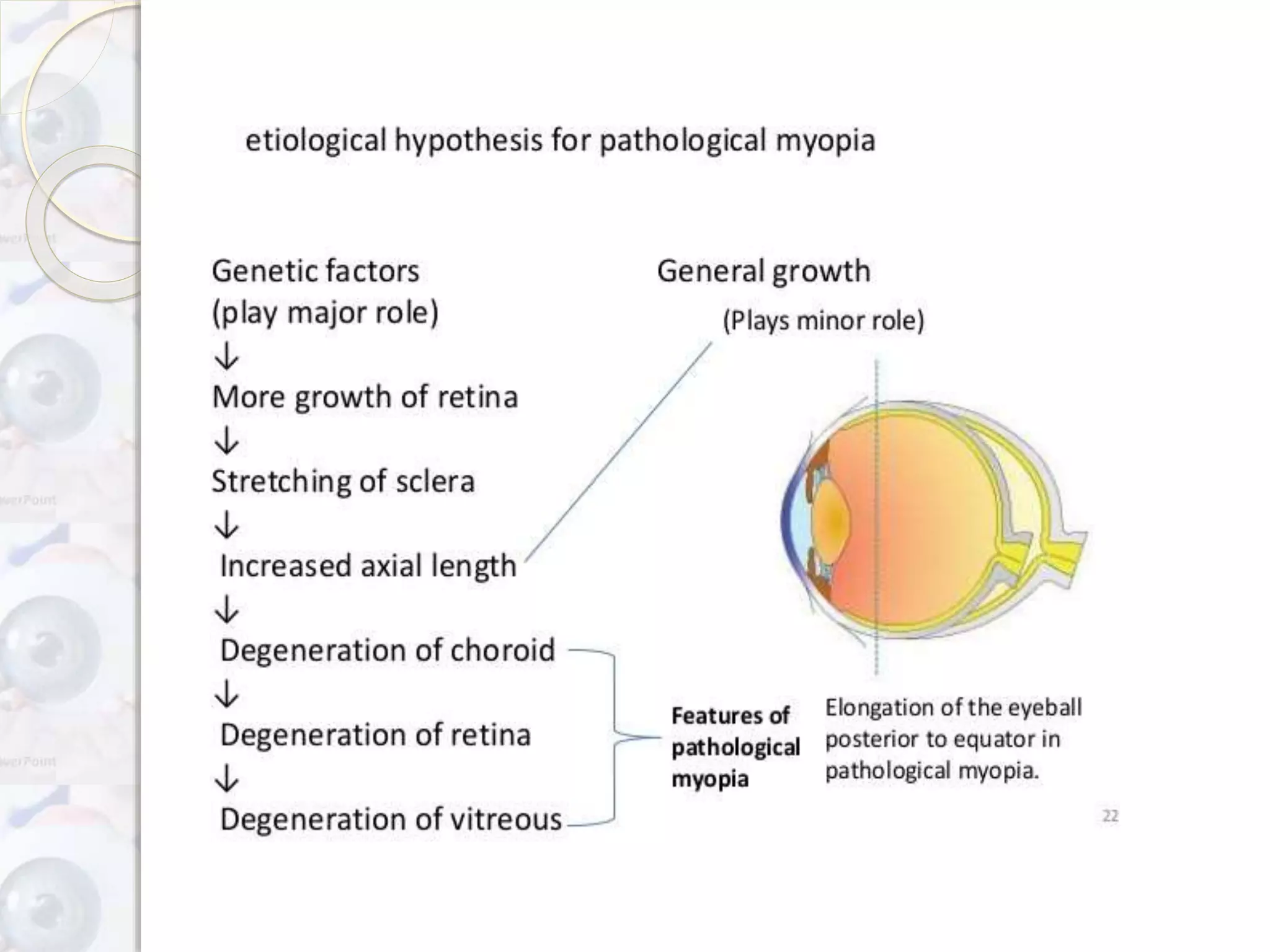
MYOPIA
BY
AYUSHI R. PATEL
MYOPIA
Shortsightedness/nearsightedn
ess
Greek word meaning:- “close
the eye”.
Definition:- it is a type of refractive error in
which parallel rays of light coming from
infinity are focused infront of the retina when
the accommodation being at rest.
AETIOLOGICAL TYPES
1. Axial myopia:- increase in antero-posterior length of
the eyeball.
2. Curvatural myopia:-increased curvature of cornea or
lens or both.
3. Index myopia:-increased in refractive index of
crystalline lens associated with nuclear sclerosis.
4. Myopia due to excessive accommodation:-occurs in
patients with spasm of accommodation.
5. Positional myopia:-anterior placement of crystalline
lens in the eyeball.
Clinical types
1. Congenital myopia
2. Simple or developmental
myopia
3. Pathological or degenerative
myopia
4. Acquired myopia
Congenital myopia
Present since birth.
Seen more frequently in children who were born
prematurely or with various birth defects like marfan
syndrome and homocystinuria.
Usually error is about 8-10D, which mostly remains
constant.
Diagnosed at age 2-3 years.
If unilateral,as anisometropia,may develop amblyopi
strabismus.
Increase in axial length and in overall globe size.
Associated conditions
Convergent squint
Cataract
Microphthalmos
Aniridia
Megalocornea
Congenital separation of
retina
Diagnosis:-if the myopia is
bilateral,the child will generally
display some noticeable difficulty
in seeing distance objects and will
tend to hold things very close for
viewing.
Management:-
early correction is desirable.
retinoscopy under full cycloplegia.
early full correction desirable
prognosis.
Simple or developmental myopia
Physiological or school
myopia.
Physiological error not
associated with any
disease of eye.
Aetiology
Result from normal biological variation
in the development of eye.
Axial:- physiological variation in the
length of the eyeball.
Curvatural:-underdevelopment of
eyeball.
Role of diet in early childhood
Theory of excessive near work.
Role of genetic plays some role.
symptoms
Poor vision for distance.
Asthenopic symptoms:- eye straib due to
dissociation between convergence and
accommodation.
May develop convergence weakness and
exophoria and supression in one eye.
Change in psychological out look.
Half shutting of the eye may by complained
by parents of the child.
signs
1. Eyes are large and predominant.
2. Slight deep anterior chamber.
3. Fundus is normal: rarely temporal myopic
crescent may be seen.
4. Usually error does not exceed 6-8D.
5. Pupil is large and react sluggishly.
diagnosis
Confirmed by performing
retinoscopy.
PATHOLOGICAL MYOPIA
Also known as degenerative
/progressive myopia.
Rapidly progressive associated with
degenerative changes in the eye.
Which starts in childhood at 5-10
years of age and results in high
myopia(7-6 D).
Aetiology
Rapid axial growth of the eyeball
outside the normal biological
variations of development.
Role of heredity.
Role of general growth process.
symptoms
Defective vision
Night blindness
Muscae volitantes/floating
black opacities.
signs
1. Prominent eyeballs.
2. Large cornea.
3. Anterior chamber is deep.
4. Pupils are large,react sluggishly.
5. Refractive error increases mid-30s and
results in myopia of 10-20D,which may
even progress to 30-40D.
6. Visual field show contaction and in
some areas ring scotoma may be
seen.
Fundus signs
1. A pale tessellate:-appearance due to
diffuse attenuation of the retinal
pigmented epithelium with visibility of
large choroidal vessels.
2. Focal chorioretinal atrophy:-
charactrised by visibility of larger
choroidal vessels and eventually
the sclera.
3. Lacquer cracks:-ruptures in the RPE
bruch membrane chorioretinal complex
charactrised by fine irregular yellow lines
branching and criss crossing at the
posterior pole.
4. CNV(choroidal
neovascularization):- which may
develop in accociation with lacquer
cracks and area of patchy atrophy.
5. Subretinal coin haemorrhages
which may develop from lacquer
cracks in the absence of cnv.
6. Foster fuchs’s pot:-is a
raised,circular,pigmented lesion
thant may develop after a macular
haemorrhage has absorbed.
complications
Retinal tears and retinal detachment
may occur.
Complicated cataract.
Nuclear sclerosis.
Vitreous haemorrhages.
Choroidal haemorrhages and
thrombosis
Primary open angle glaucoma.
Acquired myopia
1. Index myopia:-
seen in nuclear sclerosis and incipient cataract.
Diabetic myopia occurs dur to decrease in refractive
index of cortex.
2. Curvatural myopia:-
Increase of corneal curvature in diseased conditions
like corneal ectasias and conical cornea.
3. Positional myopia:-
Conditions producing anterior subluxation of lens.
4. Consecutive myopia:-
surgical overcorrection of
hypermetropia.
Pseudophakia with overcorrecting
IOL.
5. Pseudomyopia:-
also called artifical myopia.
Produced in a conditions such as
excessive accommodation and spasm
of accommodation.
May develop after too full a
hypermetropic correction in chidren.
6. Space myopia:-
experienced when the individual
has no stimulation for distance
fixation.
7. Night myopia/twilight myopia:-
Shift from photopic to scotopic vision at
twilight
Increased sensitivity to shorter
wavelength of light
The emmetropic eye,if accommodation
for the middle range of visual spectrum
will be slightly myopic for the shorter
wavelengths.
8. Drug induced myopia:-
Cholinergic drugs such as
pilocarpine,echothiopate,di-
isopropyl fluorophosphate.
Sulphonamides
Steroid induced myopia
Treatment of myopia
Optical treatment:-
Appropriate concave
lenses(spectacles and contact
lens)
Minimum acceptance
providing maximum vision.
Guidelines
1. Low degree of myopia:-
In young people:-in children above 3 years
of age,myopia should be fully corrected and
instructed to use their glasses constantly,for
avoid developing the habit of squinting and
accommodation-convergence reflex.
Never overcorrect myopia.
In adults people:-older than 30years
patients are not able to tolerate a full
correction because ciliary muscle are not
accustomed to accommodate.
So,prescribed less than full correction.
2. High degree of myopia
Full correction can rarely be tolerated.
Undercorrection as little is complicated
with comfort for binocular near vision.
Undercorrection to the tune of 1-3D or
even more may be required.
Undercorrection is always better to avoid
the problem of near vision and minification
of image.
Surgical treatment:-
Radial keratotomy, photorefractive
keratectomy,LASIK(laser in situ
keratomileusis)
Visual hygiene:-
To avoid asthenopic symptoms.
Adequate illumination during close
work.
The clarity of the print should be good
to avoid undue ocular fatigue.
Preventive measures
Therapeutic inteventions:-atropine and
pirenzepine both drugs slow downs the
progression of myopia.
General measures:-balanced diet in
vitamins and protiens. Early management of
associated debiliatating diseases.
Genetic counselling:- people having
pathological myopia,not to marriage with
pathological myopic peoples.
Low vision aids
Indicated in patients of progressive
myopia with advanced degenerative
changes.
Where useful vision can’t be
obtained with spectacles and contact
lenses.
prognosis
1. Simple myopia:-
Prognosis is good.
Error usually does not progress beyond 6-8D.
Stabilized by the age of 21.
2. Pathological myopia:-
Visual prognosis is always guarded.
Possibility of progressive visual loss due to
degenerative changes and danger of complications
such as retinal detachment should be borne in
mind.
MYOPIA A REFRACTIVE ERROR OF AN EYE

MYOPIA A REFRACTIVE ERROR OF AN EYE

  • 1.
  • 2.
  • 3.
    Definition:- it isa type of refractive error in which parallel rays of light coming from infinity are focused infront of the retina when the accommodation being at rest.
  • 4.
    AETIOLOGICAL TYPES 1. Axialmyopia:- increase in antero-posterior length of the eyeball. 2. Curvatural myopia:-increased curvature of cornea or lens or both. 3. Index myopia:-increased in refractive index of crystalline lens associated with nuclear sclerosis. 4. Myopia due to excessive accommodation:-occurs in patients with spasm of accommodation. 5. Positional myopia:-anterior placement of crystalline lens in the eyeball.
  • 5.
    Clinical types 1. Congenitalmyopia 2. Simple or developmental myopia 3. Pathological or degenerative myopia 4. Acquired myopia
  • 6.
    Congenital myopia Present sincebirth. Seen more frequently in children who were born prematurely or with various birth defects like marfan syndrome and homocystinuria. Usually error is about 8-10D, which mostly remains constant. Diagnosed at age 2-3 years. If unilateral,as anisometropia,may develop amblyopi strabismus. Increase in axial length and in overall globe size.
  • 7.
  • 8.
    Diagnosis:-if the myopiais bilateral,the child will generally display some noticeable difficulty in seeing distance objects and will tend to hold things very close for viewing. Management:- early correction is desirable. retinoscopy under full cycloplegia. early full correction desirable prognosis.
  • 9.
    Simple or developmentalmyopia Physiological or school myopia. Physiological error not associated with any disease of eye.
  • 10.
    Aetiology Result from normalbiological variation in the development of eye. Axial:- physiological variation in the length of the eyeball. Curvatural:-underdevelopment of eyeball. Role of diet in early childhood Theory of excessive near work. Role of genetic plays some role.
  • 11.
    symptoms Poor vision fordistance. Asthenopic symptoms:- eye straib due to dissociation between convergence and accommodation. May develop convergence weakness and exophoria and supression in one eye. Change in psychological out look. Half shutting of the eye may by complained by parents of the child.
  • 12.
    signs 1. Eyes arelarge and predominant. 2. Slight deep anterior chamber. 3. Fundus is normal: rarely temporal myopic crescent may be seen. 4. Usually error does not exceed 6-8D. 5. Pupil is large and react sluggishly.
  • 13.
  • 14.
    PATHOLOGICAL MYOPIA Also knownas degenerative /progressive myopia. Rapidly progressive associated with degenerative changes in the eye. Which starts in childhood at 5-10 years of age and results in high myopia(7-6 D).
  • 15.
    Aetiology Rapid axial growthof the eyeball outside the normal biological variations of development. Role of heredity. Role of general growth process.
  • 17.
  • 18.
    signs 1. Prominent eyeballs. 2.Large cornea. 3. Anterior chamber is deep. 4. Pupils are large,react sluggishly. 5. Refractive error increases mid-30s and results in myopia of 10-20D,which may even progress to 30-40D. 6. Visual field show contaction and in some areas ring scotoma may be seen.
  • 19.
    Fundus signs 1. Apale tessellate:-appearance due to diffuse attenuation of the retinal pigmented epithelium with visibility of large choroidal vessels.
  • 20.
    2. Focal chorioretinalatrophy:- charactrised by visibility of larger choroidal vessels and eventually the sclera.
  • 21.
    3. Lacquer cracks:-rupturesin the RPE bruch membrane chorioretinal complex charactrised by fine irregular yellow lines branching and criss crossing at the posterior pole.
  • 22.
    4. CNV(choroidal neovascularization):- whichmay develop in accociation with lacquer cracks and area of patchy atrophy.
  • 23.
    5. Subretinal coinhaemorrhages which may develop from lacquer cracks in the absence of cnv.
  • 24.
    6. Foster fuchs’spot:-is a raised,circular,pigmented lesion thant may develop after a macular haemorrhage has absorbed.
  • 25.
    complications Retinal tears andretinal detachment may occur. Complicated cataract. Nuclear sclerosis. Vitreous haemorrhages. Choroidal haemorrhages and thrombosis Primary open angle glaucoma.
  • 26.
    Acquired myopia 1. Indexmyopia:- seen in nuclear sclerosis and incipient cataract. Diabetic myopia occurs dur to decrease in refractive index of cortex. 2. Curvatural myopia:- Increase of corneal curvature in diseased conditions like corneal ectasias and conical cornea. 3. Positional myopia:- Conditions producing anterior subluxation of lens.
  • 27.
    4. Consecutive myopia:- surgicalovercorrection of hypermetropia. Pseudophakia with overcorrecting IOL. 5. Pseudomyopia:- also called artifical myopia. Produced in a conditions such as excessive accommodation and spasm of accommodation. May develop after too full a hypermetropic correction in chidren.
  • 28.
    6. Space myopia:- experiencedwhen the individual has no stimulation for distance fixation. 7. Night myopia/twilight myopia:- Shift from photopic to scotopic vision at twilight Increased sensitivity to shorter wavelength of light The emmetropic eye,if accommodation for the middle range of visual spectrum will be slightly myopic for the shorter wavelengths.
  • 29.
    8. Drug inducedmyopia:- Cholinergic drugs such as pilocarpine,echothiopate,di- isopropyl fluorophosphate. Sulphonamides Steroid induced myopia
  • 30.
    Treatment of myopia Opticaltreatment:- Appropriate concave lenses(spectacles and contact lens) Minimum acceptance providing maximum vision.
  • 31.
    Guidelines 1. Low degreeof myopia:- In young people:-in children above 3 years of age,myopia should be fully corrected and instructed to use their glasses constantly,for avoid developing the habit of squinting and accommodation-convergence reflex. Never overcorrect myopia. In adults people:-older than 30years patients are not able to tolerate a full correction because ciliary muscle are not accustomed to accommodate. So,prescribed less than full correction.
  • 32.
    2. High degreeof myopia Full correction can rarely be tolerated. Undercorrection as little is complicated with comfort for binocular near vision. Undercorrection to the tune of 1-3D or even more may be required. Undercorrection is always better to avoid the problem of near vision and minification of image.
  • 33.
    Surgical treatment:- Radial keratotomy,photorefractive keratectomy,LASIK(laser in situ keratomileusis) Visual hygiene:- To avoid asthenopic symptoms. Adequate illumination during close work. The clarity of the print should be good to avoid undue ocular fatigue.
  • 34.
    Preventive measures Therapeutic inteventions:-atropineand pirenzepine both drugs slow downs the progression of myopia. General measures:-balanced diet in vitamins and protiens. Early management of associated debiliatating diseases. Genetic counselling:- people having pathological myopia,not to marriage with pathological myopic peoples.
  • 35.
    Low vision aids Indicatedin patients of progressive myopia with advanced degenerative changes. Where useful vision can’t be obtained with spectacles and contact lenses.
  • 36.
    prognosis 1. Simple myopia:- Prognosisis good. Error usually does not progress beyond 6-8D. Stabilized by the age of 21. 2. Pathological myopia:- Visual prognosis is always guarded. Possibility of progressive visual loss due to degenerative changes and danger of complications such as retinal detachment should be borne in mind.