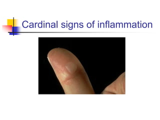
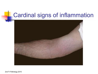
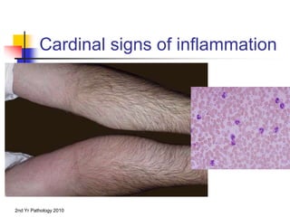
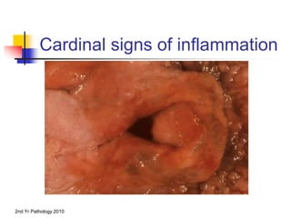
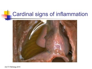
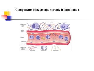
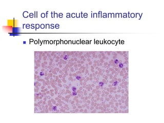
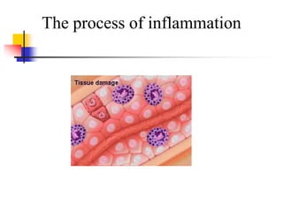
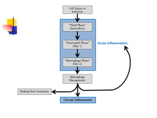
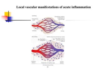
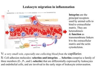
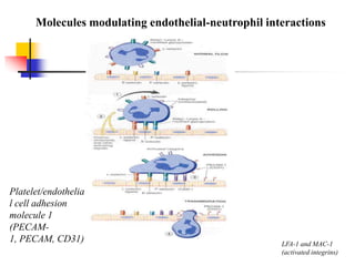
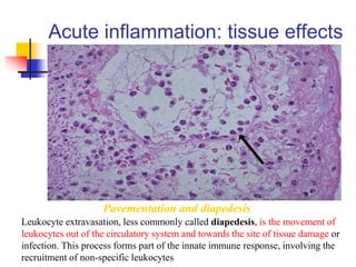
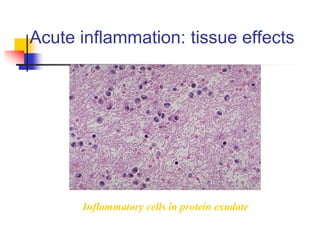
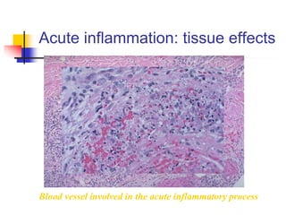
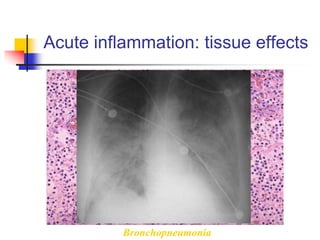
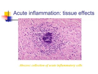
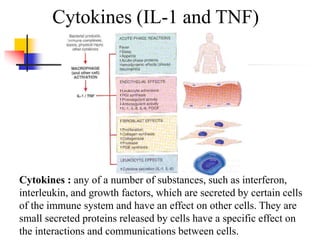
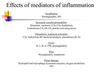
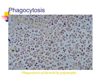
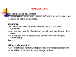
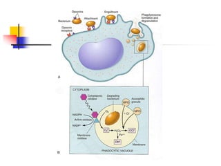
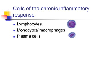
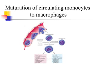
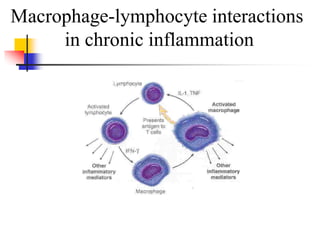
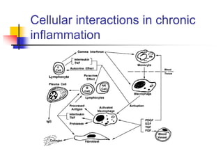
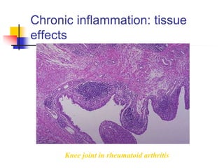
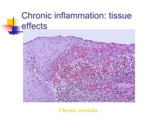
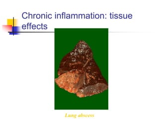
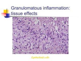
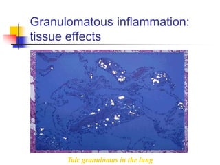
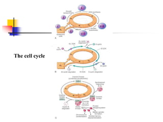
Inflammation &
Cellular responses
Inflammation
 Is a protective response
 The body’s response to injury
 Interwoven with the repair process
Inflammation
 Types
 Acute (sec, mins., hrs)
 Chronic (days, weeks, months, yrs)
Causes of inflammation
 Bacterial
 Viral
 Protozoal
 Metazoal*
 Fungal
 Immunological
 Tumours
 Chemicals, toxins etc
 Radiation
*A subdivision of the animal kingdom that includes all
multicellular animal organisms having cells that are
differentiated and form tissues and organs.
Acute inflammation
the initial response of the body to harmful stimuli,
achieved by the increased movement of plasma and
leukocytes (especially granulocytes) from the blood into
the injured tissues.
Inflammation is not a synonym for infection.
Inflammation
 The Cardinal Signs of Acute
Inflammation
RUBOR
CALOR
TUMOR
FUNCTIO LAESA
1. calor - heat
2. rubor - redness
3. tumor - swelling
4. dolor - pain
5. functio laesa - loss (or
impairment) of function (Galen)
Cardinal signs of inflammation
2nd Yr Pathology 2010
Cardinal signs of inflammation
2nd Yr Pathology 2010
Cardinal signs of inflammation
2nd Yr Pathology 2010
Cardinal signs of inflammation
2nd Yr Pathology 2010
Cardinal signs of inflammation
2nd Yr Pathology 2010
Cardinal signs of inflammation
Inflammation
The basis of the five cardinal signs
 Increased blood flow due to vascular dilatation gives
redness and heat.
 Increased vascular permeability gives oedema causing
tissue swelling.
 Certain chemical mediators stimulate sensory nerve
endings giving pain. Nerves are also stimulated by
stretching due to oedema.
 Pain and swelling result in loss of function.
Components of acute and chronic inflammation
Cell of the acute inflammatory
response
 Polymorphonuclear leukocyte
The process of inflammation
The phases of inflammation
 FIRST THERE IS VASCULAR DILATATION
followed by exudation of protein-rich oedema
fluid which floods the area, dilutes toxins, allows
immunoglobulins to opsonise bacteria and
provides substrate (fibrinogen) for fibrin
scaffold*.
 SECOND THERE IS ACTIVE EMIGRATION OF
POLYMORPHS through vessel wall and along the
chemotactic gradient to the site of injury
make (a foreign cell) more susceptible to phagocytosis
*a temporary or movable platform
the movement of a microorganism or cell in response to a chemical
stimulus.
 THE VASCULAR PHASE OF INFLAMMATION
Fluid escapes from vessels because of endothelial cell (EC)
retraction, opening up gap-junctions.
The vessels which are normally involved are the post-capillary
venules where the EC have high affinity receptors for histamine.
Severe EC injury leads to leakiness of all vessels capillaries,
venules and arterioles - giving acute local oedema,
e.g. blister formation after a burn.
The phases of inflammation
Local vascular manifestations of acute inflammation
Leukocyte migration in inflammation
V: a very small vein, especially one collecting blood from the capillaries
S: Cell adhesion molecules: selectins and integrins. ... Selectins comprise a family of
three members (E-, P-, and L-selectin) that are differentially expressed by leukocytes
and endothelial cells, and are involved in the early steps of leukocyte extravasation.
Integrins are the
principal receptors
used by animal cells to
bind to extracellular
matrix. They are
heterodimers
& function as
transmembrane linkers
b/w the extracellular
matrix and the actin
cytoskeleton.
Molecules modulating endothelial-neutrophil interactions
LFA-1 and MAC-1
(activated integrins)
Platelet/endothelia
l cell adhesion
molecule 1
(PECAM-
1, PECAM, CD31)
Acute inflammation: tissue effects
Pavementation and diapedesis
Leukocyte extravasation, less commonly called diapedesis, is the movement of
leukocytes out of the circulatory system and towards the site of tissue damage or
infection. This process forms part of the innate immune response, involving the
recruitment of non-specific leukocytes
Acute inflammation: tissue effects
Inflammatory cells in protein exudate
Acute inflammation: tissue effects
Blood vessel involved in the acute inflammatory process
Acute inflammation: tissue effects
Bronchopneumonia
Acute inflammation: tissue effects
Abscess: collection of acute inflammatory cells
Acute inflammation: tissue effects
Multiple splenic abscesses
 Vasoactive amines
 Histamine
 Serotonin (5-HT)
 Neuropeptides
 Substance P
 Plasma proteases and the complement system
 Action of Hageman factor
 Arachidonic acid metabolites
 Prostaglandins
 Leukotrienes
 Lipoxins
 Cytokines
 IL-1, TNF etc.
 Chemokines (CXC and CC)
 Nitric oxide and oxygen-derived free radicals
Chemical mediators of inflammation
 PREFORMED
Histamine, Serotonin
 NEWLY SYNTHESISED
Prostaglandins
Leucotrienes
Platelet activating factor
Cytokines
Nitric oxide
 LOCAL AND SYSTEMIC*
*mechanisms by which local immune stimulation is transmitted to
the periphery, leading to systemic inflammation. We concentrate
on the role of three signalling molecules: interleukin-1,
interleukin-6, and tumour necrosis factor alpha. These cytokines
provide a direct link between local and systemic inflammation
Chemical mediators of inflammation
(local and systemic)
Plasma proteases
Kinins are polypeptides and cause vasodilation and smooth muscle contraction. These
structurally related polypeptides, such as bradykinin and kallidin are members of the
autacoid family. They act locally to induce vasodilation and contraction of smooth
Kinins: any of a
group of
substances
formed in body
tissue in
response to
injury.
The complement system
Arachidonic acid metabolites
HETE =
hydroxyeicosatetraen
oic acid
HPETE =
hydroperoxyeicosatet
raenoic acid
Arachidonic acid
Arachidonic acid; 506-32-1; Arachidonate; Immunocytophyte;
(5Z,8Z,11Z,14Z)-icosa-5,8,11,14-tetraenoic acid; (all-Z)-
5,8,11,14-Eicosatetraenoic acid
Molecular Formula: C20H32O2
Molecular Weight: 304.474 g/mol
Arachidonic Acid is an unsaturated, essential fatty acid. It is found in animal and human
fat as well as in the liver, brain, and glandular organs, and is a constituent of animal
phosphatides. It is formed by the synthesis from dietary linoleic acid and is a precursor
in the biosynthesis of prostaglandins, thromboxanes, and leukotrienes.(ChemID Plus)
The beneficial effects of Omega-3 fatty acids are believed to be due in part to selective alteration
of arachidonate metabolism that involves cyclooxygenase (COX) enzymes. (PMID: 23371504)
Here, 9-oxononanoic acid (9-ONA), one of the major products of peroxidized fatty acids, was
found to stimulate the activity of phospholipase A2 (PLA2), the key enzyme to
initiate arachidonate cascade and eicosanoid production.
(PMID: 23704812) Arachidonate lipoxygenase (ALOX) enzymes metabolize arachidonic acid to
generate potent inflammatory mediators and play an important role in inflammation-associated
diseases. (PMID: 23404351)
Cytokines (IL-1 and TNF)
Cytokines : any of a number of substances, such as interferon,
interleukin, and growth factors, which are secreted by certain cells
of the immune system and have an effect on other cells. They are
small secreted proteins released by cells have a specific effect on
the interactions and communications between cells.
Nitric oxide (NO)
Effects of mediators of inflammation
Vasodilation:
Prostaglandins, NO
Increased vascular permeability:
Histamine, serotonin, C3a, C5a, bradykinin,
Leukotrienes C4, D4, E4, platelet activating factor
Chemotaxis, leukocyte activation:
C5a, leukotriene B4, bacterial products, chemokines (IL-8)
Fever:
IL-1, IL-6, TNF, prostaglandins
Pain:
Prostaglandins, bradykinin
Tissue damage:
Neutrophil and macrophage lysosomal enzymes, oxygen metabolites
NO
Phagocytosis
Phagocytosis of bacteria by polymorphs
PHAGOCYTOSIS
Recognition and attachment
Foreign objects coated with opsonins IgG and C3b which attach to
receptors on polymorph surface.
Engulfment
Cell membrane fuses around an object: at the some time
lysosomes
empty into the vacuole, often before vacuole has time to seal - this
gives
rise to 'regurgitation during feeding' and enzymatic damage to
surrounding
tissue.
Killing or degradation
H2O2, hypohalous acid (HOC1) produced by myeloperoxidase and
superoxides kill bacteria. Lysozyme digests them.
Chronic inflammation
Cells of the chronic inflammatory
response
 Lymphocytes
 Monocytes/ macrophages
 Plasma cells
Maturation of circulating monocytes
to macrophages
Macrophage-lymphocyte interactions
in chronic inflammation
Cellular interactions in chronic
inflammation
Chronic inflammation: tissue
effects
Knee joint in rheumatoid arthritis
Chronic inflammation: tissue
effects
Chronic cervicitis
Chronic inflammation: tissue
effects
Lung abscess
Granulomatous
inflammation:
a special form of
chronic inflammation
Granuloma
 Definition
 A collection of macrophages, lymphocytes,
mononuclear cells and fibroblasts with or
without giant cell formation and constitutes
a special form of chronic inflammation
Granulomatous inflammation
Bacterial:
TB, Leprosy, Syphillis, cat-scratch disease
Parasitic:
Schistosomiasis
Fungal:
Histoplasma, blastomycosis, cryptococcus
Inorganics, metals, dusts:
Silicosis, berrylliosis
Foreign body
Unknown:
Sarcoidosis
Granulomatous inflammation:
tissue effects
Granulomatous inflammation:
tissue effects
Granulomatous inflammation:
tissue effects
Granulomatous inflammation:
tissue effects
Epithelioid cells
Granulomatous inflammation:
tissue effects
Talc granulomas in the lung
Healing and repair
Wound healing: critical steps
The cell cycle
Cyclins, cyclin dependent kinases and
cyclin dependent kinase inhibitors
Cell-cell interactions in repair
Cell surface receptors in healing
and repair
The major components of the
extracellular matrix (ECM) required for
healing and repair
Extracellular matrix re-modelling occurs by the action of
Matrix metalloproteinases
Matrix metalloproteinase
regulation
Critical steps in angiogenesis
Major growth factors in
wound healing
Wound healing: critical steps
Granulation tissue
Granulation tissue
Scar tissue
Skin
Scar tissue
Lung
Outcome of healing and repair
When healing goes wrong
Keloid scar

Inflammation & cellular response

  • 1.
  • 2.
    Inflammation  Is aprotective response  The body’s response to injury  Interwoven with the repair process
  • 3.
    Inflammation  Types  Acute(sec, mins., hrs)  Chronic (days, weeks, months, yrs)
  • 4.
    Causes of inflammation Bacterial  Viral  Protozoal  Metazoal*  Fungal  Immunological  Tumours  Chemicals, toxins etc  Radiation *A subdivision of the animal kingdom that includes all multicellular animal organisms having cells that are differentiated and form tissues and organs.
  • 5.
    Acute inflammation the initialresponse of the body to harmful stimuli, achieved by the increased movement of plasma and leukocytes (especially granulocytes) from the blood into the injured tissues. Inflammation is not a synonym for infection.
  • 6.
    Inflammation  The CardinalSigns of Acute Inflammation RUBOR CALOR TUMOR FUNCTIO LAESA
  • 7.
    1. calor -heat 2. rubor - redness 3. tumor - swelling 4. dolor - pain 5. functio laesa - loss (or impairment) of function (Galen)
  • 8.
    Cardinal signs ofinflammation
  • 9.
    2nd Yr Pathology2010 Cardinal signs of inflammation
  • 10.
    2nd Yr Pathology2010 Cardinal signs of inflammation
  • 11.
    2nd Yr Pathology2010 Cardinal signs of inflammation
  • 12.
    2nd Yr Pathology2010 Cardinal signs of inflammation
  • 13.
    2nd Yr Pathology2010 Cardinal signs of inflammation
  • 14.
    Inflammation The basis ofthe five cardinal signs  Increased blood flow due to vascular dilatation gives redness and heat.  Increased vascular permeability gives oedema causing tissue swelling.  Certain chemical mediators stimulate sensory nerve endings giving pain. Nerves are also stimulated by stretching due to oedema.  Pain and swelling result in loss of function.
  • 15.
    Components of acuteand chronic inflammation
  • 16.
    Cell of theacute inflammatory response  Polymorphonuclear leukocyte
  • 17.
    The process ofinflammation
  • 19.
    The phases ofinflammation  FIRST THERE IS VASCULAR DILATATION followed by exudation of protein-rich oedema fluid which floods the area, dilutes toxins, allows immunoglobulins to opsonise bacteria and provides substrate (fibrinogen) for fibrin scaffold*.  SECOND THERE IS ACTIVE EMIGRATION OF POLYMORPHS through vessel wall and along the chemotactic gradient to the site of injury make (a foreign cell) more susceptible to phagocytosis *a temporary or movable platform the movement of a microorganism or cell in response to a chemical stimulus.
  • 20.
     THE VASCULARPHASE OF INFLAMMATION Fluid escapes from vessels because of endothelial cell (EC) retraction, opening up gap-junctions. The vessels which are normally involved are the post-capillary venules where the EC have high affinity receptors for histamine. Severe EC injury leads to leakiness of all vessels capillaries, venules and arterioles - giving acute local oedema, e.g. blister formation after a burn. The phases of inflammation
  • 21.
    Local vascular manifestationsof acute inflammation
  • 22.
    Leukocyte migration ininflammation V: a very small vein, especially one collecting blood from the capillaries S: Cell adhesion molecules: selectins and integrins. ... Selectins comprise a family of three members (E-, P-, and L-selectin) that are differentially expressed by leukocytes and endothelial cells, and are involved in the early steps of leukocyte extravasation. Integrins are the principal receptors used by animal cells to bind to extracellular matrix. They are heterodimers & function as transmembrane linkers b/w the extracellular matrix and the actin cytoskeleton.
  • 23.
    Molecules modulating endothelial-neutrophilinteractions LFA-1 and MAC-1 (activated integrins) Platelet/endothelia l cell adhesion molecule 1 (PECAM- 1, PECAM, CD31)
  • 24.
    Acute inflammation: tissueeffects Pavementation and diapedesis Leukocyte extravasation, less commonly called diapedesis, is the movement of leukocytes out of the circulatory system and towards the site of tissue damage or infection. This process forms part of the innate immune response, involving the recruitment of non-specific leukocytes
  • 25.
    Acute inflammation: tissueeffects Inflammatory cells in protein exudate
  • 26.
    Acute inflammation: tissueeffects Blood vessel involved in the acute inflammatory process
  • 27.
    Acute inflammation: tissueeffects Bronchopneumonia
  • 28.
    Acute inflammation: tissueeffects Abscess: collection of acute inflammatory cells
  • 29.
    Acute inflammation: tissueeffects Multiple splenic abscesses
  • 30.
     Vasoactive amines Histamine  Serotonin (5-HT)  Neuropeptides  Substance P  Plasma proteases and the complement system  Action of Hageman factor  Arachidonic acid metabolites  Prostaglandins  Leukotrienes  Lipoxins  Cytokines  IL-1, TNF etc.  Chemokines (CXC and CC)  Nitric oxide and oxygen-derived free radicals Chemical mediators of inflammation
  • 31.
    Chemical mediators ofinflammation  PREFORMED Histamine, Serotonin  NEWLY SYNTHESISED Prostaglandins Leucotrienes Platelet activating factor Cytokines Nitric oxide  LOCAL AND SYSTEMIC* *mechanisms by which local immune stimulation is transmitted to the periphery, leading to systemic inflammation. We concentrate on the role of three signalling molecules: interleukin-1, interleukin-6, and tumour necrosis factor alpha. These cytokines provide a direct link between local and systemic inflammation
  • 32.
    Chemical mediators ofinflammation (local and systemic)
  • 33.
    Plasma proteases Kinins arepolypeptides and cause vasodilation and smooth muscle contraction. These structurally related polypeptides, such as bradykinin and kallidin are members of the autacoid family. They act locally to induce vasodilation and contraction of smooth Kinins: any of a group of substances formed in body tissue in response to injury.
  • 34.
  • 35.
    Arachidonic acid metabolites HETE= hydroxyeicosatetraen oic acid HPETE = hydroperoxyeicosatet raenoic acid
  • 36.
    Arachidonic acid Arachidonic acid;506-32-1; Arachidonate; Immunocytophyte; (5Z,8Z,11Z,14Z)-icosa-5,8,11,14-tetraenoic acid; (all-Z)- 5,8,11,14-Eicosatetraenoic acid Molecular Formula: C20H32O2 Molecular Weight: 304.474 g/mol Arachidonic Acid is an unsaturated, essential fatty acid. It is found in animal and human fat as well as in the liver, brain, and glandular organs, and is a constituent of animal phosphatides. It is formed by the synthesis from dietary linoleic acid and is a precursor in the biosynthesis of prostaglandins, thromboxanes, and leukotrienes.(ChemID Plus) The beneficial effects of Omega-3 fatty acids are believed to be due in part to selective alteration of arachidonate metabolism that involves cyclooxygenase (COX) enzymes. (PMID: 23371504) Here, 9-oxononanoic acid (9-ONA), one of the major products of peroxidized fatty acids, was found to stimulate the activity of phospholipase A2 (PLA2), the key enzyme to initiate arachidonate cascade and eicosanoid production. (PMID: 23704812) Arachidonate lipoxygenase (ALOX) enzymes metabolize arachidonic acid to generate potent inflammatory mediators and play an important role in inflammation-associated diseases. (PMID: 23404351)
  • 37.
    Cytokines (IL-1 andTNF) Cytokines : any of a number of substances, such as interferon, interleukin, and growth factors, which are secreted by certain cells of the immune system and have an effect on other cells. They are small secreted proteins released by cells have a specific effect on the interactions and communications between cells.
  • 38.
  • 39.
    Effects of mediatorsof inflammation Vasodilation: Prostaglandins, NO Increased vascular permeability: Histamine, serotonin, C3a, C5a, bradykinin, Leukotrienes C4, D4, E4, platelet activating factor Chemotaxis, leukocyte activation: C5a, leukotriene B4, bacterial products, chemokines (IL-8) Fever: IL-1, IL-6, TNF, prostaglandins Pain: Prostaglandins, bradykinin Tissue damage: Neutrophil and macrophage lysosomal enzymes, oxygen metabolites NO
  • 40.
  • 41.
    PHAGOCYTOSIS Recognition and attachment Foreignobjects coated with opsonins IgG and C3b which attach to receptors on polymorph surface. Engulfment Cell membrane fuses around an object: at the some time lysosomes empty into the vacuole, often before vacuole has time to seal - this gives rise to 'regurgitation during feeding' and enzymatic damage to surrounding tissue. Killing or degradation H2O2, hypohalous acid (HOC1) produced by myeloperoxidase and superoxides kill bacteria. Lysozyme digests them.
  • 43.
  • 44.
    Cells of thechronic inflammatory response  Lymphocytes  Monocytes/ macrophages  Plasma cells
  • 45.
    Maturation of circulatingmonocytes to macrophages
  • 46.
  • 47.
    Cellular interactions inchronic inflammation
  • 48.
    Chronic inflammation: tissue effects Kneejoint in rheumatoid arthritis
  • 49.
  • 50.
  • 51.
  • 52.
    Granuloma  Definition  Acollection of macrophages, lymphocytes, mononuclear cells and fibroblasts with or without giant cell formation and constitutes a special form of chronic inflammation
  • 53.
    Granulomatous inflammation Bacterial: TB, Leprosy,Syphillis, cat-scratch disease Parasitic: Schistosomiasis Fungal: Histoplasma, blastomycosis, cryptococcus Inorganics, metals, dusts: Silicosis, berrylliosis Foreign body Unknown: Sarcoidosis
  • 54.
  • 55.
  • 56.
  • 57.
  • 58.
  • 59.
  • 60.
  • 61.
  • 62.
    Cyclins, cyclin dependentkinases and cyclin dependent kinase inhibitors
  • 63.
  • 64.
    Cell surface receptorsin healing and repair
  • 65.
    The major componentsof the extracellular matrix (ECM) required for healing and repair
  • 66.
    Extracellular matrix re-modellingoccurs by the action of Matrix metalloproteinases
  • 67.
  • 68.
    Critical steps inangiogenesis
  • 69.
    Major growth factorsin wound healing
  • 70.
  • 71.
  • 72.
  • 73.
  • 74.
  • 75.
  • 76.
    When healing goeswrong Keloid scar