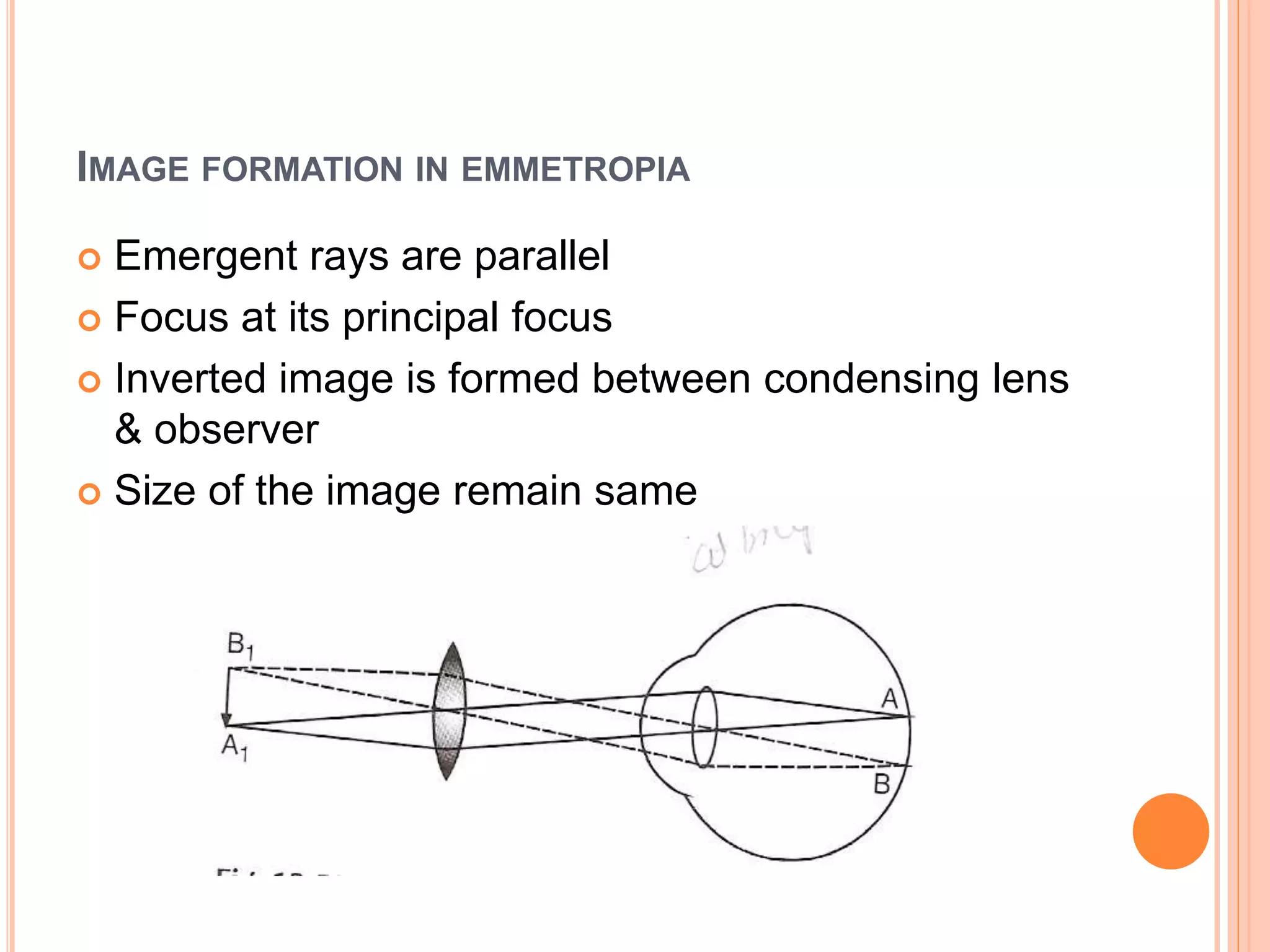
This document provides an overview of indirect ophthalmoscopy. It describes how indirect ophthalmoscopy works by using a strong convex lens to make the eye highly myopic and form a real, inverted image of the fundus. The key components of an indirect ophthalmoscope include binocular viewing systems and adjustable lighting. Indirect ophthalmoscopy allows examination of the posterior segment up to the ora serrata and provides a larger field of view compared to direct ophthalmoscopy. While it provides less magnification, indirect ophthalmoscopy is useful for examining patients with hazy media and during retinal surgery.