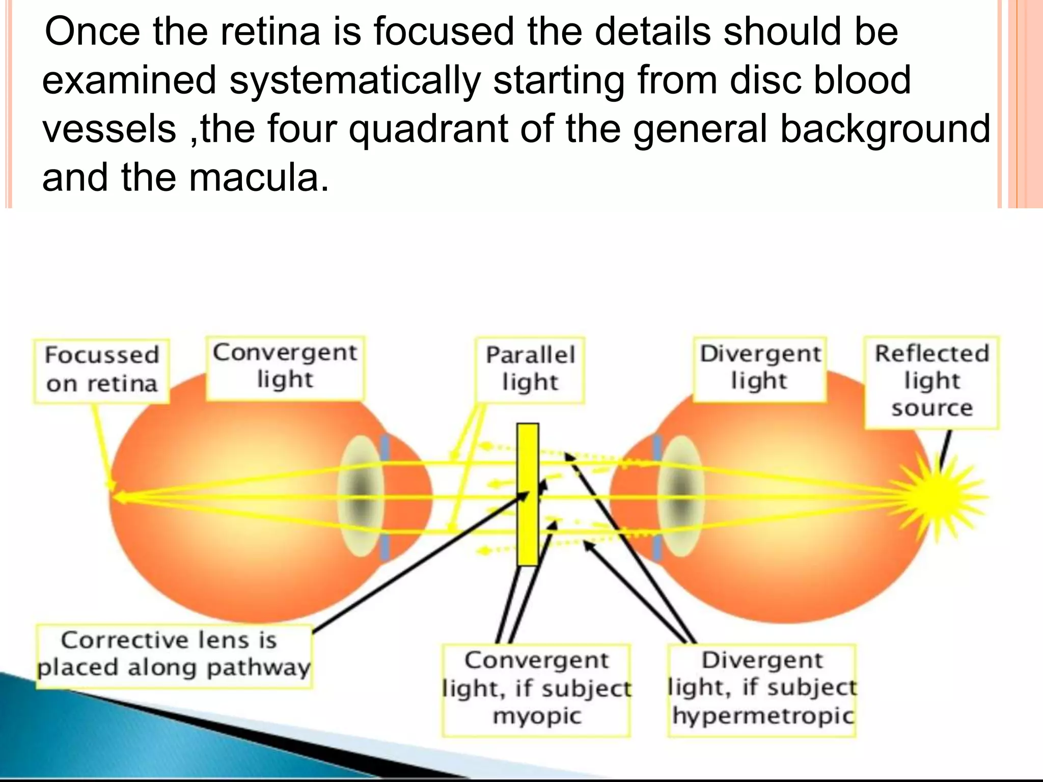
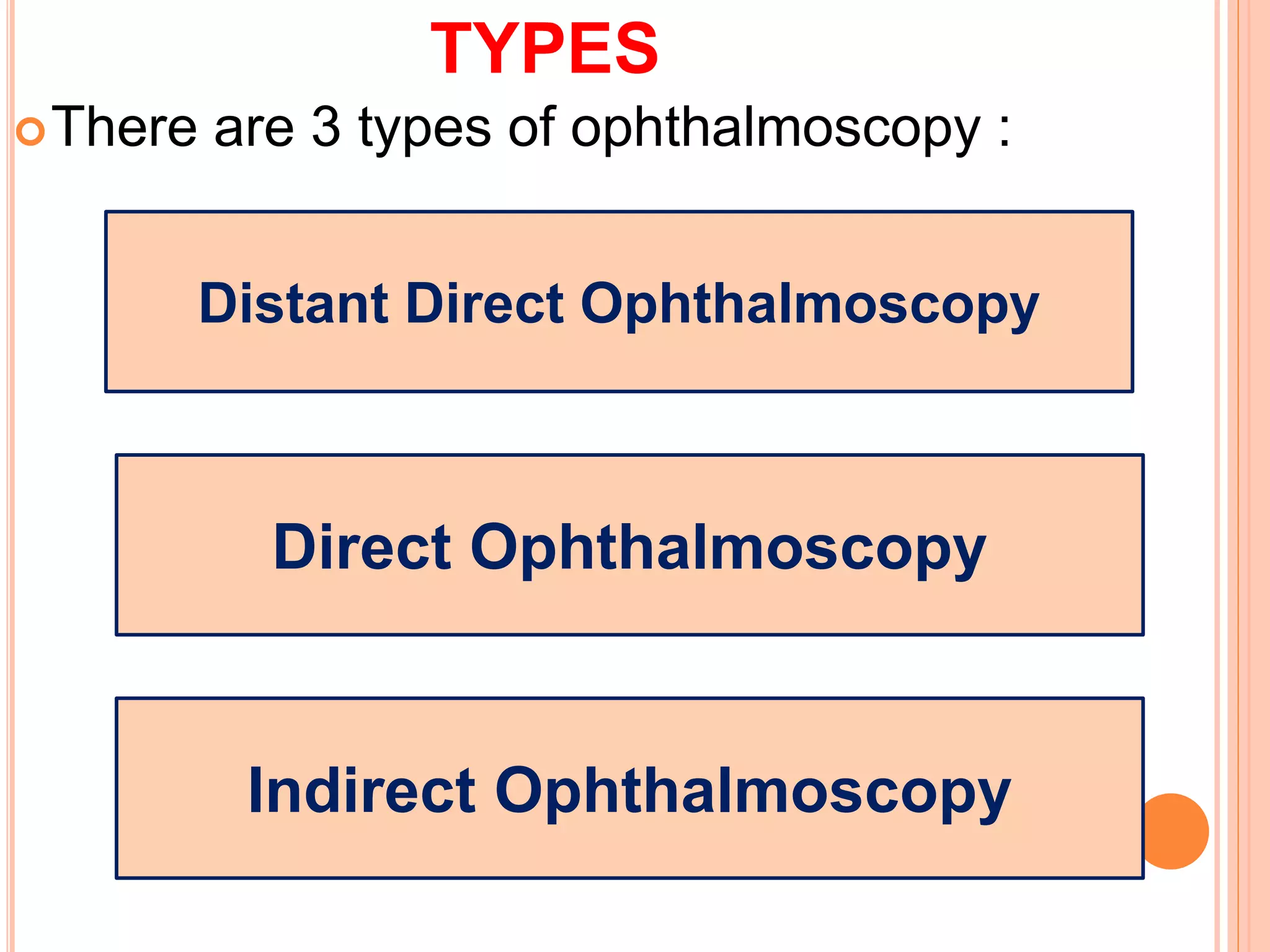
U.P UNIVERSITY OF MEDICAL SCIENCE
SALAL MOHAMMAD
B.OPTOM 2ND YEAR
OPHTHALMOSCOPY
CONTENTS
Introduction
History
Principle
Optics
Types
 Distant Direct Ophthalmoscopy ( DDO )
 Direct Ophthalmoscopy ( DO )
 Indirect Ophthalmoscopy ( IDO )
Technique
INTRODUCTION
Ophthalmoscopy is a clinical examination of the
interior of eye by means of an ophthalmoscope.
It is primarily done to assess the state of fundus
and detect the opacities of ocular media.
It is done as part of an eye examination and
may be done as part of a routine physical
examination.
HISTORY
The ophthalmoscope was invented by Babbage
,1848.
In 1850,Hermann von Helmholz reinvented the
ophhalmoscope and revolutionized
ophthalmoscopy.
Helmholtz could place his eye in the path of the
light rays entering and leaving the patient’s eye
,by looking through the source of light ,thus
allowing the patient’s retina to be seen.
PRINCIPLE
 Helmholtz instrument operated by using a mirror to
shine a beam of light into the eye.
 The observer would look through a tiny aperature
(opening ) in the mirror.
 Helmholtz found that looking through the lens into the
back of the eye only produced a red reflection.
 By attaching a condenser lens he obtained a clearer
inverted image ,which was then magnified five times.
 He c/d this combination of a mirror and condenser lens
an indirect ophthalmoscope.
OPTICS
 The image is erect ,virtual and about 15 times
magnified in emmetropes.
 Technique – Should be performed in a semidark room
with the patient seated and looking straight ahead
,while the observer standing or seated slightly over to
the side of the eye to be examined by the observer
with his/her right eye and left with the left eye.
 The observer should reflect beam of light from the
ophthalmoscope into patient pupil .once the red reflex
is seen the observer should move as close to the
patient eye as possible.
Once the retina is focused the details should be
examined systematically starting from disc blood
vessels ,the four quadrant of the general background
and the macula.
TYPES
There are 3 types of ophthalmoscopy :
Distant Direct Ophthalmoscopy
Direct Ophthalmoscopy
Indirect Ophthalmoscopy
DISTANT DIRECT OPHTHALMOSCOPY ( DDO )
It should be performed routinely before the
direct ophthalmoscope ,as it gives a lot of useful
information .
It can be performed with the help of a self
illuminated ophthalmoscope or a simple plane
mirror with a hole in the centre.
PROCDURE –
The light is thrown into the patient’s eye –with
the patient sitting in semidark room –from a
distance of 20-25 cm and the features of the red
glow in the pupillary area are noted.
APPLICATION
To diagnose opacities in the refractive
media.
To differentiate between a mole and hole
of the iris.
To recognize detached retina or a tumour
arising from the fundus.
DIRECT OPHTHALMOSCOPY ( DO )
 It is the most commonly practised method for routine
fundus examination.
 It is done as close to the patient as possible .
OPTICS
 The modern DO works on the basic optical principle of
glass plate ophthalmoscope introduced by von
Helmholtz .
 A convergent beam of light is reflected into the
patient’s pupil .
 The emergent rays from any point on the patient’s
fundus reach the observer’s retina through the viewing
hole in the ophthalmoscope.
 The emergent rays from the patient’s eye are parallel
and brought to focus on the retina of the emmetropic
observer when accommodation is relaxed.
 In hypermetropic patient ,the emergent ray from the
retina will be divergent,and thus can be brought to
focus on the obsever’s retina ,if latter accommodates
or by the help of convex lens.
 In myopic patient ,the emergent
ray from the retina will be
convergent,and thus can be
brought to focus on the obsever’s
retina by the help of concave
lens.
Characterstics of Image –
Erect ,virtual
14-15 times magnified in emmetropes ( more in
myopes and less in hypermetropes ).
50-70 % fundus seen
Field of vision : smaller the sight hole better the
field of vision.
Directly proportional to the size of pupil of
observed eye and Axial length of the eyeball.
Inversely proportional to distance between
observed and observer’s eye.
TECHNIQUE
 In semidark room with the patient seated and looking
straight ahead ,while the observer standing or seated
slightly over to the side of the eye to be examined.
 The patient eye should be examined by the observer
with his/her Rt. eye and left with the left.
 The observer should reflect beam of the light from the
ophthalmoscope into patient’s pupil. Once the red
reflex is seen ,the observer should move as close to
the patient’s eye as possible.
 DO should then be focused by twirling the dial for the
Reskoss disc ,which has several plus and minus
powered lasers.
 Once the retina is focused ,the details should be
examined systematically starting from disc ,blood
vessels ,the four quadrants of the general background
and the macula by utilizing the various illumination
option and aperatures provided in the DO.
INDIRECT OPHTHALMOSCOPY ( IDO )
 IDO introduced by Nagel ,1864, is now a very popular
method for examination of the post. Segment.
 There are two types :
 Monocular
 Binocular
MONOCULAR INDIRECT OPHTHALMOSCOPY
Structural features :
Illumination rheostat at its base.
Focussing lever for image refinement.
Filter dial with red free and yellow filter.
Forhead rest for steady proper observer head
positioning .
Iris diaphragm lever to adjust the illumination
beam diameter.
OPTICS –
An internal lens system re-inverts the initially
inverted image to a real erect one,which is then
magnified .this image is focusable using the
focusing lever/eyepiece system.
Extent of view-
Vitreous base views also.
Peripheral equatorial region.
Advantages :
Increased field of view.
Erect ,real image similar to DO>
Disadvantages :
Lack of stereopsis
Limited illumination
Fixed magnification
Fair to good resolution
BINOCULAR INDIRECT OPHTHALMOSCOPY
 In this modern era ,IDO is of great general use in
ophthalmology and requires much effort and practice
by the ant. As well as the post. Segment surgeons.
OPTICS –
 The principle IDO is to make the eye highly myopic by
placing a strong convex lens in front of patient’s eye so
that the emergent rays from an area of the fundus are
brought to focus as a real inverted image b/w the lens
and the observer’s eye.
 Field of Illumination :
 More in myopia and less in hypermetropia.
IMAGE FORMATION
Image formation in emmetropia
Image formation in hypermetropia
Image formation in myopia
CHARACTERSTICS OF THE IMAGE
 The image formed in IDO is real ,inverted and
magnified.
 Magnification of image D/o the dioptric power of the
convex lens ,positon of the lens in relation to the
eyeball and refractive state of the eyeball.
 The important characterstics of the image formed by
an IDO are as follows:
 Relative position of images formed in emmetropic,
myopic and hypermetropic eye.
 Size of the image vis-a vis refractive condition of the
eye.
 Image magnification in IDO.
PREREQUISITES :
Indirect Ophthalmoscope ( IDO ).
Dark room
Convex lens ( +14D/+20D/+28D/+30D.)
Pupils of the patient’s should be dilated
Couch
TECHNIQUE
 The procedure is explained to patient.
 He/she is made to lie in the supine position,with one
pillow on a couch/bed.
 Instructed to both eyes open.
 The examiner throws the light into the patient’s eye
from an arm’s distance.
 Binocular ophthalmoscope with head band or that
mounted on the spectacles frame is employed most
frequently.
 Keeping the eyes on the reflex ,the examiner then
interpose the condensing.
 lens (+20D routinely),in the path of beam of light close
to patient’s eye ,and then slowly moves the lens away
from the eye until the image of the retina is clearly
seen.
 He/she has to stand opposite the clock hour position to
be examined .Exp-to examine inf. Quadrant (around
6’o clock meridian ),the examiner stands towards
patient’s head (12’o clock meridian) and so on.
 By asking the patient’s to look in extreme gaze ,and
using scleral indenter the whole peripheral retina upto
ora serrata can be examined.
Difficulties
The technique is difficult and can be mastered
by hours of practice.
Reflexes from the corneal surface can be
decreased by holding the condensing lens at a
distance equal to its focal length from the ant.
Focus of the eye.
Formation of reflexes by the two surfaces of
convex lens can be eliminated by slightly tilting
the lens and the use of aspheric lens.
Advantages
Larger field of retina is visible.
Lesser distortion of the image of the retina.
Easier to examine,if the patient’s eye movement
are present and with high spherical or
astigmatic error.
Easy visualization of the retina ant. to the
equator.
It gives a 3D stereoscopic view of the retina.
It is helpful in hazy media bcz of its bright light
and optical property.
Disadvantages
 Magnification in IDO is 5 times.
 IDO is impossible with small pupil.
 The patient is usually more uncomfortable with the
intense light of IDO and with scleral indentation.
 The procedure is more cumbersome.
 Reflex sneezing can occur on exposure to bright light.
Ophthalmoscopy
Ophthalmoscopy
Ophthalmoscopy

Ophthalmoscopy

  • 1.
    U.P UNIVERSITY OFMEDICAL SCIENCE SALAL MOHAMMAD B.OPTOM 2ND YEAR
  • 2.
  • 3.
    CONTENTS Introduction History Principle Optics Types  Distant DirectOphthalmoscopy ( DDO )  Direct Ophthalmoscopy ( DO )  Indirect Ophthalmoscopy ( IDO ) Technique
  • 4.
    INTRODUCTION Ophthalmoscopy is aclinical examination of the interior of eye by means of an ophthalmoscope. It is primarily done to assess the state of fundus and detect the opacities of ocular media. It is done as part of an eye examination and may be done as part of a routine physical examination.
  • 5.
    HISTORY The ophthalmoscope wasinvented by Babbage ,1848. In 1850,Hermann von Helmholz reinvented the ophhalmoscope and revolutionized ophthalmoscopy. Helmholtz could place his eye in the path of the light rays entering and leaving the patient’s eye ,by looking through the source of light ,thus allowing the patient’s retina to be seen.
  • 6.
    PRINCIPLE  Helmholtz instrumentoperated by using a mirror to shine a beam of light into the eye.  The observer would look through a tiny aperature (opening ) in the mirror.  Helmholtz found that looking through the lens into the back of the eye only produced a red reflection.  By attaching a condenser lens he obtained a clearer inverted image ,which was then magnified five times.  He c/d this combination of a mirror and condenser lens an indirect ophthalmoscope.
  • 7.
    OPTICS  The imageis erect ,virtual and about 15 times magnified in emmetropes.  Technique – Should be performed in a semidark room with the patient seated and looking straight ahead ,while the observer standing or seated slightly over to the side of the eye to be examined by the observer with his/her right eye and left with the left eye.  The observer should reflect beam of light from the ophthalmoscope into patient pupil .once the red reflex is seen the observer should move as close to the patient eye as possible.
  • 8.
    Once the retinais focused the details should be examined systematically starting from disc blood vessels ,the four quadrant of the general background and the macula.
  • 9.
    TYPES There are 3types of ophthalmoscopy : Distant Direct Ophthalmoscopy Direct Ophthalmoscopy Indirect Ophthalmoscopy
  • 10.
    DISTANT DIRECT OPHTHALMOSCOPY( DDO ) It should be performed routinely before the direct ophthalmoscope ,as it gives a lot of useful information . It can be performed with the help of a self illuminated ophthalmoscope or a simple plane mirror with a hole in the centre. PROCDURE – The light is thrown into the patient’s eye –with the patient sitting in semidark room –from a distance of 20-25 cm and the features of the red glow in the pupillary area are noted.
  • 11.
    APPLICATION To diagnose opacitiesin the refractive media. To differentiate between a mole and hole of the iris. To recognize detached retina or a tumour arising from the fundus.
  • 12.
    DIRECT OPHTHALMOSCOPY (DO )  It is the most commonly practised method for routine fundus examination.  It is done as close to the patient as possible .
  • 13.
    OPTICS  The modernDO works on the basic optical principle of glass plate ophthalmoscope introduced by von Helmholtz .  A convergent beam of light is reflected into the patient’s pupil .
  • 14.
     The emergentrays from any point on the patient’s fundus reach the observer’s retina through the viewing hole in the ophthalmoscope.  The emergent rays from the patient’s eye are parallel and brought to focus on the retina of the emmetropic observer when accommodation is relaxed.  In hypermetropic patient ,the emergent ray from the retina will be divergent,and thus can be brought to focus on the obsever’s retina ,if latter accommodates or by the help of convex lens.
  • 15.
     In myopicpatient ,the emergent ray from the retina will be convergent,and thus can be brought to focus on the obsever’s retina by the help of concave lens.
  • 16.
    Characterstics of Image– Erect ,virtual 14-15 times magnified in emmetropes ( more in myopes and less in hypermetropes ). 50-70 % fundus seen Field of vision : smaller the sight hole better the field of vision. Directly proportional to the size of pupil of observed eye and Axial length of the eyeball. Inversely proportional to distance between observed and observer’s eye.
  • 17.
    TECHNIQUE  In semidarkroom with the patient seated and looking straight ahead ,while the observer standing or seated slightly over to the side of the eye to be examined.  The patient eye should be examined by the observer with his/her Rt. eye and left with the left.  The observer should reflect beam of the light from the ophthalmoscope into patient’s pupil. Once the red reflex is seen ,the observer should move as close to the patient’s eye as possible.
  • 18.
     DO shouldthen be focused by twirling the dial for the Reskoss disc ,which has several plus and minus powered lasers.  Once the retina is focused ,the details should be examined systematically starting from disc ,blood vessels ,the four quadrants of the general background and the macula by utilizing the various illumination option and aperatures provided in the DO.
  • 19.
    INDIRECT OPHTHALMOSCOPY (IDO )  IDO introduced by Nagel ,1864, is now a very popular method for examination of the post. Segment.  There are two types :  Monocular  Binocular
  • 20.
    MONOCULAR INDIRECT OPHTHALMOSCOPY Structuralfeatures : Illumination rheostat at its base. Focussing lever for image refinement. Filter dial with red free and yellow filter. Forhead rest for steady proper observer head positioning . Iris diaphragm lever to adjust the illumination beam diameter.
  • 21.
    OPTICS – An internallens system re-inverts the initially inverted image to a real erect one,which is then magnified .this image is focusable using the focusing lever/eyepiece system. Extent of view- Vitreous base views also. Peripheral equatorial region.
  • 23.
    Advantages : Increased fieldof view. Erect ,real image similar to DO> Disadvantages : Lack of stereopsis Limited illumination Fixed magnification Fair to good resolution
  • 24.
    BINOCULAR INDIRECT OPHTHALMOSCOPY In this modern era ,IDO is of great general use in ophthalmology and requires much effort and practice by the ant. As well as the post. Segment surgeons. OPTICS –  The principle IDO is to make the eye highly myopic by placing a strong convex lens in front of patient’s eye so that the emergent rays from an area of the fundus are brought to focus as a real inverted image b/w the lens and the observer’s eye.  Field of Illumination :  More in myopia and less in hypermetropia.
  • 25.
  • 26.
    Image formation inhypermetropia
  • 27.
  • 28.
    CHARACTERSTICS OF THEIMAGE  The image formed in IDO is real ,inverted and magnified.  Magnification of image D/o the dioptric power of the convex lens ,positon of the lens in relation to the eyeball and refractive state of the eyeball.  The important characterstics of the image formed by an IDO are as follows:  Relative position of images formed in emmetropic, myopic and hypermetropic eye.  Size of the image vis-a vis refractive condition of the eye.  Image magnification in IDO.
  • 30.
    PREREQUISITES : Indirect Ophthalmoscope( IDO ). Dark room Convex lens ( +14D/+20D/+28D/+30D.) Pupils of the patient’s should be dilated Couch
  • 31.
    TECHNIQUE  The procedureis explained to patient.  He/she is made to lie in the supine position,with one pillow on a couch/bed.  Instructed to both eyes open.  The examiner throws the light into the patient’s eye from an arm’s distance.
  • 32.
     Binocular ophthalmoscopewith head band or that mounted on the spectacles frame is employed most frequently.  Keeping the eyes on the reflex ,the examiner then interpose the condensing.  lens (+20D routinely),in the path of beam of light close to patient’s eye ,and then slowly moves the lens away from the eye until the image of the retina is clearly seen.
  • 33.
     He/she hasto stand opposite the clock hour position to be examined .Exp-to examine inf. Quadrant (around 6’o clock meridian ),the examiner stands towards patient’s head (12’o clock meridian) and so on.  By asking the patient’s to look in extreme gaze ,and using scleral indenter the whole peripheral retina upto ora serrata can be examined.
  • 35.
    Difficulties The technique isdifficult and can be mastered by hours of practice. Reflexes from the corneal surface can be decreased by holding the condensing lens at a distance equal to its focal length from the ant. Focus of the eye. Formation of reflexes by the two surfaces of convex lens can be eliminated by slightly tilting the lens and the use of aspheric lens.
  • 36.
    Advantages Larger field ofretina is visible. Lesser distortion of the image of the retina. Easier to examine,if the patient’s eye movement are present and with high spherical or astigmatic error. Easy visualization of the retina ant. to the equator. It gives a 3D stereoscopic view of the retina. It is helpful in hazy media bcz of its bright light and optical property.
  • 37.
    Disadvantages  Magnification inIDO is 5 times.  IDO is impossible with small pupil.  The patient is usually more uncomfortable with the intense light of IDO and with scleral indentation.  The procedure is more cumbersome.  Reflex sneezing can occur on exposure to bright light.