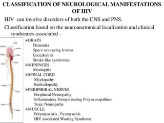
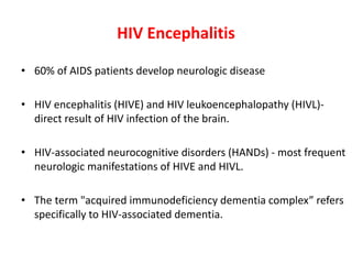
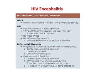
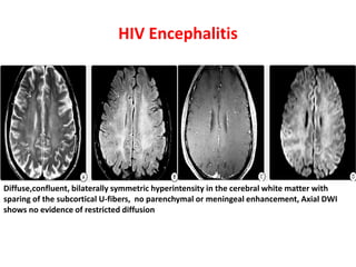
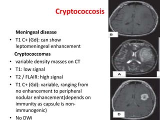
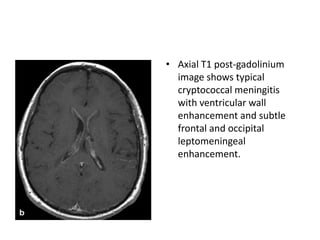
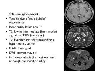
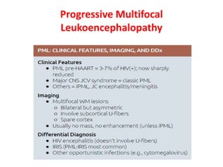
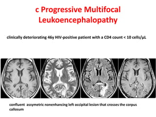
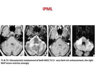
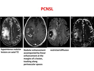
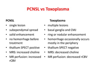
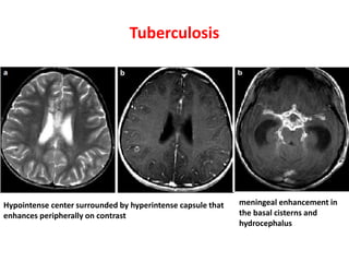
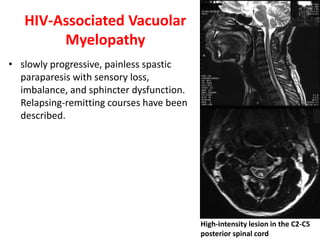
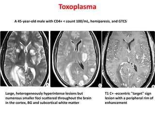
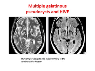
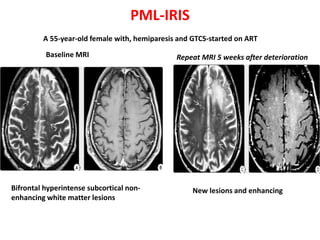
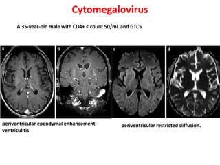
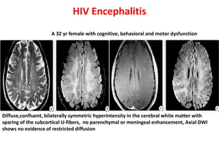
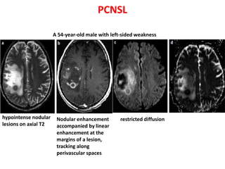
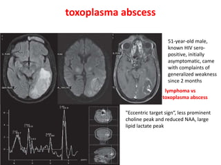
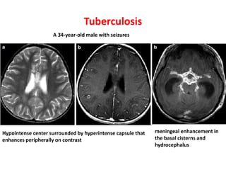
Radiology of Neurological
Manifestations of HIV
Dr. Rahi kiran.B
SR Neurology
GMC, KOTA
HIV Encephalitis
• 60% of AIDS patients develop neurologic disease
• HIV encephalitis (HIVE) and HIV leukoencephalopathy (HIVL)-
direct result of HIV infection of the brain.
• HIV-associated neurocognitive disorders (HANDs) - most frequent
neurologic manifestations of HIVE and HIVL.
• The term "acquired immunodeficiency dementia complex” refers
specifically to HIV-associated dementia.
HIV Encephalitis
HIV Encephalitis
Diffuse,confluent, bilaterally symmetric hyperintensity in the cerebral white matter with
sparing of the subcortical U-fibers, no parenchymal or meningeal enhancement, Axial DWI
shows no evidence of restricted diffusion
Toxoplasma
• most commonly involves the basal ganglia, thalami, CMJ and
cerebellum
• Multifocal lesions > solitary
• average SIZE 2-3 cm in diameter.
Toxoplasma
NECT – hypodense in left BG and frontal lobe with marked peripheral edema.T2WI- three
separate heterogeneous masses surrounded by marked edema, T1 C+ -eccentric "target" sign
lesion with a peripheral rim of enhancement
Cryptococcosis
Meningeal disease
• T1 C+ (Gd): can show
leptomeningeal enhancement
Cryptococcomas
• variable density masses on CT
• T1: low signal
• T2 / FLAIR: high signal
• T1 C+ (Gd): variable, ranging from
no enhancement to peripheral
nodular enhancement(depends on
immunity as capsule is non-
immunogenic)
• No DWI
• Immunocompetent - more likely to present with
cryptococcomas.
• Enhancement of these lesions might occur as a result of an
immunologic reaction by the host.
• Immediate and delayed imaging with a double dose of
contrast has been reported to reduce the false negative
studies by showing meningeal enhancement in
immunocompromised patients.
• Axial T1 post-gadolinium
image shows typical
cryptococcal meningitis
with ventricular wall
enhancement and subtle
frontal and occipital
leptomeningeal
enhancement.
Gelatinous pseudocysts
• Tend to give a "soap bubble"
appearance.
• low-density lesions on CT
• T1: low to intermediate (from mucin)
signal , no T1C+ (avascular)
• T2: hypointense ring surrounding a
hyperintense center
• FLAIR: low signal
• DWI - may or may not
• Hydrocephalus is the most common,
although nonspecific finding.
Progressive Multifocal
Leukoencephalopathy
c Progressive Multifocal
Leukoencephalopathy
clinically deteriorating 46y HIV-positive patient with a CD4 count < 10 cells/μL
confluent assymetric nonenhancing left occipital lesion that crosses the corpus
callosum
iPML
T1 & T2 -Characteristic involvement of both MCP, T1 C+ very faint rim enhancement, the right
MCP lesion restricts strongly
Cytomegalovirus
periventricular ependymal enhancement-
ventriculitis
periventricular restricted diffusion.
PCNSL
• 2-6% of HIV patients.
• 70% of all solitary brain parenchymal lesions in HIV/AIDS
patients.
• 90% are supratentorial - BG and deep white matter
• cross the corpus callosum
• highly specific - Linear enhancement at the margins of a
lesion, tracking along Virchow-Robin spaces
PCNSL
hypointense nodular
lesions on axial T2
Nodular enhancement
accompanied by linear
enhancement at the
margins of a lesion,
tracking along
perivascular spaces
restricted diffusion
PCNSL vs Toxoplasma
PCNSL
• single lesion
• subependymal spread
• solid enhancement
• no hemorrhage before
treatment
• thallium SPECT positive
• MRS: increased choline
• MR perfusion: increased
rCBV
Toxoplasma
• multiple lesions
• basal ganglia and CMJ
• ring or nodular enhancement
• hemorrhage occasionally occurs
mostly in the periphery
• thallium SPECT negative
• MRS: decreased choline
• MR perfusion: decreased rCBV
Tuberculosis
Hypointense center surrounded by hyperintense capsule that
enhances peripherally on contrast
meningeal enhancement in
the basal cisterns and
hydrocephalus
HIV-Associated Vacuolar
Myelopathy
• slowly progressive, painless spastic
paraparesis with sensory loss,
imbalance, and sphincter dysfunction.
Relapsing-remitting courses have been
described.
High-intensity lesion in the C2-C5
posterior spinal cord
Toxoplasma
T1 C+ -eccentric "target" sign
lesion with a peripheral rim of
enhancement
Large, heterogeneously hyperintense lesions but
numerous smaller foci scattered throughout the brain
in the cortex, BG and subcortical white matter
A 45-year-old male with CD4+ < count 100/mL, hemiparesis, and GTCS
Multiple gelatinous
pseudocysts and HIVE
Multiple pseudocysts and hyperintensity in the
cerebral white matter
PML-IRIS
Baseline MRI Repeat MRI 5 weeks after deterioration
Bifrontal hyperintense subcortical non-
enhancing white matter lesions
New lesions and enhancing
A 55-year-old female with, hemiparesis and GTCS-started on ART
Cytomegalovirus
periventricular ependymal enhancement-
ventriculitis
periventricular restricted diffusion.
A 35-year-old male with CD4+ < count 50/mL and GTCS
HIV Encephalitis
Diffuse,confluent, bilaterally symmetric hyperintensity in the cerebral white matter with
sparing of the subcortical U-fibers, no parenchymal or meningeal enhancement, Axial DWI
shows no evidence of restricted diffusion
A 32 yr female with cognitive, behavioral and motor dysfunction
PCNSL
hypointense nodular
lesions on axial T2
Nodular enhancement
accompanied by linear
enhancement at the
margins of a lesion,
tracking along
perivascular spaces
restricted diffusion
A 54-year-old male with left-sided weakness
toxoplasma abscess
"Eccentric target sign“, less prominent
choline peak and reduced NAA, large
lipid lactate peak
51-year-old male,
known HIV sero-
positive, initially
asymptomatic, came
with complaints of
generalized weakness
since 2 months
lymphoma vs
toxoplasma abscess
Tuberculosis
Hypointense center surrounded by hyperintense capsule that
enhances peripherally on contrast
meningeal enhancement in
the basal cisterns and
hydrocephalus
A 34-year-old male with seizures
THANK YOU

Imaging hiv

  • 1.
    Radiology of Neurological Manifestationsof HIV Dr. Rahi kiran.B SR Neurology GMC, KOTA
  • 3.
    HIV Encephalitis • 60%of AIDS patients develop neurologic disease • HIV encephalitis (HIVE) and HIV leukoencephalopathy (HIVL)- direct result of HIV infection of the brain. • HIV-associated neurocognitive disorders (HANDs) - most frequent neurologic manifestations of HIVE and HIVL. • The term "acquired immunodeficiency dementia complex” refers specifically to HIV-associated dementia.
  • 4.
  • 5.
    HIV Encephalitis Diffuse,confluent, bilaterallysymmetric hyperintensity in the cerebral white matter with sparing of the subcortical U-fibers, no parenchymal or meningeal enhancement, Axial DWI shows no evidence of restricted diffusion
  • 6.
    Toxoplasma • most commonlyinvolves the basal ganglia, thalami, CMJ and cerebellum • Multifocal lesions > solitary • average SIZE 2-3 cm in diameter.
  • 7.
    Toxoplasma NECT – hypodensein left BG and frontal lobe with marked peripheral edema.T2WI- three separate heterogeneous masses surrounded by marked edema, T1 C+ -eccentric "target" sign lesion with a peripheral rim of enhancement
  • 8.
    Cryptococcosis Meningeal disease • T1C+ (Gd): can show leptomeningeal enhancement Cryptococcomas • variable density masses on CT • T1: low signal • T2 / FLAIR: high signal • T1 C+ (Gd): variable, ranging from no enhancement to peripheral nodular enhancement(depends on immunity as capsule is non- immunogenic) • No DWI
  • 9.
    • Immunocompetent -more likely to present with cryptococcomas. • Enhancement of these lesions might occur as a result of an immunologic reaction by the host. • Immediate and delayed imaging with a double dose of contrast has been reported to reduce the false negative studies by showing meningeal enhancement in immunocompromised patients.
  • 10.
    • Axial T1post-gadolinium image shows typical cryptococcal meningitis with ventricular wall enhancement and subtle frontal and occipital leptomeningeal enhancement.
  • 11.
    Gelatinous pseudocysts • Tendto give a "soap bubble" appearance. • low-density lesions on CT • T1: low to intermediate (from mucin) signal , no T1C+ (avascular) • T2: hypointense ring surrounding a hyperintense center • FLAIR: low signal • DWI - may or may not • Hydrocephalus is the most common, although nonspecific finding.
  • 12.
  • 13.
    c Progressive Multifocal Leukoencephalopathy clinicallydeteriorating 46y HIV-positive patient with a CD4 count < 10 cells/μL confluent assymetric nonenhancing left occipital lesion that crosses the corpus callosum
  • 14.
    iPML T1 & T2-Characteristic involvement of both MCP, T1 C+ very faint rim enhancement, the right MCP lesion restricts strongly
  • 15.
  • 16.
    PCNSL • 2-6% ofHIV patients. • 70% of all solitary brain parenchymal lesions in HIV/AIDS patients. • 90% are supratentorial - BG and deep white matter • cross the corpus callosum • highly specific - Linear enhancement at the margins of a lesion, tracking along Virchow-Robin spaces
  • 17.
    PCNSL hypointense nodular lesions onaxial T2 Nodular enhancement accompanied by linear enhancement at the margins of a lesion, tracking along perivascular spaces restricted diffusion
  • 18.
    PCNSL vs Toxoplasma PCNSL •single lesion • subependymal spread • solid enhancement • no hemorrhage before treatment • thallium SPECT positive • MRS: increased choline • MR perfusion: increased rCBV Toxoplasma • multiple lesions • basal ganglia and CMJ • ring or nodular enhancement • hemorrhage occasionally occurs mostly in the periphery • thallium SPECT negative • MRS: decreased choline • MR perfusion: decreased rCBV
  • 19.
    Tuberculosis Hypointense center surroundedby hyperintense capsule that enhances peripherally on contrast meningeal enhancement in the basal cisterns and hydrocephalus
  • 20.
    HIV-Associated Vacuolar Myelopathy • slowlyprogressive, painless spastic paraparesis with sensory loss, imbalance, and sphincter dysfunction. Relapsing-remitting courses have been described. High-intensity lesion in the C2-C5 posterior spinal cord
  • 22.
    Toxoplasma T1 C+ -eccentric"target" sign lesion with a peripheral rim of enhancement Large, heterogeneously hyperintense lesions but numerous smaller foci scattered throughout the brain in the cortex, BG and subcortical white matter A 45-year-old male with CD4+ < count 100/mL, hemiparesis, and GTCS
  • 23.
    Multiple gelatinous pseudocysts andHIVE Multiple pseudocysts and hyperintensity in the cerebral white matter
  • 24.
    PML-IRIS Baseline MRI RepeatMRI 5 weeks after deterioration Bifrontal hyperintense subcortical non- enhancing white matter lesions New lesions and enhancing A 55-year-old female with, hemiparesis and GTCS-started on ART
  • 25.
    Cytomegalovirus periventricular ependymal enhancement- ventriculitis periventricularrestricted diffusion. A 35-year-old male with CD4+ < count 50/mL and GTCS
  • 26.
    HIV Encephalitis Diffuse,confluent, bilaterallysymmetric hyperintensity in the cerebral white matter with sparing of the subcortical U-fibers, no parenchymal or meningeal enhancement, Axial DWI shows no evidence of restricted diffusion A 32 yr female with cognitive, behavioral and motor dysfunction
  • 27.
    PCNSL hypointense nodular lesions onaxial T2 Nodular enhancement accompanied by linear enhancement at the margins of a lesion, tracking along perivascular spaces restricted diffusion A 54-year-old male with left-sided weakness
  • 28.
    toxoplasma abscess "Eccentric targetsign“, less prominent choline peak and reduced NAA, large lipid lactate peak 51-year-old male, known HIV sero- positive, initially asymptomatic, came with complaints of generalized weakness since 2 months lymphoma vs toxoplasma abscess
  • 29.
    Tuberculosis Hypointense center surroundedby hyperintense capsule that enhances peripherally on contrast meningeal enhancement in the basal cisterns and hydrocephalus A 34-year-old male with seizures
  • 30.