Digital Images
• Digitalimages are images that have been converted into discrete
numerical values for transmission or processing
• They are usually described in terms of the number of values
displayed given number of rows and columns known as a matrix.
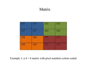
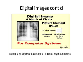
• A matrix is a square series of boxes and it gives rise to the image.
Each box in the matrix is known as a pixel.
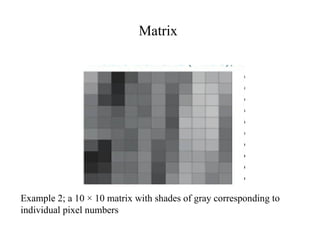
• The pixel displays a numerical value which is transformed into
visual brightness or optical density level.
• The matrix is usually expressed in terms of the number of pixels in
two orthogonal directions eg 512 × 512, 256 ×256 etc.
Image Processing
• Digitalimage processing involves analyzing the characteristics of
image signals or modifying an image in some way to enhance or
remove certain features.
• Applications of image processing in medicine are partly to
surmount the challenges posed by the limitations of the human
visual system.
• Image processing tools also help to achieve objective
measurements of what people only estimate subjectively and
visualize things not perceivable or visible
7.
Preprocessing
• Low level(pre) image processing refers to those manual or
automatic techniques that can be designed without prior
knowledge of the specific content of an image.
• Preprocessing operations apply appropriate corrections to the
raw data.
8.
Pre-processing
Fourier transformation
• Thisis the primary mathematical method used in the creation of
computerized images and involves the conversion of data into more
useful forms
• It is used to mathematically add several data representing image
intensities at different locations coming from the image receptor
Convolution
• This is the process of modifying pixel values by mathematical
formula
• Modifying the pixel values can help enhance or suppress a visual
characteristic of the image.
9.
Determinants of Qualityin Digital Images
• Frequency – this applies to the raw data from the receptor and refers
to the quantity of signal to be subjected to fourier transform. It
determines the density of the image hence its contrast
• Contrast – this refers to the differences between the data values and
is majorly determined by the subject contrast (inherent density or
thickness differences in object imaged). The smallest exposure
change that can be detected indicates the contrast of an imaging
system. In digital systems unlike conventional film screen systems, a
direct relationship exists between subject and image contrast ie
almost all tissue or subject differences are represented on the image.
10.
Determinants of Qualityin Digital Images
• Resolution - Spatial resolution is a measure of the ability of an image to
show fine detail. In digital images it is greatly influenced by the matrix size.
The greater the matrix size the finer the resolution hence procedures that
require fine resolution like mammography images are produced using matrix
size of as large as 4000 × 4000, while procedures like nuclear medicine
where the interest is only gross distribution of emissions, the resolution used
is 64 × 64.
• Signal to noise ratio (SNR) – the SNR quantifies in terms of a quotient the
amount of signal to noise in an image. Signal refers to the important
information that will be used to form the image while noise is the random
background information that does not contribute rather limits the amount of
image that can be seen and could arise due to electrical components of the
imaging system. A high SNR indicates less noise hence better image quality
11.
Image postprocessing
• Imagepostprocessing simply refers to processing of
image using a digital computer.
• It mainly aims to alter an image to enhance diagnostic
interpretation by transforming input images into an output
that suits the viewing needs of the observer in making a
diagnosis.
• Postprocessing is also important in compensating for
acquired images that lack sufficient contrast or density
12.
Postprocessing
• Image restoration:this attempts to improve the quality of the
images by reversing or correcting for degradations occurring
in the imaging system using algorithms like inverse filtering
etc. These degradations can arise from distortions, motion,
significant noise etc
• Image enhancement: this is used to make images look better to
the observer or prepare it for further processing. It involves
operations like windowing which is used to adjust the level of
brightness or density of an image, image smoothening, edge
enhancements etc
• Image analysis: this is used to estimate, detect or make
inference from images. It allows for measurements and
statistics to be performed as well as image segmentation,
shape or density classifications which are used in pattern
recognition in computed aided detection (CAD)
13.
Postprocessing
• Image synthesis:this involves the bringing or fusing together
images (projections) or non imaging information in order to create a
new one . Examples of image synthesis operations include
reconstructions from axially acquired images to multiple planes
(sagittal, coronal) in CT, 3-D reconstruction etc
• Image compression – this is generally carried out to reduce the size
of images, their transmission time as well as the the amount of space
they will require for archiving (memory or internet). There are two
(2) forms; the lossy and lossless compressions. Lossy compression
is capable of reducing an image to 1:100 of its original size and as
the name implies,there is a loss of some information,. Thus images
that have gone through lossy compression are no longer useful for
diagnosis due to loss of details. While the lossless compression can
resize an image to about 1:10 of its original size and is not
accompanied by much loss of important information, hence can still
be used for diagnosis.