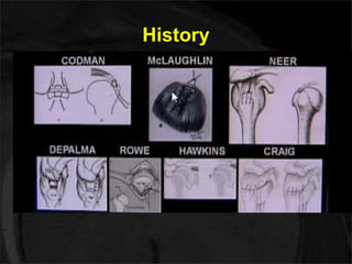
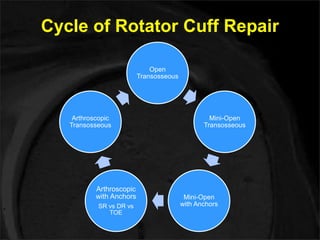
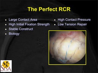
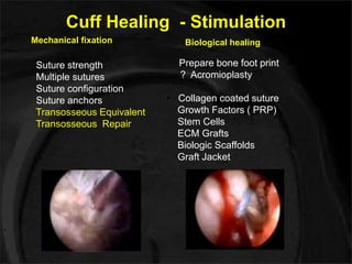
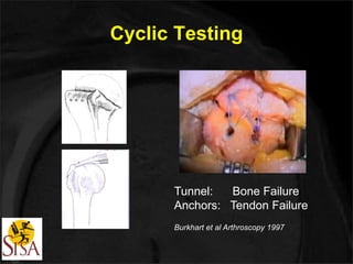
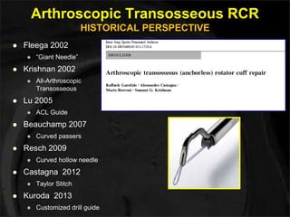
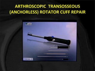
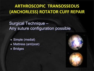
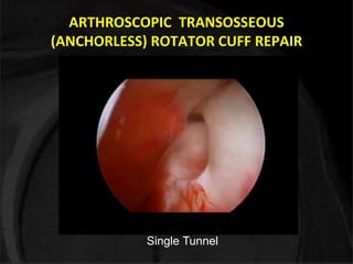
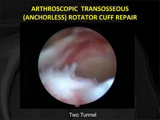
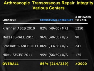
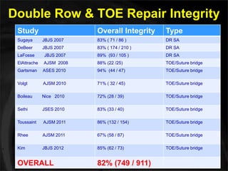
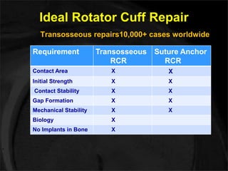
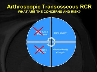
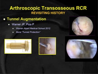
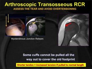
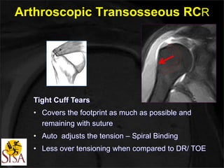
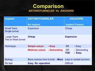
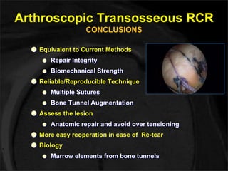
IAS 2014 
ARTHROSCOPIC TRANSOSSEOUS 
(No implant) ROTATOR CUFF REPAIR 
Dr. Raghuveer Reddy. K 
Sai Institute of Sports Injury & Arthroscopy 
Shoulder & Knee Specialty Centre, Hyderabad
I am Thankful to Dr. Sumant G. Krishnan 
for providing with Biomechanical work 
& Clinical outcome statistics of his 
study done in U.S.
History
Cycle of Rotator Cuff Repair 
Open 
Transosseous 
Mini-Open 
Transosseous 
Mini-Open 
with Anchors 
Arthroscopic 
Transosseous 
Arthroscopic 
with Anchors 
SR vs DR vs 
TOE
The Perfect RCR 
 Large Contact Area 
 High Initial Fixation Strength 
 Stable Construct 
 Biology 
 High Contact Pressure 
 Low Tension Repair
Cuff Healing - Stimulation 
Mechanical fixation Biological healing 
Suture strength 
Multiple sutures 
Suture configuration 
Suture anchors 
Transosseous Equivalent 
Transosseous Repair 
Prepare bone foot print 
? Acromioplasty?? 
• Collagen coated suture 
Growth Factors ( PRP) 
Stem Cells 
ECM Grafts 
Biologic Scaffolds 
Graft Jacket
Cyclic Testing 
Tunnel: Bone Failure 
Anchors: Tendon Failure 
Burkhart et al Arthroscopy 1997
Design Arthrotunneller
Arthroscopic Transosseous RCR 
HISTORICAL PERSPECTIVE 
 Fleega 2002 
 “Giant Needle” 
 Krishnan 2002 
 All-Arthroscopic 
Transosseous 
 Lu 2005 
 ACL Guide 
 Beauchamp 2007 
 Curved passers 
 Resch 2009 
 Curved hollow needle 
 Castagna 2012 
 Taylor Stitch 
 Kuroda 2013 
 Customized drill guide
ATRCR 
The Surgical Technique
ARTHROSCOPIC TRANSOSSEOUS 
(ANCHORLESS) ROTATOR CUFF REPAIR
ARTHROSCOPIC TRANSOSSEOUS 
(ANCHORLESS) ROTATOR CUFF REPAIR 
Surgical Technique – 
Any suture configuration possible 
 Simple (medial) 
 Mattress (ant/post) 
 Bridges
ARTHROSCOPIC TRANSOSSEOUS 
(ANCHORLESS) ROTATOR CUFF REPAIR 
Single Tunnel
ARTHROSCOPIC TRANSOSSEOUS 
(ANCHORLESS) ROTATOR CUFF REPAIR 
Two Tunnel
ATRCR 
The Science 
BIOMECHANICAL EVALUATION
Ideal Rotator Cuff Repair 
● High initial fixation strength 
● Minimal gap formation 
● Mechanical stability till tendon bone healing 
Gerber JBJS (Br) 1994 
Sugaya JBJS 2007
Arthroscopic Transosseous RCR 
REVISITING HISTORY 
● Burkhart et al. - Arthroscopy, 2000 
● Barber et al. - Arthroscopy, 2010 
● Jost et al. - JBJS, 2012 
“Increasing the number of sutures crossing the repair 
site increases the load to failure and decreases gap 
formation under cyclic loading”
ATRCR 
The Outcomes of 
Our Prospective Clinical Study & 
Sumant’s Randomized Study
My Experience 
PROSPECTIVE CLINICAL STUDY 
Material 2013 - 14 20 cases
Our Prospective Clinical Study 
 Primary 18, Revision 2 
 Posterior superior tears 14, Superior 6 tears 
 Single tunnel 11Pts. Simple Suture 
Two tunnel 9Pts. Mattress Suture 
 10 Cases evaluated. 6 - 12 months follow up 
 Functional evaluation (VAS, ASES) 
One pt. Had ASES < 70 
 MRI Evaluation – Sugaya criteria for cuff healing 
5 pts. Type I Three pts , Type II Two pts
MRI evaluation using Sugaya 
Criteria for Cuff Healing Arthroscopy 2005 
Type I: Sufficient thickness with homogeneously low intensity 
Type II: Sufficient thickness with partial high intensity 
Type III: Insufficient thickness without discontinuity 
Type IV: Presence of a minor discontinuity 
Type V: Presence of a major discontinuity
Case 1 Post op MRI 
Type I: Sufficient thickness with homogeneously low intensity 
Type II: Sufficient thickness with partial high intensity 
Type III: Insufficient thickness without discontinuity 
Type IV: Presence of a minor discontinuity 
Type V: Presence of a major discontinuity
Case 2 Post op MRI 
Type I: Sufficient thickness with homogeneously low intensity 
Type II: Sufficient thickness with partial high intensity 
Type III: Insufficient thickness without discontinuity 
Type IV: Presence of a minor discontinuity 
Type V: Presence of a major discontinuity
Case 3 Post op MRI 
Type I: Sufficient thickness with homogeneously low intensity 
Type II: Sufficient thickness with partial high intensity 
Type III: Insufficient thickness without discontinuity 
Type IV: Presence of a minor discontinuity 
Type V: Presence of a major discontinuity
Prospective Randomized Study - Sumant 
INCLUSION CRITERIA 
Posterosuperior rotator cuff tear amenable 
to GT footprint repair without tension (L , 
Crescent, reverse L) 
• No subscapularis tendon involvement 
• Grade I, II, III (Goutallier) FI 
• One single surgeon 
• Prospective Randomized allocation 
• MRI evaluation at 1 year postop from 3 
independent radiologists using Sugaya 
Criteria for cuff healing
TECHNIQUE AT SUTURE ANCHORS 
N cases 28 24 
Retear (NH) 4 (14%) 4 (16%) 
Grade I 10 (38%) 6 (26%) 
Grade II 13 (48%) 8 (34%) 
Grade III (PT) 1 (3%) 6 (26%) 
OVERALL 86% 84%
Type I healing Type III healing 
Type I: Sufficient thickness with homogeneously low intensity 
Type II: Sufficient thickness with partial high intensity 
Type III: Insufficient thickness without discontinuity 
Type IV: Presence of a minor discontinuity 
Type V: Presence of a major discontinuity
Arthroscopic Transosseous Repair Integrity 
Various Centers 
LOCATION STRUCTURAL INTEGRITY 
# OF CASES 
TO DATE 
Krishnan ASES 2010 82% (49/60) MRI 1350 
Mozes ISRAEL 2011 96% (48/50) U/S 98 
Brassart FRANCE 2011 86% (33/38) U/S 241 
Mikek SECEC 2011 95% (56/59) U/S 175 
OVERALL 86% (214/239) >2000
Double Row & TOE Repair Integrity 
Study Overall Integrity Type 
Sugaya JBJS 2007 83% ( 71 / 86 ) DR SA 
DeBeer JBJS 2007 83% ( 174 / 210 ) DR SA 
LaFosse JBJS 2007 89% (93 / 105 ) DR SA 
ElAttrache AJSM 2008 88% (22 /25) TOE/Suture bridge 
Gartsman ASES 2010 94% (44 / 47) TOE/Suture bridge 
Volgt AJSM 2010 71% ( 32 / 45) TOE/Suture bridge 
Boileau Nice 2010 72% (28 / 39) TOE/Suture bridge 
Sethi JSES 2010 83% (33 / 40) TOE/Suture bridge 
Toussaint AJSM 2011 86% (132 / 154) TOE/Suture bridge 
Rhee AJSM 2011 67% (58 / 87) TOE/Suture bridge 
Kim JBJS 2012 85% (62 / 73) TOE/Suture bridge 
OVERALL 82% (749 / 911)
Ideal Rotator Cuff Repair 
Transosseous repairs10,000+ cases worldwide 
Requirement Transosseous 
RCR 
Suture Anchor 
RCR 
Contact Area X X 
Initial Strength X X 
Contact Stability X X 
Gap Formation X X 
Mechanical Stability X X 
Biology X 
No Implants in Bone X
Arthroscopic Transosseous RCR 
WHAT ARE THE CONCERNS AND RISK? 
Bone Tunnel 
Placement 
Bone Quality 
Overtensioning 
Of repair 
Number of 
Tunnels
Arthroscopic Transosseous RCR 
REVISITING HISTORY 
 Tunnel Augmentation 
 Warner JP, Piza P 
 Warren Alpert Medical School 2012 
 Bone “Tunnel Protection” 
Courtesy: Warner JP
Arthroscopic Transosseous RCR 
ASSESS THE TEAR AND AVOID OVERTENSIONING 
Myotendinous Junction Retears 
Some cuffs cannot be pulled all the 
way out to cover the old footprint 
Shorter tendon = increased tension if pulled to normal length
Arthroscopic Transosseous RCR 
Tight Cuff Tears 
• Covers the footprint as much as possible and 
remaining with suture 
• Auto adjusts the tension – Spiral Binding 
• Less over tensioning when compared to DR/ TOE
Comparison 
ARTHROTUNNELER Vs ANCHORS 
Implant ARTHROTUNNELER 
No Implant 
ANCHORS 
Implant Present 
Small Tears 
Single tunnel 
Expensive Cheap 
Large Tears 
Two or three tunnel 
Cheap Expensive 
Technique Simple suture - Easy SR - Easy 
Mattress suture - Demanding DR - Demanding 
TOE - Easy 
Biology Bone marrow from tunnel - More Less in vented anchors 
Re tear Easy Re -operation Difficult
Arthroscopic Transosseous RCR 
CONCLUSIONS 
● Equivalent to Current Methods 
● Repair Integrity 
● Biomechanical Strength 
● Reliable/Reproducible Technique 
● Multiple Sutures 
● Bone Tunnel Augmentation 
● Assess the lesion 
● Anatomic repair and avoid over tensioning 
● More easy reoperation in case of Re-tear 
● Biology 
● Marrow elements from bone tunnels
IAS 2014 
ARTHROSCOPIC TRANSOSSEOUS 
(ANCHORLESS) ROTATOR CUFF REPAIR 
Dr. Raghuveer Reddy. K 
Sai Institute of Sports Injury & Arthroscopy 
Shoulder & Knee Specialty Centre, Hyderabad
06 Hrs
Recent Advances 
Rotator Cuff Repair 
OASIS 2014 
ARTHROSCOPIC TRANSOSSEOUS 
(ANCHORLESS) ROTATOR CUFF REPAIR 
Dr. Raghuveer Reddy. K 
Sai Institute of Sports Injury & Arthroscopy 
Shoulder & Knee Specialty Centre, Hyderabad
TOE Concerns 
Myotendinous Junction Retears 
Lill, et al. Arthroscopic Supraspinatus Tendon Repair with 
Suture Bridging Technique: Functional Outcome 
and MRI. - AJSM 2010 
Retear rate by MRI at 12 mos: 28.9% 
Cho, et al. Retear Patterns After Arthroscopic Cuff Repair: 
Single Row vs. Suture Bridge Technique. - AJSM 
2010 
27 cases of failed suture bridge technique 
74% failure at myotendinous junction 
Gerhardt et 
al. 
Arthroscopic Single-Row Modified Mason-Allen 
Repair vs. Double-Row SutureBridge 
Reconstruction for Supraspinatus Tendon Tears - 
AJSM Dec. 2012 
20 patients/5 retears 
80% retears at myotendinous junction
TOE Concerns 
Myotendinous Junction Retears 
Hayashida et al. Characteristic re-tear pattern after arthroscopic double-row 
repair. Arthroscopy, 2012 
15% retear rate at myotendinous junction 
Conclusion: 
“A new repair method, which achieves a wide 
footprint, a good initial fixation strength, and 
avoids re-tearing around the proximal suture 
anchors should be developed to obtain better 
cuff integrity and clinical results.”

Arthroscopic Transosseous(No Implant) Rotator Cuff Repair-Dr. Raghuveer Reddy .K

  • 1.
    IAS 2014 ARTHROSCOPICTRANSOSSEOUS (No implant) ROTATOR CUFF REPAIR Dr. Raghuveer Reddy. K Sai Institute of Sports Injury & Arthroscopy Shoulder & Knee Specialty Centre, Hyderabad
  • 2.
    I am Thankfulto Dr. Sumant G. Krishnan for providing with Biomechanical work & Clinical outcome statistics of his study done in U.S.
  • 3.
  • 4.
    Cycle of RotatorCuff Repair Open Transosseous Mini-Open Transosseous Mini-Open with Anchors Arthroscopic Transosseous Arthroscopic with Anchors SR vs DR vs TOE
  • 5.
    The Perfect RCR  Large Contact Area  High Initial Fixation Strength  Stable Construct  Biology  High Contact Pressure  Low Tension Repair
  • 6.
    Cuff Healing -Stimulation Mechanical fixation Biological healing Suture strength Multiple sutures Suture configuration Suture anchors Transosseous Equivalent Transosseous Repair Prepare bone foot print ? Acromioplasty?? • Collagen coated suture Growth Factors ( PRP) Stem Cells ECM Grafts Biologic Scaffolds Graft Jacket
  • 7.
    Cyclic Testing Tunnel:Bone Failure Anchors: Tendon Failure Burkhart et al Arthroscopy 1997
  • 8.
  • 9.
    Arthroscopic Transosseous RCR HISTORICAL PERSPECTIVE  Fleega 2002  “Giant Needle”  Krishnan 2002  All-Arthroscopic Transosseous  Lu 2005  ACL Guide  Beauchamp 2007  Curved passers  Resch 2009  Curved hollow needle  Castagna 2012  Taylor Stitch  Kuroda 2013  Customized drill guide
  • 10.
  • 11.
  • 13.
    ARTHROSCOPIC TRANSOSSEOUS (ANCHORLESS)ROTATOR CUFF REPAIR Surgical Technique – Any suture configuration possible  Simple (medial)  Mattress (ant/post)  Bridges
  • 14.
    ARTHROSCOPIC TRANSOSSEOUS (ANCHORLESS)ROTATOR CUFF REPAIR Single Tunnel
  • 15.
    ARTHROSCOPIC TRANSOSSEOUS (ANCHORLESS)ROTATOR CUFF REPAIR Two Tunnel
  • 16.
    ATRCR The Science BIOMECHANICAL EVALUATION
  • 17.
    Ideal Rotator CuffRepair ● High initial fixation strength ● Minimal gap formation ● Mechanical stability till tendon bone healing Gerber JBJS (Br) 1994 Sugaya JBJS 2007
  • 18.
    Arthroscopic Transosseous RCR REVISITING HISTORY ● Burkhart et al. - Arthroscopy, 2000 ● Barber et al. - Arthroscopy, 2010 ● Jost et al. - JBJS, 2012 “Increasing the number of sutures crossing the repair site increases the load to failure and decreases gap formation under cyclic loading”
  • 21.
    ATRCR The Outcomesof Our Prospective Clinical Study & Sumant’s Randomized Study
  • 22.
    My Experience PROSPECTIVECLINICAL STUDY Material 2013 - 14 20 cases
  • 23.
    Our Prospective ClinicalStudy  Primary 18, Revision 2  Posterior superior tears 14, Superior 6 tears  Single tunnel 11Pts. Simple Suture Two tunnel 9Pts. Mattress Suture  10 Cases evaluated. 6 - 12 months follow up  Functional evaluation (VAS, ASES) One pt. Had ASES < 70  MRI Evaluation – Sugaya criteria for cuff healing 5 pts. Type I Three pts , Type II Two pts
  • 24.
    MRI evaluation usingSugaya Criteria for Cuff Healing Arthroscopy 2005 Type I: Sufficient thickness with homogeneously low intensity Type II: Sufficient thickness with partial high intensity Type III: Insufficient thickness without discontinuity Type IV: Presence of a minor discontinuity Type V: Presence of a major discontinuity
  • 25.
    Case 1 Postop MRI Type I: Sufficient thickness with homogeneously low intensity Type II: Sufficient thickness with partial high intensity Type III: Insufficient thickness without discontinuity Type IV: Presence of a minor discontinuity Type V: Presence of a major discontinuity
  • 26.
    Case 2 Postop MRI Type I: Sufficient thickness with homogeneously low intensity Type II: Sufficient thickness with partial high intensity Type III: Insufficient thickness without discontinuity Type IV: Presence of a minor discontinuity Type V: Presence of a major discontinuity
  • 27.
    Case 3 Postop MRI Type I: Sufficient thickness with homogeneously low intensity Type II: Sufficient thickness with partial high intensity Type III: Insufficient thickness without discontinuity Type IV: Presence of a minor discontinuity Type V: Presence of a major discontinuity
  • 28.
    Prospective Randomized Study- Sumant INCLUSION CRITERIA Posterosuperior rotator cuff tear amenable to GT footprint repair without tension (L , Crescent, reverse L) • No subscapularis tendon involvement • Grade I, II, III (Goutallier) FI • One single surgeon • Prospective Randomized allocation • MRI evaluation at 1 year postop from 3 independent radiologists using Sugaya Criteria for cuff healing
  • 29.
    TECHNIQUE AT SUTUREANCHORS N cases 28 24 Retear (NH) 4 (14%) 4 (16%) Grade I 10 (38%) 6 (26%) Grade II 13 (48%) 8 (34%) Grade III (PT) 1 (3%) 6 (26%) OVERALL 86% 84%
  • 30.
    Type I healingType III healing Type I: Sufficient thickness with homogeneously low intensity Type II: Sufficient thickness with partial high intensity Type III: Insufficient thickness without discontinuity Type IV: Presence of a minor discontinuity Type V: Presence of a major discontinuity
  • 31.
    Arthroscopic Transosseous RepairIntegrity Various Centers LOCATION STRUCTURAL INTEGRITY # OF CASES TO DATE Krishnan ASES 2010 82% (49/60) MRI 1350 Mozes ISRAEL 2011 96% (48/50) U/S 98 Brassart FRANCE 2011 86% (33/38) U/S 241 Mikek SECEC 2011 95% (56/59) U/S 175 OVERALL 86% (214/239) >2000
  • 32.
    Double Row &TOE Repair Integrity Study Overall Integrity Type Sugaya JBJS 2007 83% ( 71 / 86 ) DR SA DeBeer JBJS 2007 83% ( 174 / 210 ) DR SA LaFosse JBJS 2007 89% (93 / 105 ) DR SA ElAttrache AJSM 2008 88% (22 /25) TOE/Suture bridge Gartsman ASES 2010 94% (44 / 47) TOE/Suture bridge Volgt AJSM 2010 71% ( 32 / 45) TOE/Suture bridge Boileau Nice 2010 72% (28 / 39) TOE/Suture bridge Sethi JSES 2010 83% (33 / 40) TOE/Suture bridge Toussaint AJSM 2011 86% (132 / 154) TOE/Suture bridge Rhee AJSM 2011 67% (58 / 87) TOE/Suture bridge Kim JBJS 2012 85% (62 / 73) TOE/Suture bridge OVERALL 82% (749 / 911)
  • 33.
    Ideal Rotator CuffRepair Transosseous repairs10,000+ cases worldwide Requirement Transosseous RCR Suture Anchor RCR Contact Area X X Initial Strength X X Contact Stability X X Gap Formation X X Mechanical Stability X X Biology X No Implants in Bone X
  • 34.
    Arthroscopic Transosseous RCR WHAT ARE THE CONCERNS AND RISK? Bone Tunnel Placement Bone Quality Overtensioning Of repair Number of Tunnels
  • 35.
    Arthroscopic Transosseous RCR REVISITING HISTORY  Tunnel Augmentation  Warner JP, Piza P  Warren Alpert Medical School 2012  Bone “Tunnel Protection” Courtesy: Warner JP
  • 36.
    Arthroscopic Transosseous RCR ASSESS THE TEAR AND AVOID OVERTENSIONING Myotendinous Junction Retears Some cuffs cannot be pulled all the way out to cover the old footprint Shorter tendon = increased tension if pulled to normal length
  • 37.
    Arthroscopic Transosseous RCR Tight Cuff Tears • Covers the footprint as much as possible and remaining with suture • Auto adjusts the tension – Spiral Binding • Less over tensioning when compared to DR/ TOE
  • 38.
    Comparison ARTHROTUNNELER VsANCHORS Implant ARTHROTUNNELER No Implant ANCHORS Implant Present Small Tears Single tunnel Expensive Cheap Large Tears Two or three tunnel Cheap Expensive Technique Simple suture - Easy SR - Easy Mattress suture - Demanding DR - Demanding TOE - Easy Biology Bone marrow from tunnel - More Less in vented anchors Re tear Easy Re -operation Difficult
  • 39.
    Arthroscopic Transosseous RCR CONCLUSIONS ● Equivalent to Current Methods ● Repair Integrity ● Biomechanical Strength ● Reliable/Reproducible Technique ● Multiple Sutures ● Bone Tunnel Augmentation ● Assess the lesion ● Anatomic repair and avoid over tensioning ● More easy reoperation in case of Re-tear ● Biology ● Marrow elements from bone tunnels
  • 41.
    IAS 2014 ARTHROSCOPICTRANSOSSEOUS (ANCHORLESS) ROTATOR CUFF REPAIR Dr. Raghuveer Reddy. K Sai Institute of Sports Injury & Arthroscopy Shoulder & Knee Specialty Centre, Hyderabad
  • 42.
  • 43.
    Recent Advances RotatorCuff Repair OASIS 2014 ARTHROSCOPIC TRANSOSSEOUS (ANCHORLESS) ROTATOR CUFF REPAIR Dr. Raghuveer Reddy. K Sai Institute of Sports Injury & Arthroscopy Shoulder & Knee Specialty Centre, Hyderabad
  • 44.
    TOE Concerns MyotendinousJunction Retears Lill, et al. Arthroscopic Supraspinatus Tendon Repair with Suture Bridging Technique: Functional Outcome and MRI. - AJSM 2010 Retear rate by MRI at 12 mos: 28.9% Cho, et al. Retear Patterns After Arthroscopic Cuff Repair: Single Row vs. Suture Bridge Technique. - AJSM 2010 27 cases of failed suture bridge technique 74% failure at myotendinous junction Gerhardt et al. Arthroscopic Single-Row Modified Mason-Allen Repair vs. Double-Row SutureBridge Reconstruction for Supraspinatus Tendon Tears - AJSM Dec. 2012 20 patients/5 retears 80% retears at myotendinous junction
  • 45.
    TOE Concerns MyotendinousJunction Retears Hayashida et al. Characteristic re-tear pattern after arthroscopic double-row repair. Arthroscopy, 2012 15% retear rate at myotendinous junction Conclusion: “A new repair method, which achieves a wide footprint, a good initial fixation strength, and avoids re-tearing around the proximal suture anchors should be developed to obtain better cuff integrity and clinical results.”