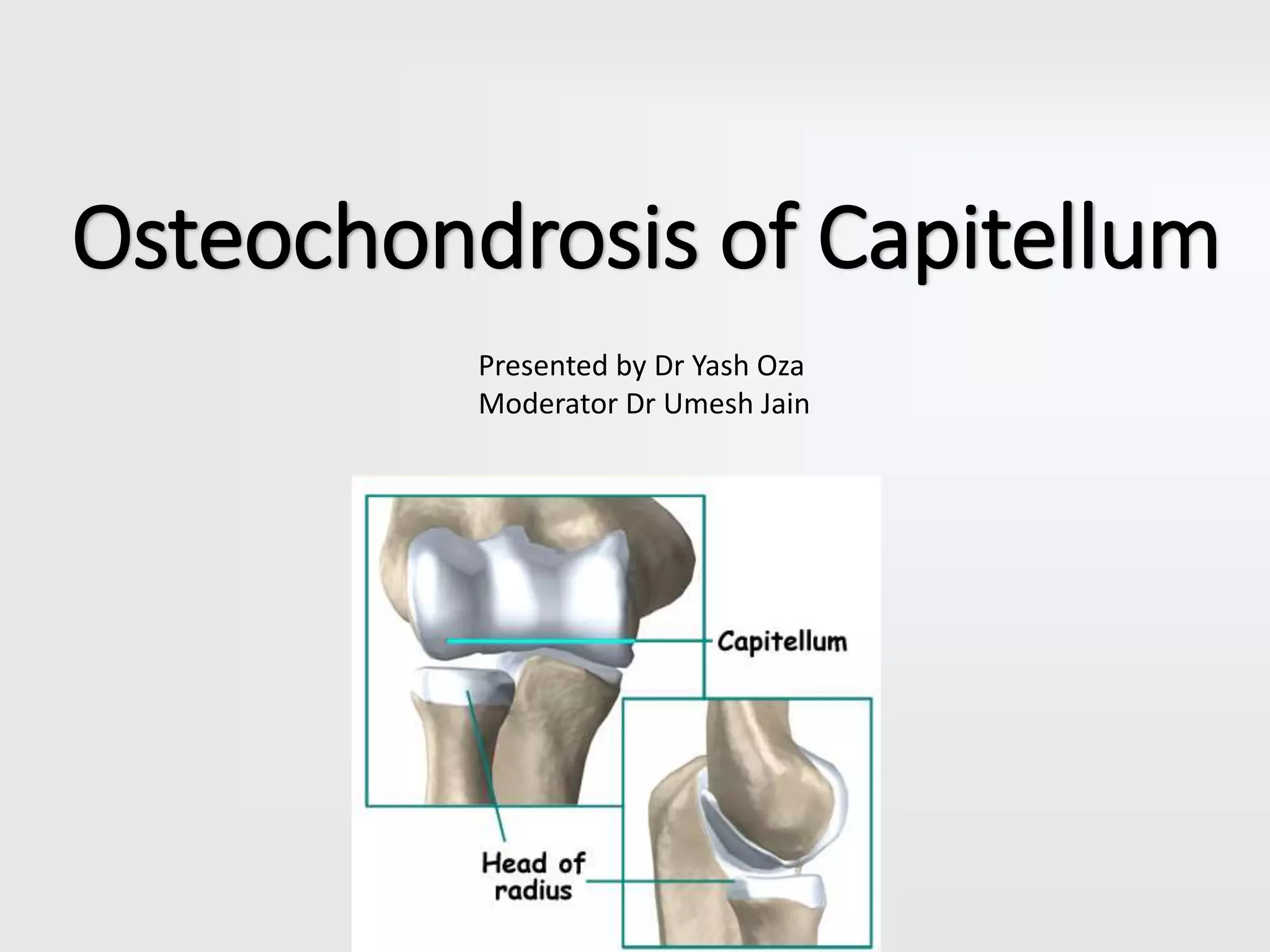
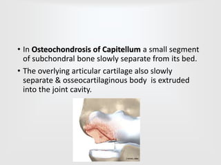
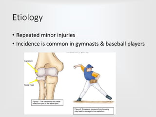
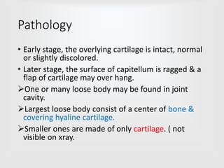
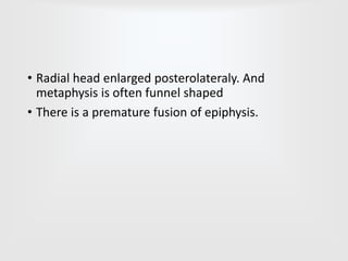
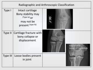
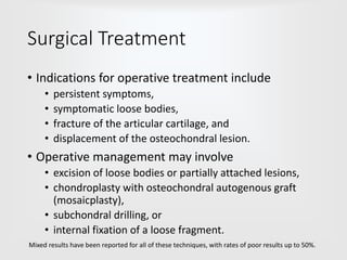
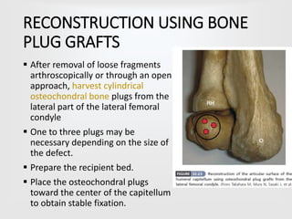
Osteochondrosis of Capitellum
Presented by Dr Yash Oza
Moderator Dr Umesh Jain
• In Osteochondrosis of Capitellum a small segment
of subchondral bone slowly separate from its bed.
• The overlying articular cartilage also slowly
separate & osseocartilaginous body is extruded
into the joint cavity.
Etiology
• Repeated minor injuries
• Incidence is common in gymnasts & baseball players
Pathology
• Early stage, the overlying cartilage is intact, normal
or slightly discolored.
• Later stage, the surface of capitellum is ragged & a
flap of cartilage may over hang.
One or many loose body may be found in joint
cavity.
Largest loose body consist of a center of bone &
covering hyaline cartilage.
Smaller ones are made of only cartilage. ( not
visible on xray.
• Radial head enlarged posterolateraly. And
metaphysis is often funnel shaped
• There is a premature fusion of epiphysis.
Clinical Presentation
• Age group : usually occurs after age 10 (typically
adolescents)
• boys more common than girls
• Right elbow more often involved, also may be
bilateral
• Insidious in onset
• First symptom include – dull ache and little stiffness
in elbow.
• History of repetitive trauma is also there.
• Pain subsides by rest.
• Locking or catching of the joint suggest the
presence of loose bodies within the joint.
• Condition may be silent and discovered accidently
by limitation of elbow extension & large radial
head.
• After a painfree period there is sudden onset of
spontaneous locking & pain followed by effusion.
Panner's disease
• It is a similar condition with slightly different
presentation
• osteochondrosis of the capitellum
• typically presents in first decade of life (<10 years old)
• usually benign self-limiting course
• same mechanism of injury as OCD
• surgery is contraindicated for Panner disease (unlike
OCD elbow)
Radiological finding
• Anteroposterior and lateral radiographs should be
obtained, and comparison views of the
contralateral elbow are helpful to identify subtle
changes in the capitellum.
Radiographic and Arthroscopic Classification
Type I Intact cartilage
Bony stability may
(Type Ia) or
may not be
present (type Ib)
Type II Cartilage fracture with
bony collapse or
displacement
Type III Loose bodies present
in joint
• MRI often identifies early changes of marrow
edema before changes are seen on plain
radiographs.
• Recent studies revealed that MRI could reliably
predict instability of an elbow lesion.
Conservative Treatment
• If a loose body is not present, nonsurgical
treatment usually is satisfactory, especially if the
lesion seems stable (type Ia).
• Resting the joint for 3 to 6 weeks with the use of a
hinged elbow brace to eliminate excessive stress
• Usually allows return to activity in 3 to 6 months.
Surgical Treatment
• Indications for operative treatment include
• persistent symptoms,
• symptomatic loose bodies,
• fracture of the articular cartilage, and
• displacement of the osteochondral lesion.
• Operative management may involve
• excision of loose bodies or partially attached lesions,
• chondroplasty with osteochondral autogenous graft
(mosaicplasty),
• subchondral drilling, or
• internal fixation of a loose fragment.
Mixed results have been reported for all of these techniques, with rates of poor results up to 50%.
OPEN APPROACH FOR FRAGMENT
REMOVAL
■ Done by posterolateral approach.
■ Make a 4 to 6 cm posterolateral oblique skin
incision on a line from the posterior edge of the
lateral epicondyle to the posterior aspect of the
radioulnar joint.
■ Incise the capsule over the capitellar lesion and
elongate the incision from the posterior edge of the
lateral epicondyle to the posterior aspect of the
radioulnar joint.
■ Perform a limited local synovectomy to remove
fragments.
• In more recent literature, ARTHROSCOPIC
techniques gives better results.
• Arthroscopic procedures include
• partial synovectomy,
• excision of loose bodies,
• drilling the crater or the intact lesion, microfracture,
• internally fixing the unstable viable fragment
(mosaicplasty) with bioabsorbable pins
• osteotomy of the capitellum.
• Although the arthroscopic results seem to be better
than nonarthroscopic techniques, no procedure,
ensures return to a throwing sport, such as
baseball. - Byrd and Jones
ARTHROSCOPIC FRAGMENT REMOVAL
■ Inject 10 to 20 mL of 1% lidocaine with epinephrine
into the elbow joint.
■ Flex the shoulder 90 degrees and elevate the elbow
until the upper arm is almost vertical.
■ Create posterior, posterolateral, anteromedial, and
anterolateral portals with a sharp blade.
■ Bluntly release the subcutaneous tissues avoiding the
cutaneous nerves.
■ Widen the portals with the use of step-up cannulas.
■ Insert an arthroscope to remove the loose bodies.
RECONSTRUCTION USING BONE
PLUG GRAFTS
 After removal of loose fragments
arthroscopically or through an open
approach, harvest cylindrical
osteochondral bone plugs from the
lateral part of the lateral femoral
condyle
 One to three plugs may be
necessary depending on the size of
the defect.
 Prepare the recipient bed.
 Place the osteochondral plugs
toward the center of the capitellum
to obtain stable fixation.
POSTOPERATIVE CARE.
• Immobilize the elbow for 1 to 2 weeks and
• Protect the knee from vigorous flexion for 3 weeks.
• 3 months after the procedure, gentle elbow
exercises against resistance are progressed to full
resistance at 4 months.
• Throwing is allowed at 4 to 5 months if there is no
pain and elbow range of motion has returned to
preoperative levels.
• The patient is released for full sports activity at 6 to
8 months.

Osteochondrosis of capitellum

  • 1.
    Osteochondrosis of Capitellum Presentedby Dr Yash Oza Moderator Dr Umesh Jain
  • 2.
    • In Osteochondrosisof Capitellum a small segment of subchondral bone slowly separate from its bed. • The overlying articular cartilage also slowly separate & osseocartilaginous body is extruded into the joint cavity.
  • 3.
    Etiology • Repeated minorinjuries • Incidence is common in gymnasts & baseball players
  • 4.
    Pathology • Early stage,the overlying cartilage is intact, normal or slightly discolored. • Later stage, the surface of capitellum is ragged & a flap of cartilage may over hang. One or many loose body may be found in joint cavity. Largest loose body consist of a center of bone & covering hyaline cartilage. Smaller ones are made of only cartilage. ( not visible on xray.
  • 5.
    • Radial headenlarged posterolateraly. And metaphysis is often funnel shaped • There is a premature fusion of epiphysis.
  • 6.
    Clinical Presentation • Agegroup : usually occurs after age 10 (typically adolescents) • boys more common than girls • Right elbow more often involved, also may be bilateral • Insidious in onset • First symptom include – dull ache and little stiffness in elbow. • History of repetitive trauma is also there. • Pain subsides by rest.
  • 7.
    • Locking orcatching of the joint suggest the presence of loose bodies within the joint. • Condition may be silent and discovered accidently by limitation of elbow extension & large radial head. • After a painfree period there is sudden onset of spontaneous locking & pain followed by effusion.
  • 8.
    Panner's disease • Itis a similar condition with slightly different presentation • osteochondrosis of the capitellum • typically presents in first decade of life (<10 years old) • usually benign self-limiting course • same mechanism of injury as OCD • surgery is contraindicated for Panner disease (unlike OCD elbow)
  • 9.
    Radiological finding • Anteroposteriorand lateral radiographs should be obtained, and comparison views of the contralateral elbow are helpful to identify subtle changes in the capitellum.
  • 10.
    Radiographic and ArthroscopicClassification Type I Intact cartilage Bony stability may (Type Ia) or may not be present (type Ib) Type II Cartilage fracture with bony collapse or displacement Type III Loose bodies present in joint
  • 11.
    • MRI oftenidentifies early changes of marrow edema before changes are seen on plain radiographs. • Recent studies revealed that MRI could reliably predict instability of an elbow lesion.
  • 12.
    Conservative Treatment • Ifa loose body is not present, nonsurgical treatment usually is satisfactory, especially if the lesion seems stable (type Ia). • Resting the joint for 3 to 6 weeks with the use of a hinged elbow brace to eliminate excessive stress • Usually allows return to activity in 3 to 6 months.
  • 13.
    Surgical Treatment • Indicationsfor operative treatment include • persistent symptoms, • symptomatic loose bodies, • fracture of the articular cartilage, and • displacement of the osteochondral lesion. • Operative management may involve • excision of loose bodies or partially attached lesions, • chondroplasty with osteochondral autogenous graft (mosaicplasty), • subchondral drilling, or • internal fixation of a loose fragment. Mixed results have been reported for all of these techniques, with rates of poor results up to 50%.
  • 14.
    OPEN APPROACH FORFRAGMENT REMOVAL ■ Done by posterolateral approach. ■ Make a 4 to 6 cm posterolateral oblique skin incision on a line from the posterior edge of the lateral epicondyle to the posterior aspect of the radioulnar joint. ■ Incise the capsule over the capitellar lesion and elongate the incision from the posterior edge of the lateral epicondyle to the posterior aspect of the radioulnar joint. ■ Perform a limited local synovectomy to remove fragments.
  • 15.
    • In morerecent literature, ARTHROSCOPIC techniques gives better results. • Arthroscopic procedures include • partial synovectomy, • excision of loose bodies, • drilling the crater or the intact lesion, microfracture, • internally fixing the unstable viable fragment (mosaicplasty) with bioabsorbable pins • osteotomy of the capitellum. • Although the arthroscopic results seem to be better than nonarthroscopic techniques, no procedure, ensures return to a throwing sport, such as baseball. - Byrd and Jones
  • 16.
    ARTHROSCOPIC FRAGMENT REMOVAL ■Inject 10 to 20 mL of 1% lidocaine with epinephrine into the elbow joint. ■ Flex the shoulder 90 degrees and elevate the elbow until the upper arm is almost vertical. ■ Create posterior, posterolateral, anteromedial, and anterolateral portals with a sharp blade. ■ Bluntly release the subcutaneous tissues avoiding the cutaneous nerves. ■ Widen the portals with the use of step-up cannulas. ■ Insert an arthroscope to remove the loose bodies.
  • 17.
    RECONSTRUCTION USING BONE PLUGGRAFTS  After removal of loose fragments arthroscopically or through an open approach, harvest cylindrical osteochondral bone plugs from the lateral part of the lateral femoral condyle  One to three plugs may be necessary depending on the size of the defect.  Prepare the recipient bed.  Place the osteochondral plugs toward the center of the capitellum to obtain stable fixation.
  • 18.
    POSTOPERATIVE CARE. • Immobilizethe elbow for 1 to 2 weeks and • Protect the knee from vigorous flexion for 3 weeks. • 3 months after the procedure, gentle elbow exercises against resistance are progressed to full resistance at 4 months. • Throwing is allowed at 4 to 5 months if there is no pain and elbow range of motion has returned to preoperative levels. • The patient is released for full sports activity at 6 to 8 months.