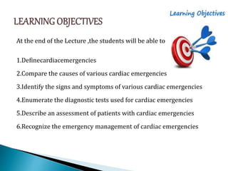
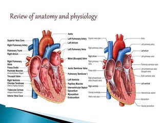
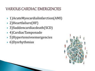
MRS.SHALINI
MSC (N),MEDICAL SURGICAL NURSING
ASSISTANT.PROFESSOR
GANGA COLLEGE OF NURSING
COIMBATORE
CARDIAC EMERGENCY
PRESENTED BY
MRS.SHALINI, MSC(NURSING),
ASST.PROFESSOR,
GANGA COLLEGE OF NURSING
At the end of the Lecture ,the students will be able to
1.Definecardiacemergencies
2.Compare the causes of various cardiac emergencies
3.Identify the signs and symptoms of various cardiac emergencies
4.Enumerate the diagnostic tests used for cardiac emergencies
5.Describe an assessment of patients with cardiac emergencies
6.Recognize the emergency management of cardiac emergencies
 Cardio vascular disease is the number one killer of
adults. Prompt recognition and initiation of
appropriate treatment can save lives during the
most deadly cardiac emergencies("Heart Disease
and Stroke Statistics",2013)
 Cardio vascular emergencies and symptoms are
one of the most common reasons for patients’
attendance in any emergency department(ED)
 1)AcuteMyocardialinfarction(AMI)
 2)Heartfailure(HF)
 3)Suddencardiacdeath(SCD)
 4)CardiacTamponade
 5)Hypertensiveemergencies
 6)Dysrhythmias
DEFINITION
•Myocardial Infarction(MI) or Heart attack
are terms used anonymously, but the preferred term
is MI
•MI is defined as cell death and necrosis
•Myocardial infarction occurs as a result of sustained
ischemia, causing a irreversible myocardial cell death
(necrosis).
•MI is usually caused by reduced or decreased blood
flow in a coronary artery due to rupture of an
atherosclerotic plaque and subsequent occlusion of
the artery by a thrombus
 Descriptions are used to further identify MI: the
type of MI (ST segment elevation myocardial
infraction STEMI and non-ST elevation MI NSTEMI
 Infarctions are usually described based on
location of damage (anterior, inferior, posterior, or
lateral wall)
 Atheroscleros is (causes luminal narrowing and
reduced blood flow)
 Eighty percent to 90% of all acute MI are
secondary to thrombus formation
 The three mechanisms that are primarily
responsible for acute reduction in O2 delivery to
the myocardium are
-Coronaryarterythrombosis
-Plaquefissureorhemorrhage
-Coronaryarteryspasm
Due to risk factor (Coronary atherosclerotic heart disease, coronary
thrombosis and embolism)
Decrease blood flow to coronary artery
Decrease myocardial oxygen supply and
ischemia
Stimulation of baroreceptor and sympathetic
receptors
Decrease myocardial contractility and
cardiac output
I Increase myocardial contractility and Heart
rate
Decrease diastolic filling and myocardial tissue
perfusion
Prolong ischemia of myocardial cell
Severe cellular damage and necrosis of cardiac
muscle
Myocardial infarction
 Chest pain
 Nausea
 Vomiting
 Diaphoresis
 History and physical examination
 12 lead ECG,
 Cardiac enzyme level
 Chest X-Ray,
 Serum cardiac markers,
 echocardiography,
 Scintigraphy
 CT angiography
ECG Changes for Acute MI
 Goal is early revascularization and reperfusion using either
fibrinolysis or primary angioplasty.
 Oxygen should be administered when blood oxygen saturation is
90% or if the patient is in respiratory distress
 Nitroglycerine:It is a coronary vasodilator and reduces
myocardial pre-load and after load
 Beta-blockers
 Calcium channel blockers(CCBs)
 ACE inhibitors
 Anti platelet therapy reduces progression
to acute infarction in patients with
non-AMIACS patients
 Anticoagulation
Reperfusion therapy
 Thrombolytics or primary Percutaneous coronary
intervention (PCI),increases the opportunity to salvage
ischemic myocardium
 Fibrinolytic therapy improves coronary flow,limits infarct
size and improves survival
- Initiated within 30minute(Door to needl etime)
- Not interventional cardiac catheterization lab
- Know the contraindication of fibrinolytic therapy
- Medication of fibrinolytic therapy
= Recombinant plasminogen activator (rPA)-
Reteplase
= Tissue plasminogen activator (tPA)-Alteplase
 Prefibrinolytic therapy care
 Post fibrinolytic therapy care
Emergency PCI (Percutaneous coronary intervention)
Percutaneous Transluminal
coronary angioplasty (PTCA)
Coronary Artery Stent
 Dysrhythmias (The most common complications
after an MI in 80% of MI cases)
 Ventricular aneurysm
 Ventricular septal defect
 Acute pulmonary edema
 Heart failure
 Cariogenic shock
 Papillary muscle dysfunction
 Pericarditis and cardiac tamponade
 Ineffective cardiac tissue perfusion related to coronary
artery occlusion
 Decrease Cardiac output related to impaired
contractility
 Acute chest pain related to decrease oxygen supply to
myocardial tissue
 Activity intolerance related to insufficient oxygenation
to perform activities of daily living
 Anxiety related to chest pain & threatening environment
 Ineffective coping related to threats to self esteem & lack
of significant support system
 Risk for injury (Bleeding) related to dissolution of
protective clots
Definition
 It is defined as the path physiologic
state in which the heart is not capable of
pumping sufficient supply of blood to meet the
body requirements or else requires elevated
ventricular filling pressures to accomplish this goal.
 The inability of the heart to pump sufficient blood to
meet the needs of the tissues for oxygen and
nutrients; signs and symptoms of pulmonary and
systemic congestion may or may not be present.
LEFT VENTRICULAR FAILURE RIGHT VENTRICULAR FAILURE
 -Tachypnea
 -Tachycardia
 -Cough,crackles
 -Hemoptysis
 -Cyanosis
 -Pulmonaryedema
 -Dyspnea orthopnea
paroxysmal nocturnal
dyspnea
 Peripheraledema
 Hepatomegaly,
splenomegaly
 Ascites,jugularvenousdiste
ntion
 -IncreasedCVP
 -Pulmonaryhypertension
 -Anorexia,weakness
 -Indigestion
 Stabilization of patient and resuscitation
 Identify the underlying and precipitating cause and
treat it,
 Control the symptoms and acute congestive state by
reducing the cardiac work load(reducing pre-and
after load),controlling excessive salt and water
retention and improving cardiac contractility
 Optimize cardio pulmonary function:-auscultate
breath sounds, administer O2, diuretics/vasodilators,
prepare for ET intubation and mechanical ventilation
and obtain daily weight
 Promote rest:-restrict activity and prescribe bed rest,
elevate the head end of the bed, monitor vital signs,
and activity intolerance.
 Pharmacologic therapy:-administer diuretics,
positive inotropes, vasodilators as ordered. Monitor
hemodynamic parameters and fluid restrictions
 Provide nutrition:-provides mall frequent meals.
Definition:
 SCD occur in patients with pre existing
ventricular dysfunction secondary to multi vessel
cardiac disease with or without history of MI
Risk factors:
 VT, VF, dilated cardiomyopathy, aortic stenosis, AV
block
Management:
 Anti dysrhythmic agents,internal cardioverter
defibrillator(ICD)
Definition
 A built-up of blood or other
fluid in the pericardial sac
puts pressure on the heart,
which may prevent it from
pumping effectively
 Pericardial layers
 •Visceral layer
 •Parietal layer
 •Fibrous pericardium
The common causes are;
 Bacterial /Mycobacterial
Fungal
 Hemopericardium
-Traumatic Iatrogenic
-Aortic Dissection
 HIV
 Uremia /Dialysis
associated
 Malignancy
 Tachycardia-Exceptions include
patients with bradycardia
during uraemia and patients
with hypothyroidism
 Tachypnea
 Jugular venous distension
 Quiet pre cordium with both
inspection and palpation
 Impure muffled heart sounds
 Rub
 Peripheral Cyanosis
 Beck Traid
Cardiac tamponade X –ray
 Oxygenation
 Increasing the volume may help only in patients with
Hypovolemic
 Bed rest and legelevation
 Inotropic drugs (Dobutamine)
 Mechanical ventilation with positive airway pressure
should be avoided in patients with tamponade,
because this further decreases cardiac output
Pericardiocentesis: The treatment of cardiac
tamponade is drainage of the pericardial contents,
preferably by needle paracentesis, with the use of
echocardiography or another type of imaging, such
as fluoroscopy or CT
In the Emergency Department hypertension presents as
one of the four varieties:
 Hypertensive emergency or crisis with acute end organ
ischemia
 Hypertensive urgency: Patients with poorly controlled
hypertension
 Mild hypertension
 Transient hypertension which is related to anxiety or
complaint
 Hypertensive crisis is characterized by arise in diastolic
BP to>120to130 mm Hg
 Only hypertension crisis requires treatment in the
emergency department within 90min of their
presentation
ETIOLOGY
 No history or non compliance to drug
 Acute renal failure
 Acute CNS events
 Drug induced hypertension
 Ingestion of tyramine containing food or beverages
during treatment with amonoamine oxidase
inhibitor (MAOI)
 Pregnancy induced eclampsia
CLINICAL MANIFESTATION
 CNS compromise
-headache, papilledema, coma
 CVScompromise
-angina ,MI
-Chestpain causes unstable angina, MI, aortic
dissection
-Acute heart failure-MI, severe HT
-Hypertension after vascular surgery-aortic
aneurysmectomy, carotid endarterectomy, CABG
DIAGNOSTIC TEST
 BP measurement in both arms, intra arterial line
monitoring
 12leadECG
MEDICAL MANAGEMENT
 Antihypertensive therapy
 Vasodilators-SNP, NTG, hydralazine
 Short acting beta blockers-betalol, esmolol
 IV ACE in hibitor
 IV Furosemide
 Hypertensive urgencies-calcium
channel blockers-nifedipine oral.
 Captoprilan ACE inhibitor,beta blocker.
Goal-To return the BP to normal
 Altered cerebral tissue perfusion related to
vasospasm or haemorrhage
 Altered myocardial tissue perfusion related to acute
myocardial ischemia
 Anxiety related to threat to biologic ,and social
integrity.
Dysrhythmias present in the emergency
department as chest pain, breathlessness,
palpitation,sweating, pre-syncope,syncope and thrombo
embolic complications
Definition:
An arrhythmiais an abnormality of rate,
regularity, or site of origin of the cardiac impulse or a
disturbance in conduction that causes an abnormal
sequence of activation.
-Irregular rhythm
-Abnormal Rate
-Conduction abnormality
Many MI patients experience
complications due to electrical dysfunction which
include bradycardia, bundle branch block, and heart
block.
Etiology of dysrhythmias in MI
 Tissue ischemia, hypoxemia
 Metabolic derangements
–acid base imbalances
 Electrolyteim balances
 Cardio myopathy
 Drugs
 Administer O2 to reduce myocardial hypoxia
 Administer bolus IV Lidocaine and infusion
 Correcting electrolyte and acid base imbalance
 History and physical examination
 ECG
 Ambulatory ECG recording: Holter recording
 Exercise ECG: tread milltest
 Trans-esophageal electro physiological study
 Invasive electrophysiological study(EPS)
DEFINITION
A run of three or more premature ventricular
contractions define VT. It is a life threatening dysrhythmia
because of decreased cardiac output and the possibility of
development of ventricular fibrillation (VF), which is a
lethal dysrhythmia
 FORMS OF VT: Monomorphic, polymorphic
 CLINICAL ASSOCIATION: VT is associated with MI, CAD,
significant electrolyte imbalances, cardiomyopathy, mitral
valve prolapse, long QT syndrome, drug toxicity, and CNS
disorders
 ECG Characteristics: ventricular rate is150 to 250 beats /minute,
rhythm may be regular or irregular. AV dissociation may be present
with p wave occurring independently of the QRS complex. The QRS
complex is distorted in appearance, with a duration >0.12 sec and with
the ST-T wave in the opposite direction of the QRS complex .The R-R
interval may be regular or irregular
 Clinical significance: VT can be stable (patient has a pulse) or
unstable (patient is pulseless). VT causes decreased cardiac out put
results in hypotension, pulmonary edema, decreased cerebral blood
flow, and cardio pulmonary arrest
Treatment:
 Identify the cause and
treat
 -If pulse is present IV
procainamide, sotalol,
amiodarone, or lidocaine
 -If Vt with out pulse-CPR
and rapid defibrillation
followed by
administration of
vasopressors and
antidysrhythmics
Definition:
VF is a severe derangement of the heart
rhythm characterized on ECG by irregular wave
forms of varying shapes and amplitude.
Clinical associations:
VF occurs in acute MI, myocardial ischemia,
and in chronic diseases such as HF and
cardiomyopathy. Other clinical associations are
accidental electrical shock, hyperkalemia, acidosis,
and drug toxicity.
ECG characteristics:
 HR not measurable, rhythm is irregular and chaotic,
the p wave is not visible, and the PR interval and the
QRS interval not measurable
Clinical significance:
VF results in an unresponsive, pulse less, and
apneic state. If not treated patient will die
Treatment:
immediate initiation of CPR and advanced
cardiac life support (ACLS) measures with the use of
defibrillation and definitive drug therapy
Definition
A systole represents the total absence of
ventricular electrical activity, occasionally p waves
are seen. Patients are unresponsive, pulse less and
apneic requires immediate treatment.
Clinical association:
Result of advanced cardiac disease, conduction
system disturbance or end stage HF
Clinical significance:
 Generally patient has a prolonged arrest and cannot be
resuscitated
Treatment:
 CPR with initiation of ACLS which includes definitive
drug therapy, intubation and possibly a transcutaneous
temporary pacemaker

Definition: no mechanical activity of the ventricles
and the patient has no pulse.The most common
cause include hypovolemia, hypoxia, metabolic
acidosis, hyper kalemia, or hypo kalemia,
hypothermia, drug over dose, cardiac tamponade,
MI,Tension pneumothorax, trauma and pulmonary
embolism. Treatment begins with CPR followed by
drug therapy (epinephrine) and intubation.
Atropine is used if the ventricular rate is slow.
Every patient diagnosed with life threatening
cardiac emergency like acute myocardial infarction,
SCD, HF, Cardiac tamponade, Dysrhythmias and
hypertensive emergencies should receive the
emergency care as early as possible including
oxygenation, ventilator support and appropriate
pharmacotherapy which would help in saving
patients life and preventing complications
 Lewis.S.I.,Hertigember.M.M.(2011).MedicalSurgical
Nursing. (6thEd). Philadelphia: Elsevier publication
 Brunner & Suddarth.S.(2012).Textbook of Medical
Surgical Nursing. Philadelphia:Lippincott Williams &
Wilkins
 Thelan et al.(2006).Critical care nursing.5th
Ed.St.Louis; Mosby
CARDIAC EMERGENCY
CARDIAC EMERGENCY

CARDIAC EMERGENCY

  • 1.
    MRS.SHALINI MSC (N),MEDICAL SURGICALNURSING ASSISTANT.PROFESSOR GANGA COLLEGE OF NURSING COIMBATORE
  • 2.
    CARDIAC EMERGENCY PRESENTED BY MRS.SHALINI,MSC(NURSING), ASST.PROFESSOR, GANGA COLLEGE OF NURSING
  • 3.
    At the endof the Lecture ,the students will be able to 1.Definecardiacemergencies 2.Compare the causes of various cardiac emergencies 3.Identify the signs and symptoms of various cardiac emergencies 4.Enumerate the diagnostic tests used for cardiac emergencies 5.Describe an assessment of patients with cardiac emergencies 6.Recognize the emergency management of cardiac emergencies
  • 4.
     Cardio vasculardisease is the number one killer of adults. Prompt recognition and initiation of appropriate treatment can save lives during the most deadly cardiac emergencies("Heart Disease and Stroke Statistics",2013)  Cardio vascular emergencies and symptoms are one of the most common reasons for patients’ attendance in any emergency department(ED)
  • 6.
     1)AcuteMyocardialinfarction(AMI)  2)Heartfailure(HF) 3)Suddencardiacdeath(SCD)  4)CardiacTamponade  5)Hypertensiveemergencies  6)Dysrhythmias
  • 7.
    DEFINITION •Myocardial Infarction(MI) orHeart attack are terms used anonymously, but the preferred term is MI •MI is defined as cell death and necrosis •Myocardial infarction occurs as a result of sustained ischemia, causing a irreversible myocardial cell death (necrosis). •MI is usually caused by reduced or decreased blood flow in a coronary artery due to rupture of an atherosclerotic plaque and subsequent occlusion of the artery by a thrombus
  • 8.
     Descriptions areused to further identify MI: the type of MI (ST segment elevation myocardial infraction STEMI and non-ST elevation MI NSTEMI  Infarctions are usually described based on location of damage (anterior, inferior, posterior, or lateral wall)
  • 9.
     Atheroscleros is(causes luminal narrowing and reduced blood flow)  Eighty percent to 90% of all acute MI are secondary to thrombus formation  The three mechanisms that are primarily responsible for acute reduction in O2 delivery to the myocardium are -Coronaryarterythrombosis -Plaquefissureorhemorrhage -Coronaryarteryspasm
  • 10.
    Due to riskfactor (Coronary atherosclerotic heart disease, coronary thrombosis and embolism) Decrease blood flow to coronary artery Decrease myocardial oxygen supply and ischemia Stimulation of baroreceptor and sympathetic receptors Decrease myocardial contractility and cardiac output
  • 11.
    I Increase myocardialcontractility and Heart rate Decrease diastolic filling and myocardial tissue perfusion Prolong ischemia of myocardial cell Severe cellular damage and necrosis of cardiac muscle Myocardial infarction
  • 12.
     Chest pain Nausea  Vomiting  Diaphoresis
  • 13.
     History andphysical examination  12 lead ECG,  Cardiac enzyme level  Chest X-Ray,  Serum cardiac markers,  echocardiography,  Scintigraphy  CT angiography ECG Changes for Acute MI
  • 14.
     Goal isearly revascularization and reperfusion using either fibrinolysis or primary angioplasty.  Oxygen should be administered when blood oxygen saturation is 90% or if the patient is in respiratory distress  Nitroglycerine:It is a coronary vasodilator and reduces myocardial pre-load and after load  Beta-blockers  Calcium channel blockers(CCBs)  ACE inhibitors  Anti platelet therapy reduces progression to acute infarction in patients with non-AMIACS patients  Anticoagulation
  • 15.
    Reperfusion therapy  Thrombolyticsor primary Percutaneous coronary intervention (PCI),increases the opportunity to salvage ischemic myocardium  Fibrinolytic therapy improves coronary flow,limits infarct size and improves survival - Initiated within 30minute(Door to needl etime) - Not interventional cardiac catheterization lab - Know the contraindication of fibrinolytic therapy - Medication of fibrinolytic therapy = Recombinant plasminogen activator (rPA)- Reteplase = Tissue plasminogen activator (tPA)-Alteplase  Prefibrinolytic therapy care  Post fibrinolytic therapy care
  • 16.
    Emergency PCI (Percutaneouscoronary intervention) Percutaneous Transluminal coronary angioplasty (PTCA) Coronary Artery Stent
  • 17.
     Dysrhythmias (Themost common complications after an MI in 80% of MI cases)  Ventricular aneurysm  Ventricular septal defect  Acute pulmonary edema  Heart failure  Cariogenic shock  Papillary muscle dysfunction  Pericarditis and cardiac tamponade
  • 18.
     Ineffective cardiactissue perfusion related to coronary artery occlusion  Decrease Cardiac output related to impaired contractility  Acute chest pain related to decrease oxygen supply to myocardial tissue  Activity intolerance related to insufficient oxygenation to perform activities of daily living  Anxiety related to chest pain & threatening environment  Ineffective coping related to threats to self esteem & lack of significant support system  Risk for injury (Bleeding) related to dissolution of protective clots
  • 19.
    Definition  It isdefined as the path physiologic state in which the heart is not capable of pumping sufficient supply of blood to meet the body requirements or else requires elevated ventricular filling pressures to accomplish this goal.  The inability of the heart to pump sufficient blood to meet the needs of the tissues for oxygen and nutrients; signs and symptoms of pulmonary and systemic congestion may or may not be present.
  • 20.
    LEFT VENTRICULAR FAILURERIGHT VENTRICULAR FAILURE  -Tachypnea  -Tachycardia  -Cough,crackles  -Hemoptysis  -Cyanosis  -Pulmonaryedema  -Dyspnea orthopnea paroxysmal nocturnal dyspnea  Peripheraledema  Hepatomegaly, splenomegaly  Ascites,jugularvenousdiste ntion  -IncreasedCVP  -Pulmonaryhypertension  -Anorexia,weakness  -Indigestion
  • 21.
     Stabilization ofpatient and resuscitation  Identify the underlying and precipitating cause and treat it,  Control the symptoms and acute congestive state by reducing the cardiac work load(reducing pre-and after load),controlling excessive salt and water retention and improving cardiac contractility
  • 22.
     Optimize cardiopulmonary function:-auscultate breath sounds, administer O2, diuretics/vasodilators, prepare for ET intubation and mechanical ventilation and obtain daily weight  Promote rest:-restrict activity and prescribe bed rest, elevate the head end of the bed, monitor vital signs, and activity intolerance.  Pharmacologic therapy:-administer diuretics, positive inotropes, vasodilators as ordered. Monitor hemodynamic parameters and fluid restrictions  Provide nutrition:-provides mall frequent meals.
  • 23.
    Definition:  SCD occurin patients with pre existing ventricular dysfunction secondary to multi vessel cardiac disease with or without history of MI Risk factors:  VT, VF, dilated cardiomyopathy, aortic stenosis, AV block Management:  Anti dysrhythmic agents,internal cardioverter defibrillator(ICD)
  • 24.
    Definition  A built-upof blood or other fluid in the pericardial sac puts pressure on the heart, which may prevent it from pumping effectively  Pericardial layers  •Visceral layer  •Parietal layer  •Fibrous pericardium
  • 25.
    The common causesare;  Bacterial /Mycobacterial Fungal  Hemopericardium -Traumatic Iatrogenic -Aortic Dissection  HIV  Uremia /Dialysis associated  Malignancy
  • 27.
     Tachycardia-Exceptions include patientswith bradycardia during uraemia and patients with hypothyroidism  Tachypnea  Jugular venous distension  Quiet pre cordium with both inspection and palpation  Impure muffled heart sounds  Rub  Peripheral Cyanosis  Beck Traid
  • 28.
  • 29.
     Oxygenation  Increasingthe volume may help only in patients with Hypovolemic  Bed rest and legelevation  Inotropic drugs (Dobutamine)  Mechanical ventilation with positive airway pressure should be avoided in patients with tamponade, because this further decreases cardiac output
  • 30.
    Pericardiocentesis: The treatmentof cardiac tamponade is drainage of the pericardial contents, preferably by needle paracentesis, with the use of echocardiography or another type of imaging, such as fluoroscopy or CT
  • 31.
    In the EmergencyDepartment hypertension presents as one of the four varieties:  Hypertensive emergency or crisis with acute end organ ischemia  Hypertensive urgency: Patients with poorly controlled hypertension  Mild hypertension  Transient hypertension which is related to anxiety or complaint  Hypertensive crisis is characterized by arise in diastolic BP to>120to130 mm Hg  Only hypertension crisis requires treatment in the emergency department within 90min of their presentation
  • 32.
    ETIOLOGY  No historyor non compliance to drug  Acute renal failure  Acute CNS events  Drug induced hypertension  Ingestion of tyramine containing food or beverages during treatment with amonoamine oxidase inhibitor (MAOI)  Pregnancy induced eclampsia
  • 33.
    CLINICAL MANIFESTATION  CNScompromise -headache, papilledema, coma  CVScompromise -angina ,MI -Chestpain causes unstable angina, MI, aortic dissection -Acute heart failure-MI, severe HT -Hypertension after vascular surgery-aortic aneurysmectomy, carotid endarterectomy, CABG
  • 34.
    DIAGNOSTIC TEST  BPmeasurement in both arms, intra arterial line monitoring  12leadECG MEDICAL MANAGEMENT  Antihypertensive therapy  Vasodilators-SNP, NTG, hydralazine  Short acting beta blockers-betalol, esmolol  IV ACE in hibitor  IV Furosemide  Hypertensive urgencies-calcium channel blockers-nifedipine oral.  Captoprilan ACE inhibitor,beta blocker.
  • 35.
    Goal-To return theBP to normal  Altered cerebral tissue perfusion related to vasospasm or haemorrhage  Altered myocardial tissue perfusion related to acute myocardial ischemia  Anxiety related to threat to biologic ,and social integrity.
  • 36.
    Dysrhythmias present inthe emergency department as chest pain, breathlessness, palpitation,sweating, pre-syncope,syncope and thrombo embolic complications Definition: An arrhythmiais an abnormality of rate, regularity, or site of origin of the cardiac impulse or a disturbance in conduction that causes an abnormal sequence of activation. -Irregular rhythm -Abnormal Rate -Conduction abnormality
  • 37.
    Many MI patientsexperience complications due to electrical dysfunction which include bradycardia, bundle branch block, and heart block. Etiology of dysrhythmias in MI  Tissue ischemia, hypoxemia  Metabolic derangements –acid base imbalances  Electrolyteim balances  Cardio myopathy  Drugs
  • 38.
     Administer O2to reduce myocardial hypoxia  Administer bolus IV Lidocaine and infusion  Correcting electrolyte and acid base imbalance
  • 39.
     History andphysical examination  ECG  Ambulatory ECG recording: Holter recording  Exercise ECG: tread milltest  Trans-esophageal electro physiological study  Invasive electrophysiological study(EPS)
  • 40.
    DEFINITION A run ofthree or more premature ventricular contractions define VT. It is a life threatening dysrhythmia because of decreased cardiac output and the possibility of development of ventricular fibrillation (VF), which is a lethal dysrhythmia  FORMS OF VT: Monomorphic, polymorphic  CLINICAL ASSOCIATION: VT is associated with MI, CAD, significant electrolyte imbalances, cardiomyopathy, mitral valve prolapse, long QT syndrome, drug toxicity, and CNS disorders
  • 41.
     ECG Characteristics:ventricular rate is150 to 250 beats /minute, rhythm may be regular or irregular. AV dissociation may be present with p wave occurring independently of the QRS complex. The QRS complex is distorted in appearance, with a duration >0.12 sec and with the ST-T wave in the opposite direction of the QRS complex .The R-R interval may be regular or irregular  Clinical significance: VT can be stable (patient has a pulse) or unstable (patient is pulseless). VT causes decreased cardiac out put results in hypotension, pulmonary edema, decreased cerebral blood flow, and cardio pulmonary arrest
  • 42.
    Treatment:  Identify thecause and treat  -If pulse is present IV procainamide, sotalol, amiodarone, or lidocaine  -If Vt with out pulse-CPR and rapid defibrillation followed by administration of vasopressors and antidysrhythmics
  • 43.
    Definition: VF is asevere derangement of the heart rhythm characterized on ECG by irregular wave forms of varying shapes and amplitude. Clinical associations: VF occurs in acute MI, myocardial ischemia, and in chronic diseases such as HF and cardiomyopathy. Other clinical associations are accidental electrical shock, hyperkalemia, acidosis, and drug toxicity.
  • 44.
    ECG characteristics:  HRnot measurable, rhythm is irregular and chaotic, the p wave is not visible, and the PR interval and the QRS interval not measurable
  • 45.
    Clinical significance: VF resultsin an unresponsive, pulse less, and apneic state. If not treated patient will die Treatment: immediate initiation of CPR and advanced cardiac life support (ACLS) measures with the use of defibrillation and definitive drug therapy
  • 46.
    Definition A systole representsthe total absence of ventricular electrical activity, occasionally p waves are seen. Patients are unresponsive, pulse less and apneic requires immediate treatment. Clinical association: Result of advanced cardiac disease, conduction system disturbance or end stage HF
  • 47.
    Clinical significance:  Generallypatient has a prolonged arrest and cannot be resuscitated Treatment:  CPR with initiation of ACLS which includes definitive drug therapy, intubation and possibly a transcutaneous temporary pacemaker 
  • 48.
    Definition: no mechanicalactivity of the ventricles and the patient has no pulse.The most common cause include hypovolemia, hypoxia, metabolic acidosis, hyper kalemia, or hypo kalemia, hypothermia, drug over dose, cardiac tamponade, MI,Tension pneumothorax, trauma and pulmonary embolism. Treatment begins with CPR followed by drug therapy (epinephrine) and intubation. Atropine is used if the ventricular rate is slow.
  • 49.
    Every patient diagnosedwith life threatening cardiac emergency like acute myocardial infarction, SCD, HF, Cardiac tamponade, Dysrhythmias and hypertensive emergencies should receive the emergency care as early as possible including oxygenation, ventilator support and appropriate pharmacotherapy which would help in saving patients life and preventing complications
  • 50.
     Lewis.S.I.,Hertigember.M.M.(2011).MedicalSurgical Nursing. (6thEd).Philadelphia: Elsevier publication  Brunner & Suddarth.S.(2012).Textbook of Medical Surgical Nursing. Philadelphia:Lippincott Williams & Wilkins  Thelan et al.(2006).Critical care nursing.5th Ed.St.Louis; Mosby