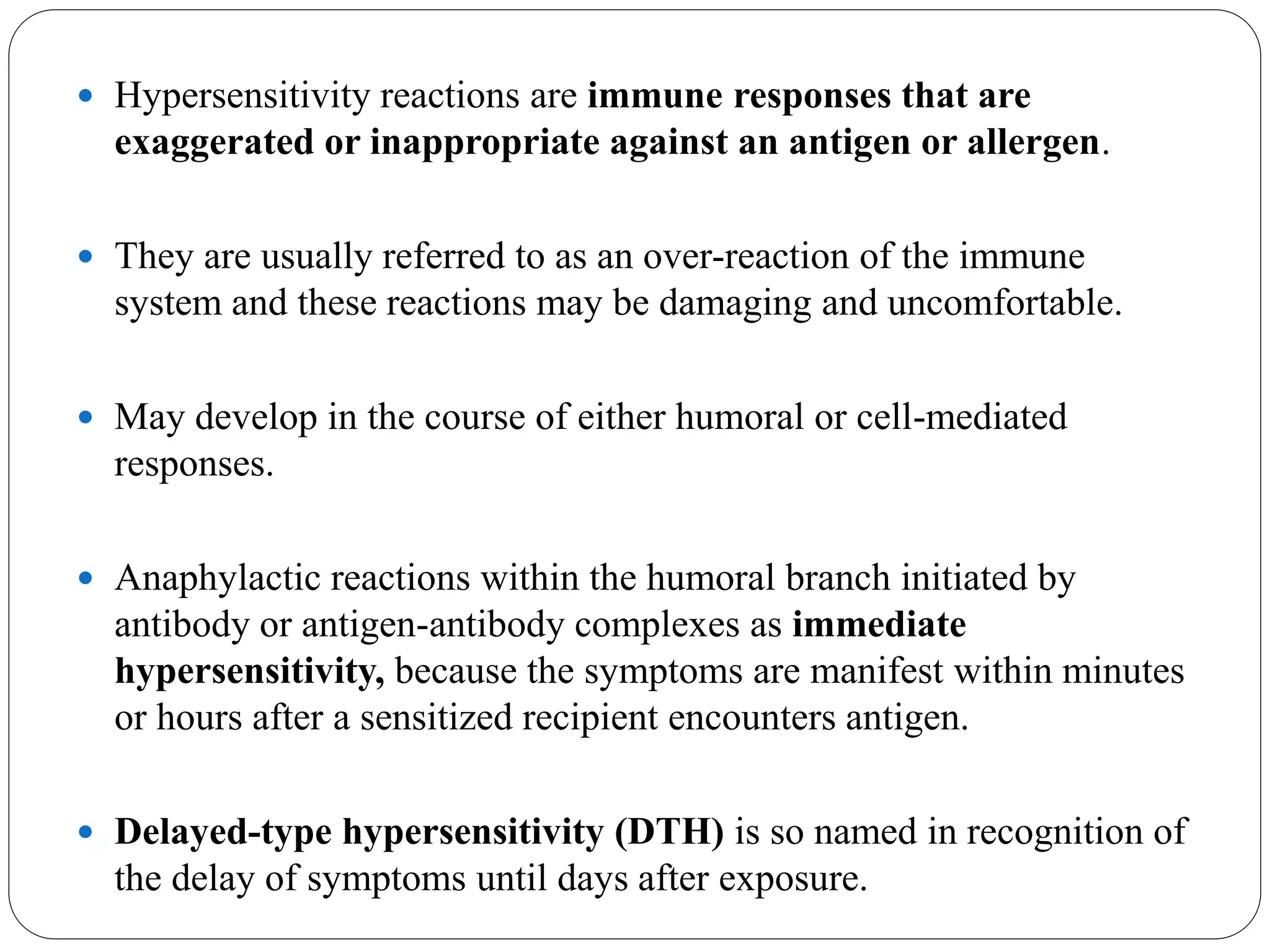
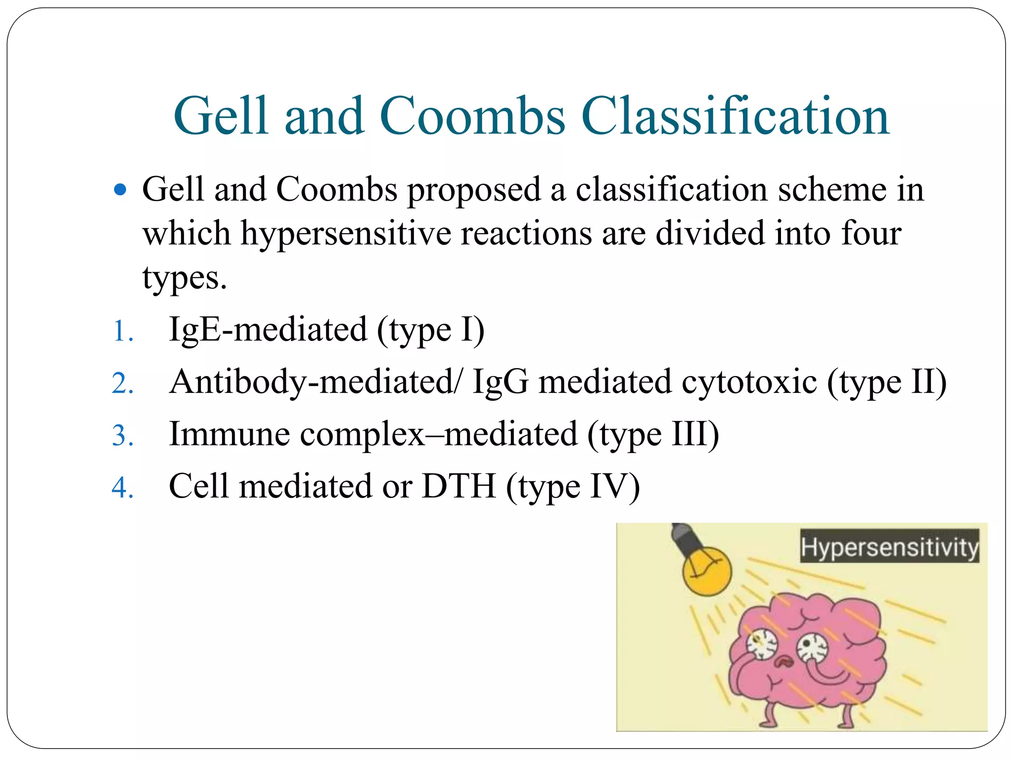
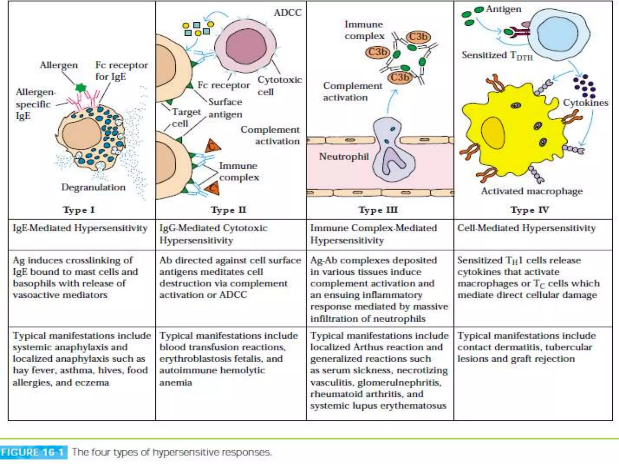
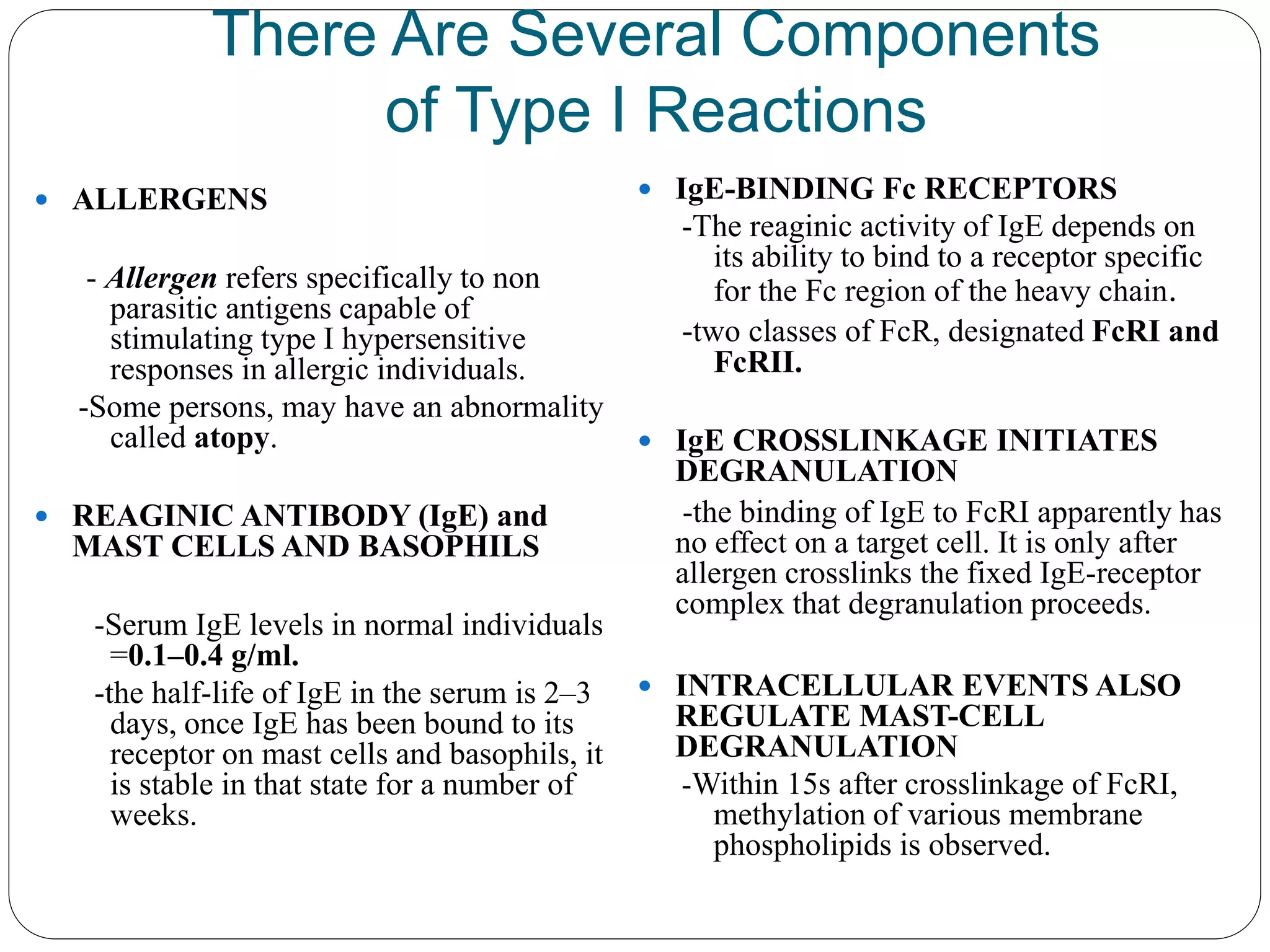
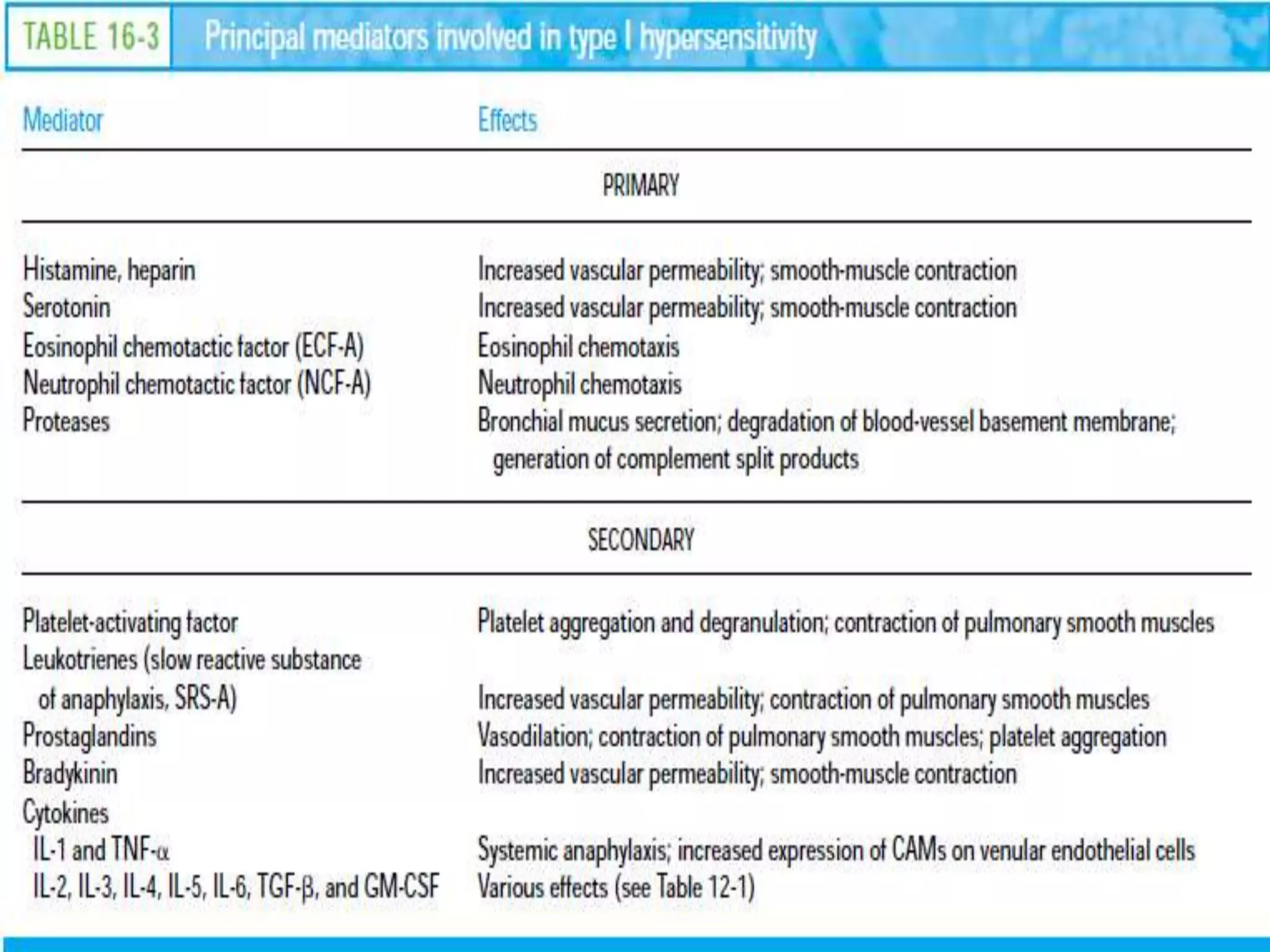
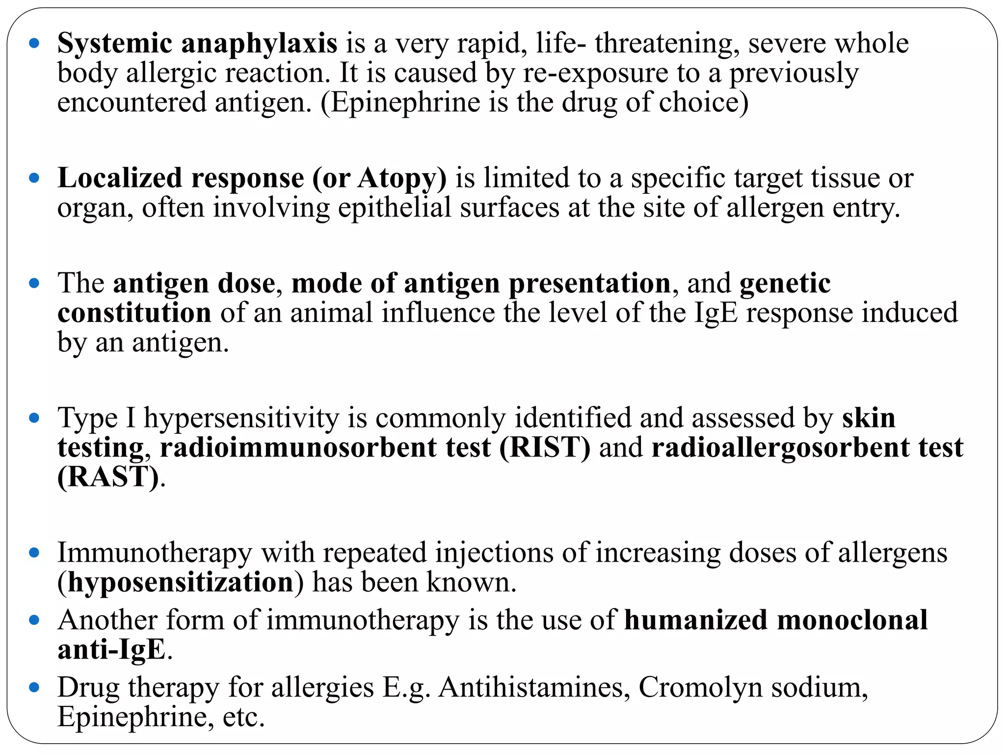
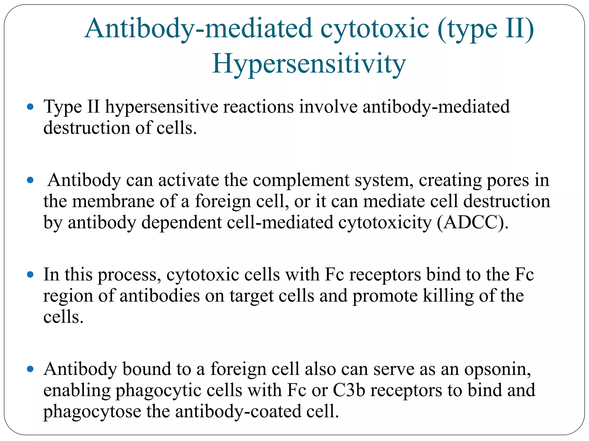
2. Hypersensitivity reactions are immune responses that are
exaggerated or inappropriate against an antigen or allergen.
They are usually referred to as an over-reaction of the immune
system and these reactions may be damaging and uncomfortable.
May develop in the course of either humoral or cell-mediated
responses.
Anaphylactic reactions within the humoral branch initiated by
antibody or antigen-antibody complexes as immediate
hypersensitivity, because the symptoms are manifest within minutes
or hours after a sensitized recipient encounters antigen.
Delayed-type hypersensitivity (DTH) is so named in recognition of
the delay of symptoms until days after exposure.
3. Gell and Coombs Classification
Gell and Coombs proposed a classification scheme in
which hypersensitive reactions are divided into four
types.
1. IgE-mediated (type I)
2. Antibody-mediated/ IgG mediated cytotoxic (type II)
3. Immune complex–mediated (type III)
4. Cell mediated or DTH (type IV)
4.
5. IgE-Mediated (Type I) Hypersensitivity
A type I hypersensitive reaction is induced by certain types of
antigens referred to as allergens.
In type I hypersensitive response, the plasma cells secrete IgE.
This class of antibody binds with high affinity to Fc receptors on
the surface of tissue mast cells and blood basophils.
A later exposure to the same allergen cross-links the membrane-
bound IgE on sensitized mast cells and basophils, causing
degranulation of these cells. (release of primary mediators,
vasoactive amines, smooth muscle contraction, etc.)
6.
7. There Are Several Components
of Type I Reactions
ALLERGENS
- Allergen refers specifically to non
parasitic antigens capable of
stimulating type I hypersensitive
responses in allergic individuals.
-Some persons, may have an abnormality
called atopy.
REAGINIC ANTIBODY (IgE) and
MAST CELLS AND BASOPHILS
-Serum IgE levels in normal individuals
=0.1–0.4 g/ml.
-the half-life of IgE in the serum is 2–3
days, once IgE has been bound to its
receptor on mast cells and basophils, it
is stable in that state for a number of
weeks.
IgE-BINDING Fc RECEPTORS
-The reaginic activity of IgE depends on
its ability to bind to a receptor specific
for the Fc region of the heavy chain.
-two classes of FcR, designated FcRI and
FcRII.
IgE CROSSLINKAGE INITIATES
DEGRANULATION
-the binding of IgE to FcRI apparently has
no effect on a target cell. It is only after
allergen crosslinks the fixed IgE-receptor
complex that degranulation proceeds.
INTRACELLULAR EVENTS ALSO
REGULATE MAST-CELL
DEGRANULATION
-Within 15s after crosslinkage of FcRI,
methylation of various membrane
phospholipids is observed.
8.
9. Systemic anaphylaxis is a very rapid, life- threatening, severe whole
body allergic reaction. It is caused by re-exposure to a previously
encountered antigen. (Epinephrine is the drug of choice)
Localized response (or Atopy) is limited to a specific target tissue or
organ, often involving epithelial surfaces at the site of allergen entry.
The antigen dose, mode of antigen presentation, and genetic
constitution of an animal influence the level of the IgE response induced
by an antigen.
Type I hypersensitivity is commonly identified and assessed by skin
testing, radioimmunosorbent test (RIST) and radioallergosorbent test
(RAST).
Immunotherapy with repeated injections of increasing doses of allergens
(hyposensitization) has been known.
Another form of immunotherapy is the use of humanized monoclonal
anti-IgE.
Drug therapy for allergies E.g. Antihistamines, Cromolyn sodium,
Epinephrine, etc.
10. Antibody-mediated cytotoxic (type II)
Hypersensitivity
Type II hypersensitive reactions involve antibody-mediated
destruction of cells.
Antibody can activate the complement system, creating pores in
the membrane of a foreign cell, or it can mediate cell destruction
by antibody dependent cell-mediated cytotoxicity (ADCC).
In this process, cytotoxic cells with Fc receptors bind to the Fc
region of antibodies on target cells and promote killing of the
cells.
Antibody bound to a foreign cell also can serve as an opsonin,
enabling phagocytic cells with Fc or C3b receptors to bind and
phagocytose the antibody-coated cell.
11. Transfusion Reactions Are Type II
Reactions
An individual possessing one allelic form of a blood-group antigen can recognize other allelic
forms on transfused blood as foreign.
In some cases, the antibodies have already been induced by natural exposure to similar
antigenic determinants. This is the case with the ABO blood-group antigens.
Antibodies to the A, B, and O antigens, called isohemagglutinins, are usually of the IgM
class.
If a type A individual is transfused with blood containing type B cells, isohemagglutinins
bind to the B blood cells and mediate their destruction by means of complement-mediated
lysis.
Typical symptoms include fever, chills, nausea, clotting within blood vessels, pain in the lower
back, and hemoglobin in the urine.
Treatment involves prompt termination of the transfusion and maintenance of urine flow with
a diuretic.
12.
13. Hemolytic Disease of the Newborn
Is Caused by Type II Reactions
Hemolytic disease of the newborn develops when maternal IgG antibodies specific
for fetal blood-group antigens cross the placenta and destroy fetal red blood cells.
Severe hemolytic disease of the newborn, called erythroblastosis fetalis, most
commonly develops when an Rh+ fetus expresses an Rh antigen on its blood cells
that the Rh– mother does not express.
The presence of maternal IgG on the surface of fetal red blood cells can be detected
by a Coombs test.
The mother can also be treated during the pregnancy by plasmapheresis.
The majority of cases (65%) of hemolytic disease of the newborn have minor
consequences and are caused by ABO blood-group incompatibility between the
mother and fetus.
Type A or B fetuses carried by type O mothers most commonly develop these
reactions.