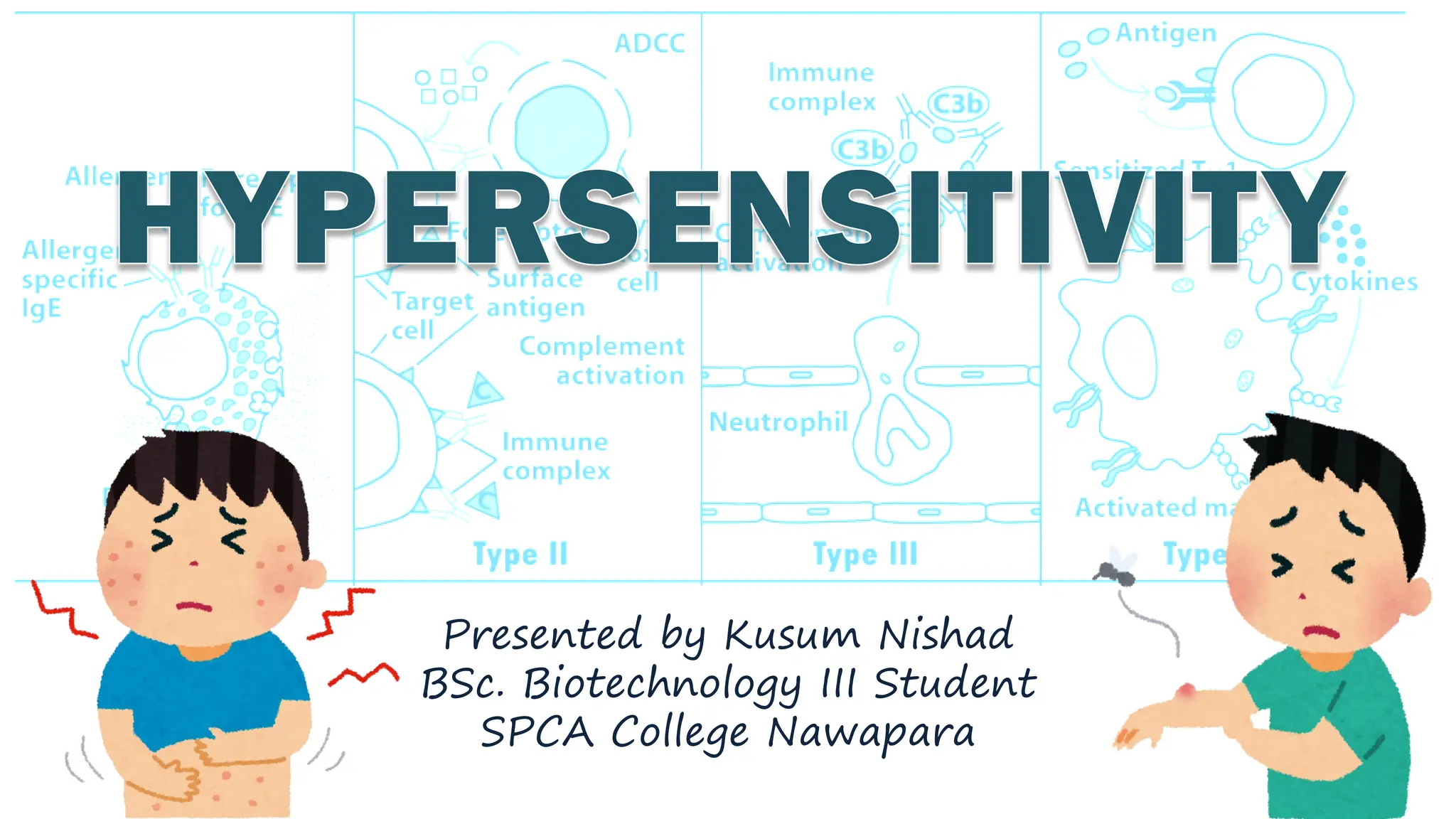
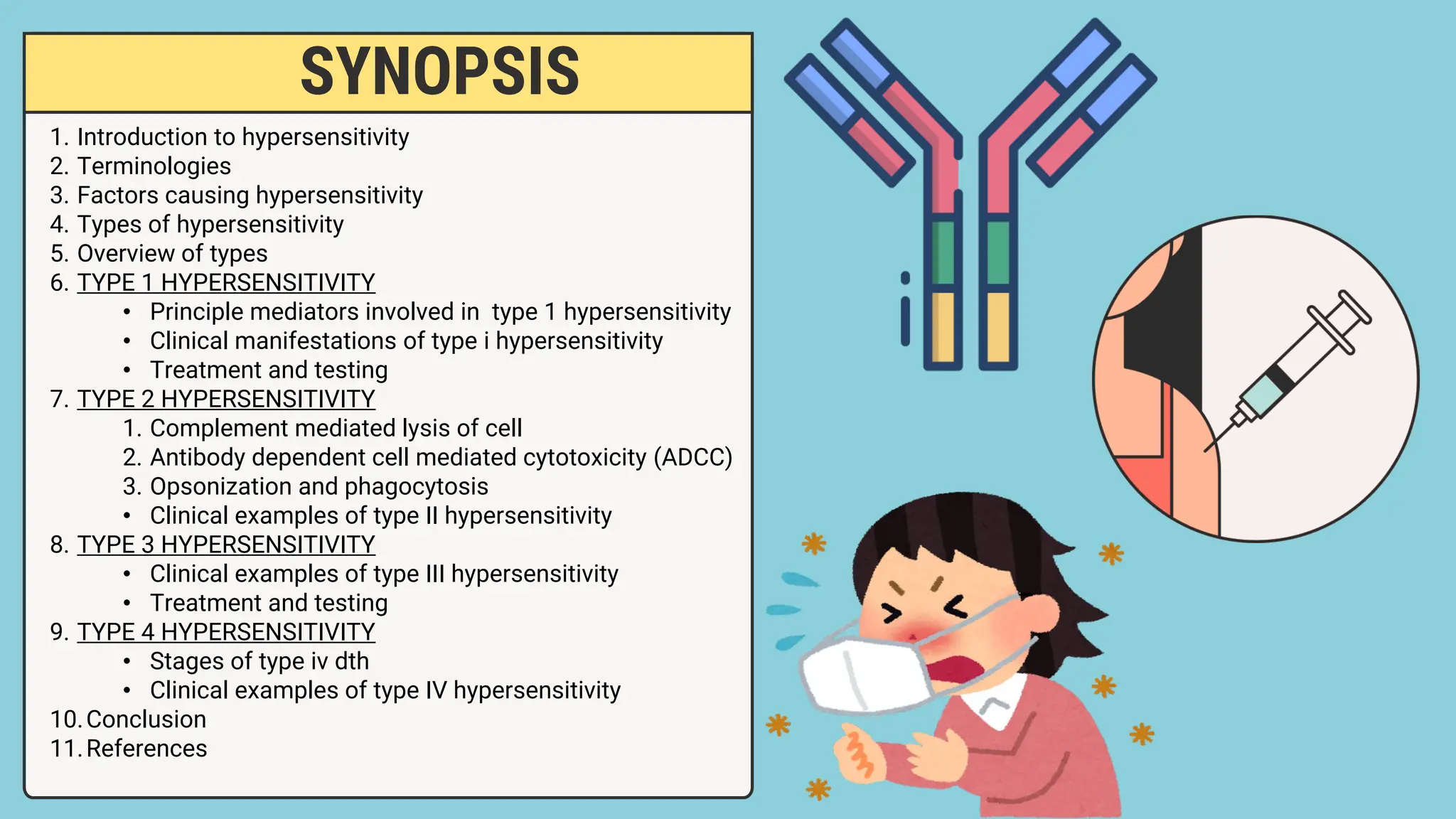
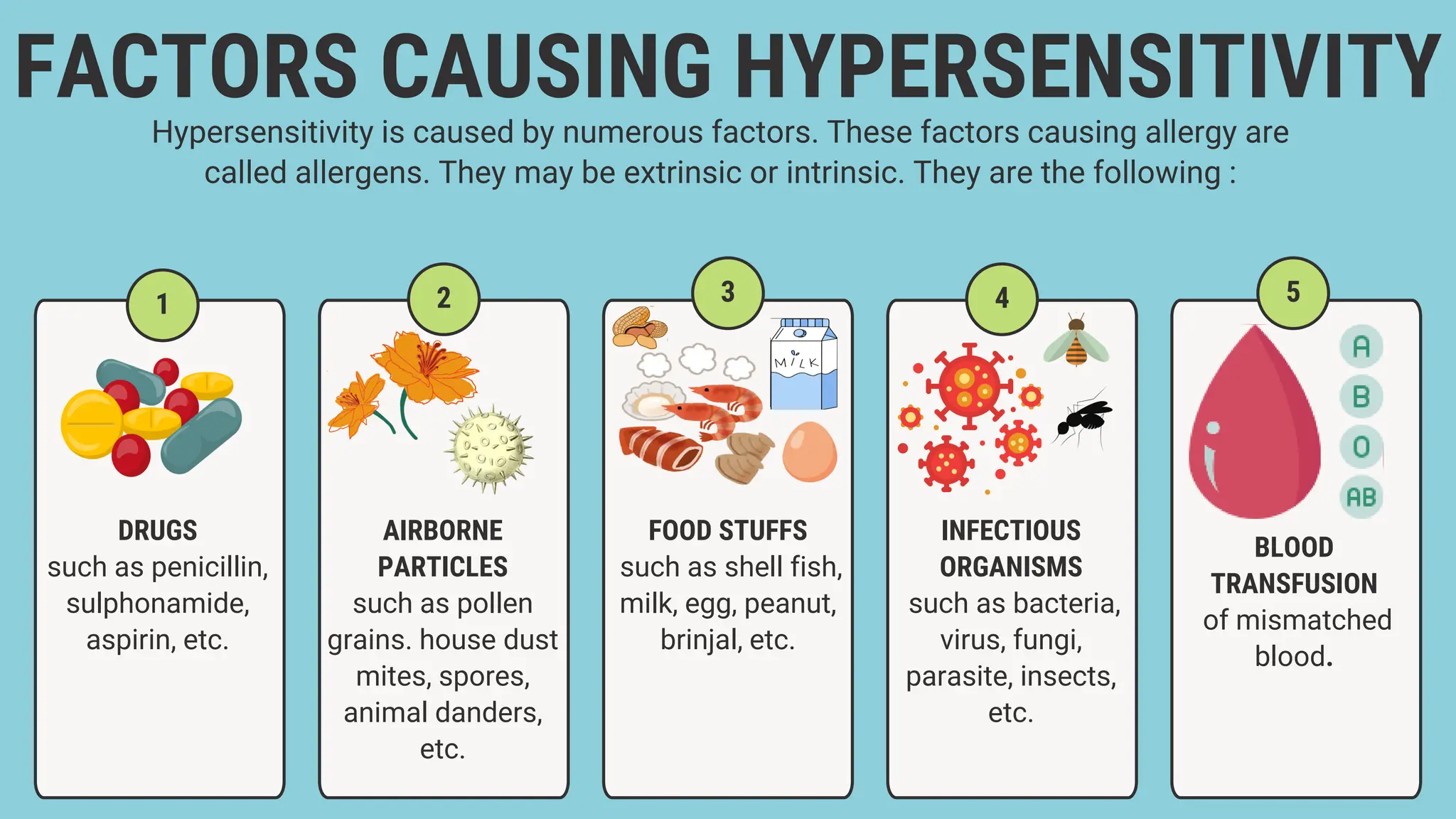
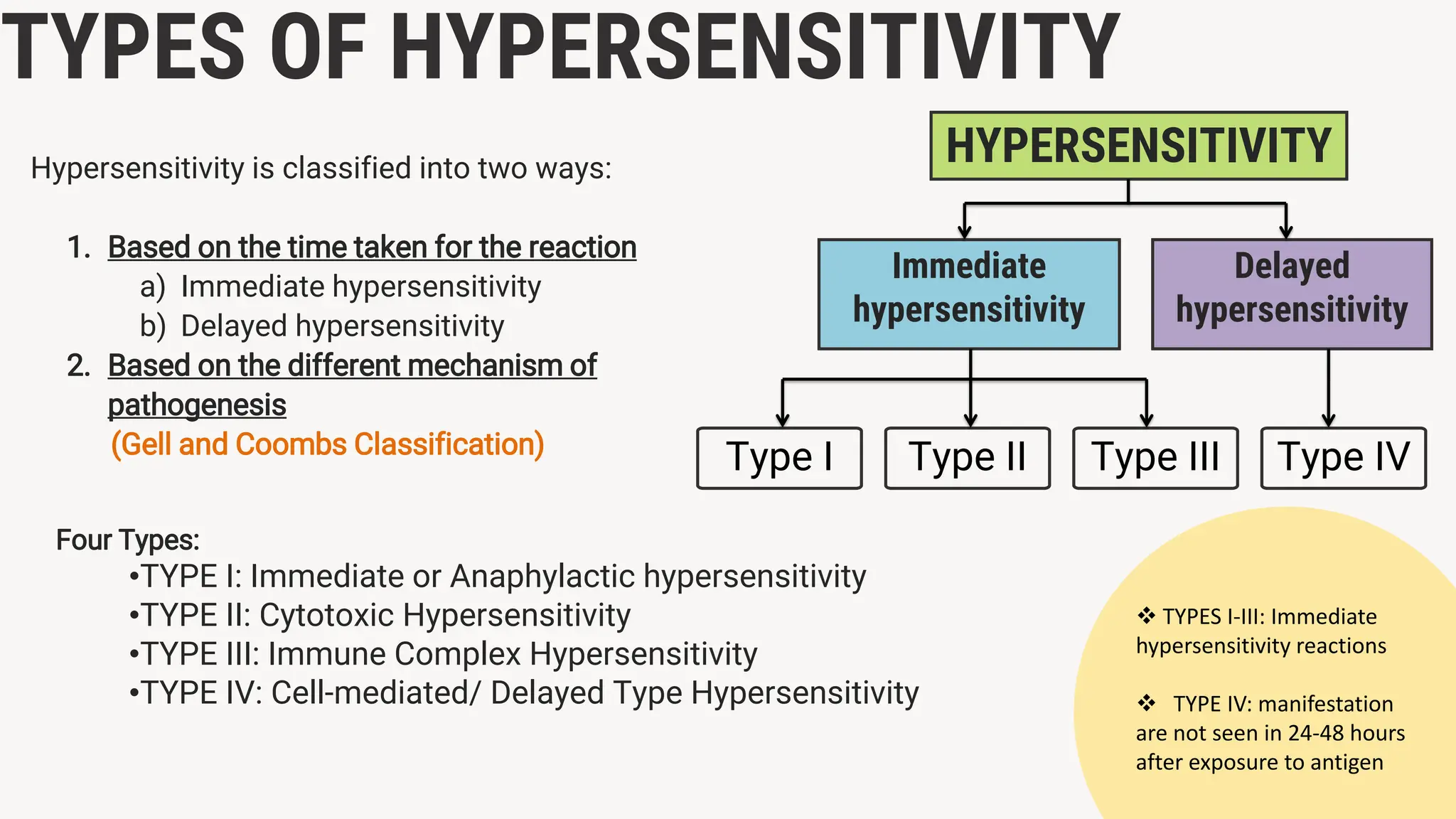
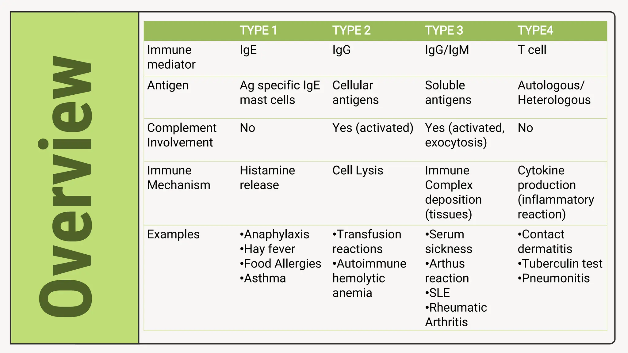
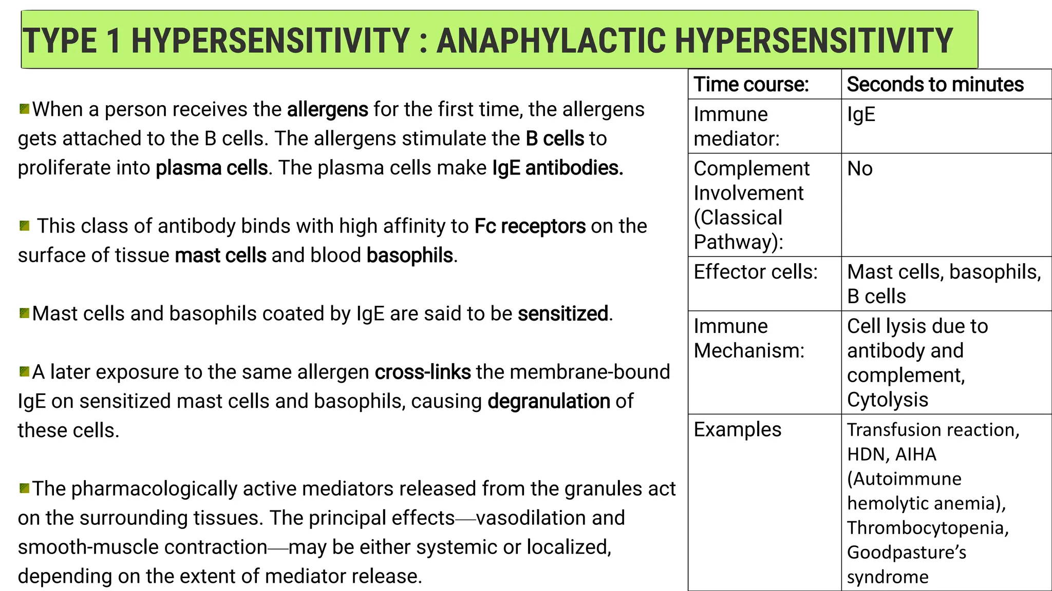
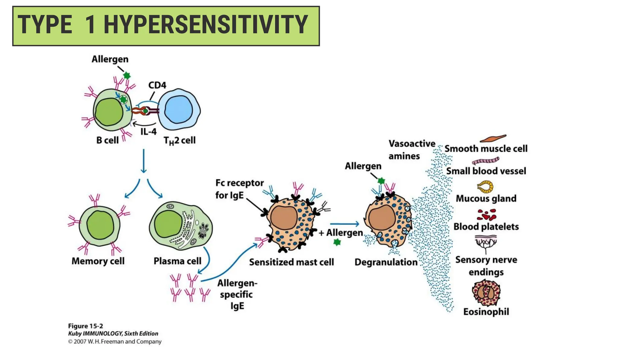
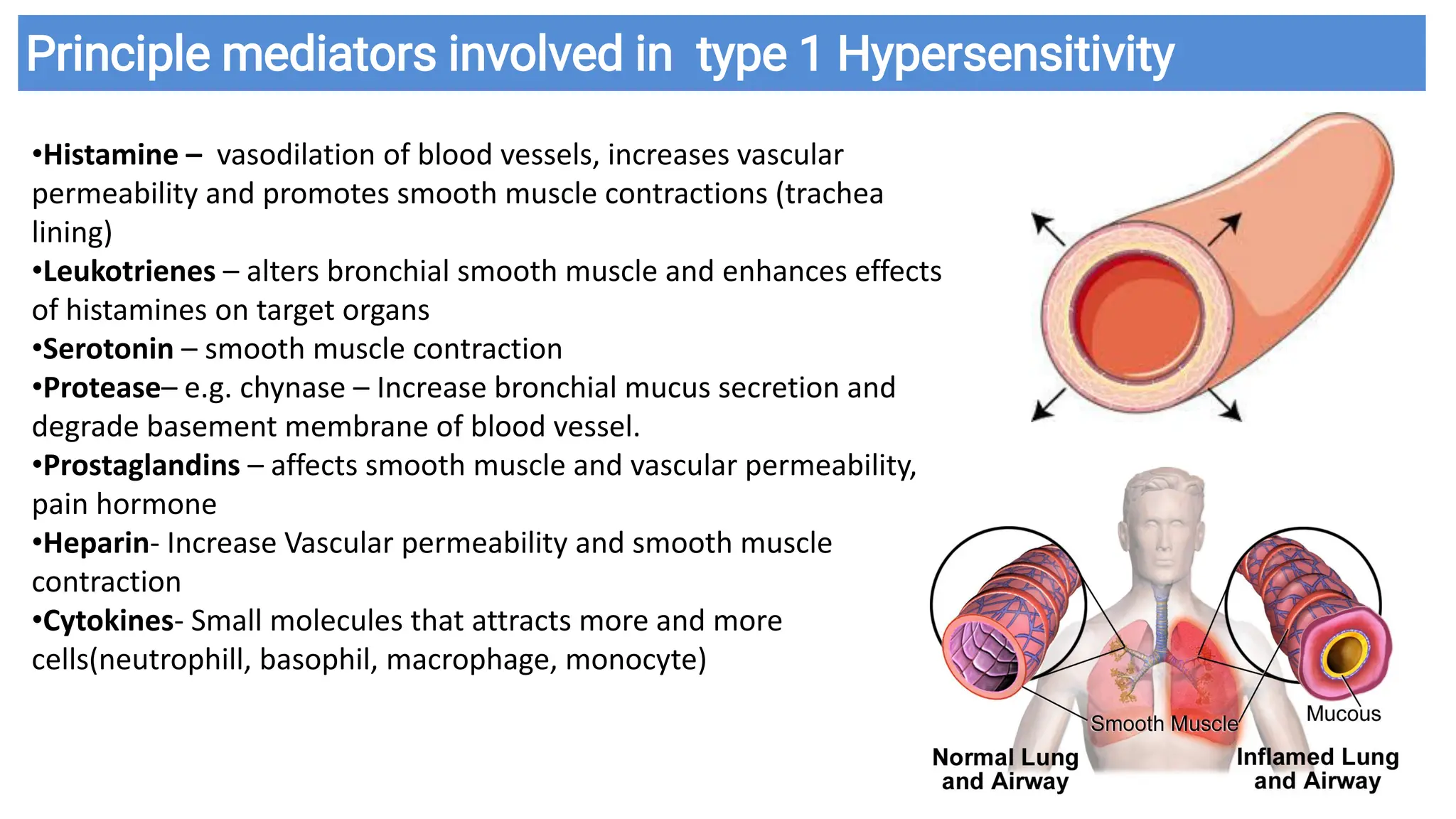
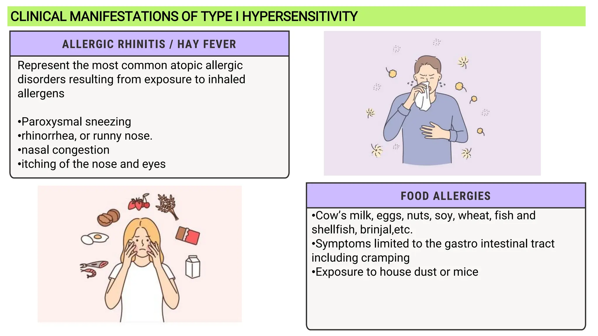
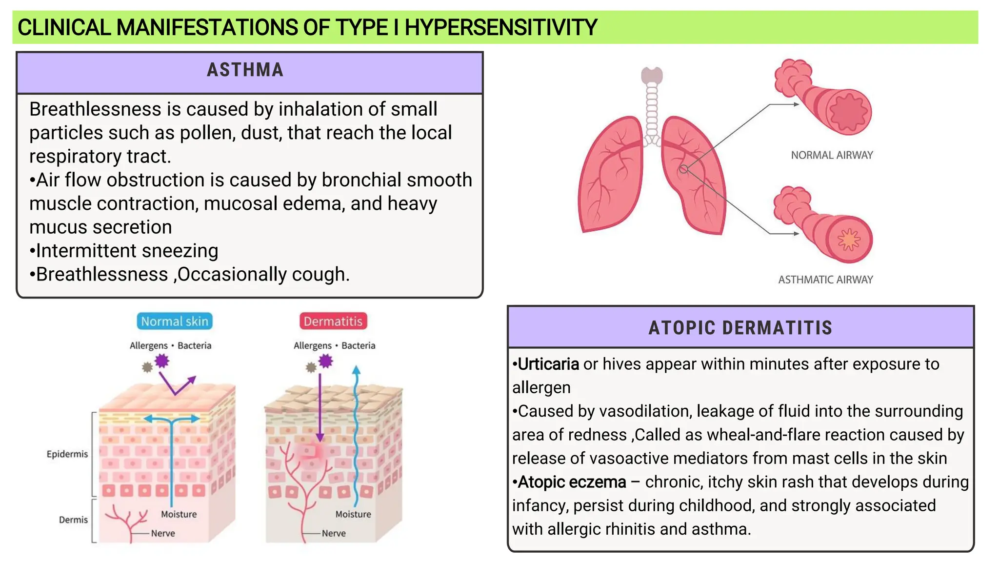
Presented by Kusum Nishad
BSc. Biotechnology III Student
SPCA College Nawapara
SYNOPSIS
1. Introduction to hypersensitivity
2. Terminologies
3. Factors causing hypersensitivity
4. Types of hypersensitivity
5. Overview of types
6. TYPE 1 HYPERSENSITIVITY
• Principle mediators involved in type 1 hypersensitivity
• Clinical manifestations of type i hypersensitivity
• Treatment and testing
7. TYPE 2 HYPERSENSITIVITY
1. Complement mediated lysis of cell
2. Antibody dependent cell mediated cytotoxicity (ADCC)
3. Opsonization and phagocytosis
• Clinical examples of type II hypersensitivity
8. TYPE 3 HYPERSENSITIVITY
• Clinical examples of type III hypersensitivity
• Treatment and testing
9. TYPE 4 HYPERSENSITIVITY
• Stages of type iv dth
• Clinical examples of type IV hypersensitivity
10.Conclusion
11.References
- immune & inflammatory responses that are harmful to the host
(von Pirquet, 1906)
HYPERSENSITIVITY
•It is defined as the violent reaction of the immune system leading to severe
symptoms and even death in a sensitized animal when it is re-exposed to the same
antigen for the second time.
•In clinical terms, hypersensitivity is called allergy.
•A familiar example for hypersensitivity is the allergy caused by penicillin injection.
•Hypersensitivity does not occur in all human beings. Only about 10% of human
population suffer from hypersensitivity.
TERMINOLOGIES
Refers to foreign substances that trigger allergic reactions •
Small molecular weight substances that could enter the body
through inhalation, ingestion, or drug administration
Allergens –
refers to immediate hypersensitivity mediated
by IgE antibodies
Atopy –
an altered and often harmful response of the
immune system to foreign substances
Allergy –
1
FACTORS CAUSING HYPERSENSITIVITY
Hypersensitivity is caused by numerous factors. These factors causing allergy are
called allergens. They may be extrinsic or intrinsic. They are the following :
DRUGS
such as penicillin,
sulphonamide,
aspirin, etc.
5
4
3
2
AIRBORNE
PARTICLES
such as pollen
grains. house dust
mites, spores,
animal danders,
etc.
BLOOD
TRANSFUSION
of mismatched
blood.
INFECTIOUS
ORGANISMS
such as bacteria,
virus, fungi,
parasite, insects,
etc.
FOOD STUFFS
such as shell fish,
milk, egg, peanut,
brinjal, etc.
HYPERSENSITIVITY
Type I
TYPES OF HYPERSENSITIVITY
Hypersensitivity is classified into two ways:
1. Based on the time taken for the reaction
a) Immediate hypersensitivity
b) Delayed hypersensitivity
2. Based on the different mechanism of
pathogenesis
(Gell and Coombs Classification)
Delayed
hypersensitivity
Immediate
hypersensitivity
Type II Type III Type IV
Four Types:
•TYPE I: Immediate or Anaphylactic hypersensitivity
•TYPE II: Cytotoxic Hypersensitivity
•TYPE III: Immune Complex Hypersensitivity
•TYPE IV: Cell-mediated/ Delayed Type Hypersensitivity
 TYPES I-III: Immediate
hypersensitivity reactions
 TYPE IV: manifestation
are not seen in 24-48 hours
after exposure to antigen
Overview
TYPE 1 TYPE 2 TYPE 3 TYPE4
Immune
mediator
IgE IgG IgG/IgM T cell
Antigen Ag specific IgE
mast cells
Cellular
antigens
Soluble
antigens
Autologous/
Heterologous
Complement
Involvement
No Yes (activated) Yes (activated,
exocytosis)
No
Immune
Mechanism
Histamine
release
Cell Lysis Immune
Complex
deposition
(tissues)
Cytokine
production
(inflammatory
reaction)
Examples •Anaphylaxis
•Hay fever
•Food Allergies
•Asthma
•Transfusion
reactions
•Autoimmune
hemolytic
anemia
•Serum
sickness
•Arthus
reaction
•SLE
•Rheumatic
Arthritis
•Contact
dermatitis
•Tuberculin test
•Pneumonitis
TYPE 1 HYPERSENSITIVITY : ANAPHYLACTIC HYPERSENSITIVITY
When a person receives the allergens for the first time, the allergens
gets attached to the B cells. The allergens stimulate the B cells to
proliferate into plasma cells. The plasma cells make IgE antibodies.
This class of antibody binds with high affinity to Fc receptors on the
surface of tissue mast cells and blood basophils.
Mast cells and basophils coated by IgE are said to be sensitized.
A later exposure to the same allergen cross-links the membrane-bound
IgE on sensitized mast cells and basophils, causing degranulation of
these cells.
The pharmacologically active mediators released from the granules act
on the surrounding tissues. The principal effects—vasodilation and
smooth-muscle contraction—may be either systemic or localized,
depending on the extent of mediator release.
Time course: Seconds to minutes
Immune
mediator:
IgE
Complement
Involvement
(Classical
Pathway):
No
Effector cells: Mast cells, basophils,
B cells
Immune
Mechanism:
Cell lysis due to
antibody and
complement,
Cytolysis
Examples Transfusion reaction,
HDN, AIHA
(Autoimmune
hemolytic anemia),
Thrombocytopenia,
Goodpasture’s
syndrome
TYPE 1 HYPERSENSITIVITY
Principle mediators involved in type 1 Hypersensitivity
•Histamine – vasodilation of blood vessels, increases vascular
permeability and promotes smooth muscle contractions (trachea
lining)
•Leukotrienes – alters bronchial smooth muscle and enhances effects
of histamines on target organs
•Serotonin – smooth muscle contraction
•Protease– e.g. chynase – Increase bronchial mucus secretion and
degrade basement membrane of blood vessel.
•Prostaglandins – affects smooth muscle and vascular permeability,
pain hormone
•Heparin- Increase Vascular permeability and smooth muscle
contraction
•Cytokines- Small molecules that attracts more and more
cells(neutrophill, basophil, macrophage, monocyte)
CLINICAL MANIFESTATIONS OF TYPE I HYPERSENSITIVITY
ALLERGIC RHINITIS / HAY FEVER
Represent the most common atopic allergic
disorders resulting from exposure to inhaled
allergens
•Paroxysmal sneezing
•rhinorrhea, or runny nose.
•nasal congestion
•itching of the nose and eyes
FOOD ALLERGIES
•Cow’s milk, eggs, nuts, soy, wheat, fish and
shellfish, brinjal,etc.
•Symptoms limited to the gastro intestinal tract
including cramping
•Exposure to house dust or mice
CLINICAL MANIFESTATIONS OF TYPE I HYPERSENSITIVITY
ATOPIC DERMATITIS
•Urticaria or hives appear within minutes after exposure to
allergen
•Caused by vasodilation, leakage of fluid into the surrounding
area of redness ,Called as wheal-and-flare reaction caused by
release of vasoactive mediators from mast cells in the skin
•Atopic eczema – chronic, itchy skin rash that develops during
infancy, persist during childhood, and strongly associated
with allergic rhinitis and asthma.
ASTHMA
Breathlessness is caused by inhalation of small
particles such as pollen, dust, that reach the local
respiratory tract.
•Air flow obstruction is caused by bronchial smooth
muscle contraction, mucosal edema, and heavy
mucus secretion
•Intermittent sneezing
•Breathlessness ,Occasionally cough.
TYPE 2 HYPERSENSITIVITY : ANTIBODY DEPENDENT CYTOTOXIC HYPERSENSITIVITY
Type II hypersensitive reactions involve antibody-mediated
destruction of cells.
Antibody can activate the complement system, creating
pores in the membrane of a foreign cell or it can mediate cell
destruction by antibody dependent cell-mediated cytotoxicity
(ADCC).
In this process, cytotoxic cells with Fc receptors bind to the
Fc region of antibodies on target cells and promote killing of
the cells.
Antibody bound to a foreign cell also can serve as an
opsonin, enabling phagocytic cells with Fc or C3b receptors to
bind and phagocytose the antibody-coated cell .
Time course: Seconds to minutes
Immune mediator: Membrane bound
IgG, IgM, and IgA
Complement
Involvement
(Classical Pathway):
Yes
Effector cells: RBC, WBC, and
platelets
Immune
Mechanism:
Release of
histamine and other
mediators
Examples Transfusion
reaction, HDN, AIHA
(Autoimmune
hemolytic anemia),
Thrombocytopenia,
Goodpasture’s
syndrome
1. Complement mediated lysis of cell:
2. Antibody dependent cell mediated cytotoxicity (ADCC):
3. Opsonization and phagocytosis
A. Hemolytic reactions
• Hemoglobinuria(level of hemoglobin in the blood rises too high)
• Chest pain
• Low back pain
• Decreased blood pressure
• Increased respiratory rate
B. Allergic reactions
a. Allergic reaction (MILD)
-- Facial flushing
-- Rash
b. Allergic reaction (SEVERE)
--Anxiety
--Decreased blood pressure
--Anaphylactic shock
C. Febrile reaction
• Sensitivity of the patient’s blood to white blood cells, platelets or plasma
proteins
• Clinical signs: fever, warm and flush skin, chills, headache, anxiety,
muscle pain
1. Transfusion Reactions
Clinical Examples of Type II Hypersensitivity
-- Cells from an incompatible
donor react with the host’s
antibody
--Occurs in the first 10-15
minutes or first 50cc of blood A
Hemolytic Disease of the Newborn Is Caused by Type II Reactions
– A medical condition where an Rh
negative mother’s Ab attack the red
blood cells of an Rh positive fetus
– Complications:
• During pregnancy :
o Mild anemia - hyperbilirubinemia
and jaundice
o Severe anemia - with
enlargement of the liver and spleen –
fast destruction of red blood cells
o Hydrops fetalis – this occurs as
the baby’s organs are unable to handle
the anemia. A fetus with hydrops is at
great risk of being stillborn
• After birth :
o Severe hyperbilirubinemia and
jaundice - the baby’s liver is unable to
handle the large amount of bilirubin that
results from red blood cell breakdown
o Kernicterus - the most severe
form of hyperbilirubinemia and results
from the buildup of bilirubin in the brain -
hyperbilirubinemia and jaundice
TYPE 3 HYPERSENSITIVITY : IMMUNE COMPLEX MEDIATED HYPERSENSITIVITY
similar to type II reactions in that IgG or IgM is involved and
destruction is complement mediated.
However, in the case of type III-associated diseases, the
antigen is soluble.
When soluble antigen combines with antibody, complexes are
formed that precipitate out of the serum.
Complexes are cleared by phagocytic cells however if immune
system is overwhelmed it deposits in the tissues where they
bind complement causing damage to tissue.
Deposition of antigen–antibody complexes is influenced by the
relative concentration of both components.
Time course: Seconds to
minutes
Immune
mediator:
Soluble antigens,
IgM, or IgG
Complement
Involvement
(Classical
Pathway):
Yes
Effector cells: Host tissue cells
Immune
Mechanism:
Deposition of
immune complex
to host tissues,
Ag-Ab complexes
Examples Serum sickness,
Arthus reaction,
SLE(systemic
lupus
erythematosus)
The Arthus reaction is a localized
inflammatory response that occurs
when an antigen is introduced into the
body of an individual who has pre-
existing antibodies against that antigen.
It is a type of Type III hypersensitivity
reaction, also known as immune
complex-mediated hypersensitivity.
Clinical Examples of Type III Hypersensitivity
1. Serum Sickness
from injection of heterologous
serum protein, usually resulting
from passive immunization with
animal serum, usually horse or
bovine, used to treat such
infections such as diphtheria,
tetanus, and gangrene
2. SLE(systemic lupus
erythematosus)
SLE - immune complex
deposition involves multiple
organs; the main damage
occurs to the joints, skin, and
glomerular basement
membrane in the kidneys
3. Rheumatoid Arthritis
Rheumatoid Arthritis - immune
complexes primarily cause
damage to the joints
TYPE 4 HYPERSENSITIVITY : CELL MEDIATED DELAYED HYPERSENSITIVITY (DTH)
When some subpopulations of activated TH cells encounter certain
types of antigens, they secrete cytokines that induce a localized
inflammatory reaction called delayed-type hypersensitivity (DTH).
The reaction is characterized by large influxes of nonspecific
inflammatory cells, in particular, macrophages.
Robert Koch observed that individuals infected with Mycobacterium
tuberculosis (Mtb) developed a localized inflammatory response after
receiving intradermal injections of a filtrate from the organism
differs from the other three types of hypersensitivity in that sensitized T
cells, rather than antibodies, play the major role in its manifestations
Specialized T cells (specially Th1 cells) play important role in its
manifestation (Involves antigen-sensitized T cells)
Antibody and complement are not directly involved.
Hallmark of occupational type IV hypersensitivity – Allergic contact
dermatitis
Time course: 24 to 72 hours
Immune
mediator:
CD4+ T cells
(Th1 cells)
Complement
Involvement
(Classical
Pathway):
No
Effector cells: Macrophages
(Antigen
presenting cells)
Immune
Mechanism:
Release
lymphokines
from T cells
Examples Contact
dermatitis,
tuberculin test,
infections
1. Sensitization stage
• Memory Th1 cells against DTH antigens are generated by
dendritic cells during the sensitization stage
• These Th1 cells can activate macrophages and trigger
inflammmatory response
STAGES OF TYPE IV DTH
2. Effector Stage
• Secondary contact yields what we call DTH
• Th1 memory cells are activated and produce cytokines
• IFN-ϒ, TNF- α and TMF-B which causes tissue destruction ,
inflammation.
• IL-2 activates T cells and CTLs
• Chemokines and macrophage recuritment
• IL-3 for increased monocyte/macrophage
Clinical Examples of Type IV Hypersensitivity
1. Contact hypersensitivity
(Poison Ivy, reactions to
metals in jewelry)
Reactions are usually caused by
low-molecular weight
compounds that touch the skin
• most common causes include
poison ivy, poison oak, and
poison sumac
• most common example is
contact dermatitis
2. Tuberculin-type (TST)
Tuberculin skin test (TST) are
administered to detect the presence
of M. tuberculosis – TB
– Mantoux – the technique for
administering the test
– Tuberculin – also called purified
protein derivatives (PPD) – the
solution used to administer the test
3. Granulomatous hypersensitivity
(Leprosy, TB, Schistosomiasis,
Chron’s disease)
It is a delayed-type hypersensitivity
reaction that occurs when T cells
are activated by organic dusts or
chemicals in the lungs.
It's an immunopathological feature
of hypersensitivity pneumonitis
(HP).
CONCLUSION
• In summary, hypersensitivity reactions represent an exaggerated immune response to harmless
substances, which can lead to significant health issues. Understanding the four main types—Type I
(immediate), Type II (cytotoxic), Type III (immune complex-mediated), and Type IV (delayed-type)—is crucial
for diagnosing and managing these conditions effectively.
•Recognizing the symptoms and triggers of hypersensitivity is essential for timely intervention and treatment,
which may include avoidance of allergens, medication management, and in some cases, immunotherapy.
•As our understanding of the immune system evolves, ongoing research into hypersensitivity mechanisms
will pave the way for more targeted and effective therapies. By educating ourselves and others about
hypersensitivity, we can better support individuals affected by these reactions and improve their quality of
life.
Thank you for your attention. If you have any questions or would like to discuss
further, I’m happy to engage!
REFERENCES
• Kuby IMMUNOLOGY, Sixth Edition
©2007 W.H. Freeman and Company
• Immunology , Saras Publication
Dulsy Fatima and N. Arumugam
• Notes extracted from Sir Diego Ofiaza RMT
• www.online biologynotes.com
• https://www.studocu.com/

Hypersensitivity:Types, Mechanism,Example

  • 1.
    Presented by KusumNishad BSc. Biotechnology III Student SPCA College Nawapara
  • 2.
    SYNOPSIS 1. Introduction tohypersensitivity 2. Terminologies 3. Factors causing hypersensitivity 4. Types of hypersensitivity 5. Overview of types 6. TYPE 1 HYPERSENSITIVITY • Principle mediators involved in type 1 hypersensitivity • Clinical manifestations of type i hypersensitivity • Treatment and testing 7. TYPE 2 HYPERSENSITIVITY 1. Complement mediated lysis of cell 2. Antibody dependent cell mediated cytotoxicity (ADCC) 3. Opsonization and phagocytosis • Clinical examples of type II hypersensitivity 8. TYPE 3 HYPERSENSITIVITY • Clinical examples of type III hypersensitivity • Treatment and testing 9. TYPE 4 HYPERSENSITIVITY • Stages of type iv dth • Clinical examples of type IV hypersensitivity 10.Conclusion 11.References
  • 3.
    - immune &inflammatory responses that are harmful to the host (von Pirquet, 1906) HYPERSENSITIVITY •It is defined as the violent reaction of the immune system leading to severe symptoms and even death in a sensitized animal when it is re-exposed to the same antigen for the second time. •In clinical terms, hypersensitivity is called allergy. •A familiar example for hypersensitivity is the allergy caused by penicillin injection. •Hypersensitivity does not occur in all human beings. Only about 10% of human population suffer from hypersensitivity.
  • 4.
    TERMINOLOGIES Refers to foreignsubstances that trigger allergic reactions • Small molecular weight substances that could enter the body through inhalation, ingestion, or drug administration Allergens – refers to immediate hypersensitivity mediated by IgE antibodies Atopy – an altered and often harmful response of the immune system to foreign substances Allergy –
  • 5.
    1 FACTORS CAUSING HYPERSENSITIVITY Hypersensitivityis caused by numerous factors. These factors causing allergy are called allergens. They may be extrinsic or intrinsic. They are the following : DRUGS such as penicillin, sulphonamide, aspirin, etc. 5 4 3 2 AIRBORNE PARTICLES such as pollen grains. house dust mites, spores, animal danders, etc. BLOOD TRANSFUSION of mismatched blood. INFECTIOUS ORGANISMS such as bacteria, virus, fungi, parasite, insects, etc. FOOD STUFFS such as shell fish, milk, egg, peanut, brinjal, etc.
  • 6.
    HYPERSENSITIVITY Type I TYPES OFHYPERSENSITIVITY Hypersensitivity is classified into two ways: 1. Based on the time taken for the reaction a) Immediate hypersensitivity b) Delayed hypersensitivity 2. Based on the different mechanism of pathogenesis (Gell and Coombs Classification) Delayed hypersensitivity Immediate hypersensitivity Type II Type III Type IV Four Types: •TYPE I: Immediate or Anaphylactic hypersensitivity •TYPE II: Cytotoxic Hypersensitivity •TYPE III: Immune Complex Hypersensitivity •TYPE IV: Cell-mediated/ Delayed Type Hypersensitivity  TYPES I-III: Immediate hypersensitivity reactions  TYPE IV: manifestation are not seen in 24-48 hours after exposure to antigen
  • 7.
    Overview TYPE 1 TYPE2 TYPE 3 TYPE4 Immune mediator IgE IgG IgG/IgM T cell Antigen Ag specific IgE mast cells Cellular antigens Soluble antigens Autologous/ Heterologous Complement Involvement No Yes (activated) Yes (activated, exocytosis) No Immune Mechanism Histamine release Cell Lysis Immune Complex deposition (tissues) Cytokine production (inflammatory reaction) Examples •Anaphylaxis •Hay fever •Food Allergies •Asthma •Transfusion reactions •Autoimmune hemolytic anemia •Serum sickness •Arthus reaction •SLE •Rheumatic Arthritis •Contact dermatitis •Tuberculin test •Pneumonitis
  • 8.
    TYPE 1 HYPERSENSITIVITY: ANAPHYLACTIC HYPERSENSITIVITY When a person receives the allergens for the first time, the allergens gets attached to the B cells. The allergens stimulate the B cells to proliferate into plasma cells. The plasma cells make IgE antibodies. This class of antibody binds with high affinity to Fc receptors on the surface of tissue mast cells and blood basophils. Mast cells and basophils coated by IgE are said to be sensitized. A later exposure to the same allergen cross-links the membrane-bound IgE on sensitized mast cells and basophils, causing degranulation of these cells. The pharmacologically active mediators released from the granules act on the surrounding tissues. The principal effects—vasodilation and smooth-muscle contraction—may be either systemic or localized, depending on the extent of mediator release. Time course: Seconds to minutes Immune mediator: IgE Complement Involvement (Classical Pathway): No Effector cells: Mast cells, basophils, B cells Immune Mechanism: Cell lysis due to antibody and complement, Cytolysis Examples Transfusion reaction, HDN, AIHA (Autoimmune hemolytic anemia), Thrombocytopenia, Goodpasture’s syndrome
  • 9.
  • 10.
    Principle mediators involvedin type 1 Hypersensitivity •Histamine – vasodilation of blood vessels, increases vascular permeability and promotes smooth muscle contractions (trachea lining) •Leukotrienes – alters bronchial smooth muscle and enhances effects of histamines on target organs •Serotonin – smooth muscle contraction •Protease– e.g. chynase – Increase bronchial mucus secretion and degrade basement membrane of blood vessel. •Prostaglandins – affects smooth muscle and vascular permeability, pain hormone •Heparin- Increase Vascular permeability and smooth muscle contraction •Cytokines- Small molecules that attracts more and more cells(neutrophill, basophil, macrophage, monocyte)
  • 11.
    CLINICAL MANIFESTATIONS OFTYPE I HYPERSENSITIVITY ALLERGIC RHINITIS / HAY FEVER Represent the most common atopic allergic disorders resulting from exposure to inhaled allergens •Paroxysmal sneezing •rhinorrhea, or runny nose. •nasal congestion •itching of the nose and eyes FOOD ALLERGIES •Cow’s milk, eggs, nuts, soy, wheat, fish and shellfish, brinjal,etc. •Symptoms limited to the gastro intestinal tract including cramping •Exposure to house dust or mice
  • 12.
    CLINICAL MANIFESTATIONS OFTYPE I HYPERSENSITIVITY ATOPIC DERMATITIS •Urticaria or hives appear within minutes after exposure to allergen •Caused by vasodilation, leakage of fluid into the surrounding area of redness ,Called as wheal-and-flare reaction caused by release of vasoactive mediators from mast cells in the skin •Atopic eczema – chronic, itchy skin rash that develops during infancy, persist during childhood, and strongly associated with allergic rhinitis and asthma. ASTHMA Breathlessness is caused by inhalation of small particles such as pollen, dust, that reach the local respiratory tract. •Air flow obstruction is caused by bronchial smooth muscle contraction, mucosal edema, and heavy mucus secretion •Intermittent sneezing •Breathlessness ,Occasionally cough.
  • 13.
    TYPE 2 HYPERSENSITIVITY: ANTIBODY DEPENDENT CYTOTOXIC HYPERSENSITIVITY Type II hypersensitive reactions involve antibody-mediated destruction of cells. Antibody can activate the complement system, creating pores in the membrane of a foreign cell or it can mediate cell destruction by antibody dependent cell-mediated cytotoxicity (ADCC). In this process, cytotoxic cells with Fc receptors bind to the Fc region of antibodies on target cells and promote killing of the cells. Antibody bound to a foreign cell also can serve as an opsonin, enabling phagocytic cells with Fc or C3b receptors to bind and phagocytose the antibody-coated cell . Time course: Seconds to minutes Immune mediator: Membrane bound IgG, IgM, and IgA Complement Involvement (Classical Pathway): Yes Effector cells: RBC, WBC, and platelets Immune Mechanism: Release of histamine and other mediators Examples Transfusion reaction, HDN, AIHA (Autoimmune hemolytic anemia), Thrombocytopenia, Goodpasture’s syndrome
  • 14.
    1. Complement mediatedlysis of cell:
  • 15.
    2. Antibody dependentcell mediated cytotoxicity (ADCC):
  • 16.
    3. Opsonization andphagocytosis
  • 17.
    A. Hemolytic reactions •Hemoglobinuria(level of hemoglobin in the blood rises too high) • Chest pain • Low back pain • Decreased blood pressure • Increased respiratory rate B. Allergic reactions a. Allergic reaction (MILD) -- Facial flushing -- Rash b. Allergic reaction (SEVERE) --Anxiety --Decreased blood pressure --Anaphylactic shock C. Febrile reaction • Sensitivity of the patient’s blood to white blood cells, platelets or plasma proteins • Clinical signs: fever, warm and flush skin, chills, headache, anxiety, muscle pain 1. Transfusion Reactions Clinical Examples of Type II Hypersensitivity -- Cells from an incompatible donor react with the host’s antibody --Occurs in the first 10-15 minutes or first 50cc of blood A
  • 18.
    Hemolytic Disease ofthe Newborn Is Caused by Type II Reactions – A medical condition where an Rh negative mother’s Ab attack the red blood cells of an Rh positive fetus – Complications: • During pregnancy : o Mild anemia - hyperbilirubinemia and jaundice o Severe anemia - with enlargement of the liver and spleen – fast destruction of red blood cells o Hydrops fetalis – this occurs as the baby’s organs are unable to handle the anemia. A fetus with hydrops is at great risk of being stillborn • After birth : o Severe hyperbilirubinemia and jaundice - the baby’s liver is unable to handle the large amount of bilirubin that results from red blood cell breakdown o Kernicterus - the most severe form of hyperbilirubinemia and results from the buildup of bilirubin in the brain - hyperbilirubinemia and jaundice
  • 19.
    TYPE 3 HYPERSENSITIVITY: IMMUNE COMPLEX MEDIATED HYPERSENSITIVITY similar to type II reactions in that IgG or IgM is involved and destruction is complement mediated. However, in the case of type III-associated diseases, the antigen is soluble. When soluble antigen combines with antibody, complexes are formed that precipitate out of the serum. Complexes are cleared by phagocytic cells however if immune system is overwhelmed it deposits in the tissues where they bind complement causing damage to tissue. Deposition of antigen–antibody complexes is influenced by the relative concentration of both components. Time course: Seconds to minutes Immune mediator: Soluble antigens, IgM, or IgG Complement Involvement (Classical Pathway): Yes Effector cells: Host tissue cells Immune Mechanism: Deposition of immune complex to host tissues, Ag-Ab complexes Examples Serum sickness, Arthus reaction, SLE(systemic lupus erythematosus)
  • 20.
    The Arthus reactionis a localized inflammatory response that occurs when an antigen is introduced into the body of an individual who has pre- existing antibodies against that antigen. It is a type of Type III hypersensitivity reaction, also known as immune complex-mediated hypersensitivity.
  • 21.
    Clinical Examples ofType III Hypersensitivity 1. Serum Sickness from injection of heterologous serum protein, usually resulting from passive immunization with animal serum, usually horse or bovine, used to treat such infections such as diphtheria, tetanus, and gangrene 2. SLE(systemic lupus erythematosus) SLE - immune complex deposition involves multiple organs; the main damage occurs to the joints, skin, and glomerular basement membrane in the kidneys 3. Rheumatoid Arthritis Rheumatoid Arthritis - immune complexes primarily cause damage to the joints
  • 22.
    TYPE 4 HYPERSENSITIVITY: CELL MEDIATED DELAYED HYPERSENSITIVITY (DTH) When some subpopulations of activated TH cells encounter certain types of antigens, they secrete cytokines that induce a localized inflammatory reaction called delayed-type hypersensitivity (DTH). The reaction is characterized by large influxes of nonspecific inflammatory cells, in particular, macrophages. Robert Koch observed that individuals infected with Mycobacterium tuberculosis (Mtb) developed a localized inflammatory response after receiving intradermal injections of a filtrate from the organism differs from the other three types of hypersensitivity in that sensitized T cells, rather than antibodies, play the major role in its manifestations Specialized T cells (specially Th1 cells) play important role in its manifestation (Involves antigen-sensitized T cells) Antibody and complement are not directly involved. Hallmark of occupational type IV hypersensitivity – Allergic contact dermatitis Time course: 24 to 72 hours Immune mediator: CD4+ T cells (Th1 cells) Complement Involvement (Classical Pathway): No Effector cells: Macrophages (Antigen presenting cells) Immune Mechanism: Release lymphokines from T cells Examples Contact dermatitis, tuberculin test, infections
  • 23.
    1. Sensitization stage •Memory Th1 cells against DTH antigens are generated by dendritic cells during the sensitization stage • These Th1 cells can activate macrophages and trigger inflammmatory response STAGES OF TYPE IV DTH 2. Effector Stage • Secondary contact yields what we call DTH • Th1 memory cells are activated and produce cytokines • IFN-ϒ, TNF- α and TMF-B which causes tissue destruction , inflammation. • IL-2 activates T cells and CTLs • Chemokines and macrophage recuritment • IL-3 for increased monocyte/macrophage
  • 24.
    Clinical Examples ofType IV Hypersensitivity 1. Contact hypersensitivity (Poison Ivy, reactions to metals in jewelry) Reactions are usually caused by low-molecular weight compounds that touch the skin • most common causes include poison ivy, poison oak, and poison sumac • most common example is contact dermatitis 2. Tuberculin-type (TST) Tuberculin skin test (TST) are administered to detect the presence of M. tuberculosis – TB – Mantoux – the technique for administering the test – Tuberculin – also called purified protein derivatives (PPD) – the solution used to administer the test 3. Granulomatous hypersensitivity (Leprosy, TB, Schistosomiasis, Chron’s disease) It is a delayed-type hypersensitivity reaction that occurs when T cells are activated by organic dusts or chemicals in the lungs. It's an immunopathological feature of hypersensitivity pneumonitis (HP).
  • 25.
    CONCLUSION • In summary,hypersensitivity reactions represent an exaggerated immune response to harmless substances, which can lead to significant health issues. Understanding the four main types—Type I (immediate), Type II (cytotoxic), Type III (immune complex-mediated), and Type IV (delayed-type)—is crucial for diagnosing and managing these conditions effectively. •Recognizing the symptoms and triggers of hypersensitivity is essential for timely intervention and treatment, which may include avoidance of allergens, medication management, and in some cases, immunotherapy. •As our understanding of the immune system evolves, ongoing research into hypersensitivity mechanisms will pave the way for more targeted and effective therapies. By educating ourselves and others about hypersensitivity, we can better support individuals affected by these reactions and improve their quality of life. Thank you for your attention. If you have any questions or would like to discuss further, I’m happy to engage!
  • 26.
    REFERENCES • Kuby IMMUNOLOGY,Sixth Edition ©2007 W.H. Freeman and Company • Immunology , Saras Publication Dulsy Fatima and N. Arumugam • Notes extracted from Sir Diego Ofiaza RMT • www.online biologynotes.com • https://www.studocu.com/