Downloaded 152 times


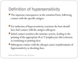


The document discusses hypersensitivity and defines it as an injurious immune response in a sensitized host following contact with a specific antigen. It describes the 4 main types of hypersensitivity reactions: Type I or immediate hypersensitivity mediated by IgE antibodies; Type II involving cytotoxic antibodies; Type III occurring via immune complex formation; and Type IV or delayed hypersensitivity mediated by T cells. It provides details on the mechanisms, mediators, examples and characteristics of each hypersensitivity type.


