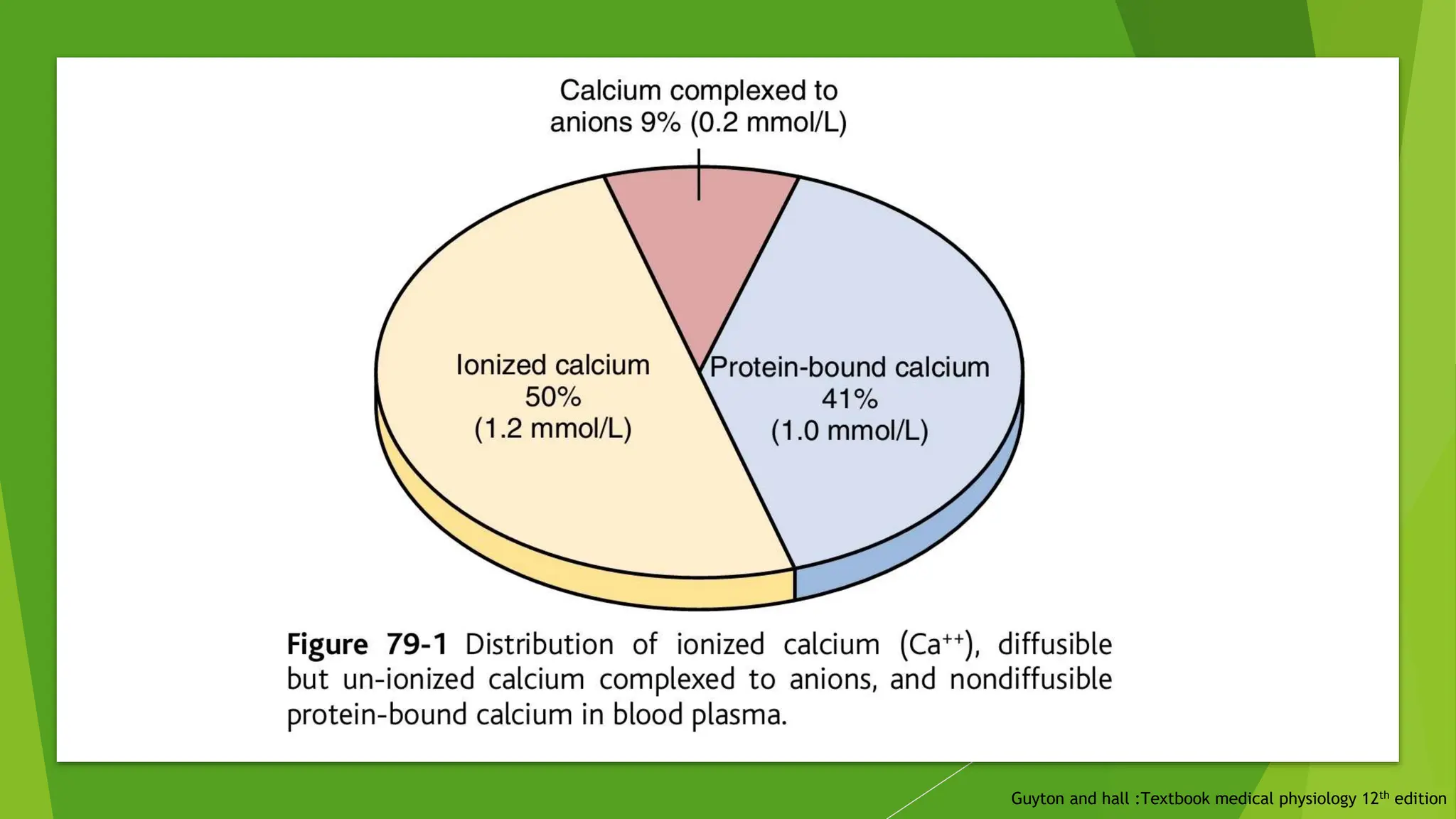
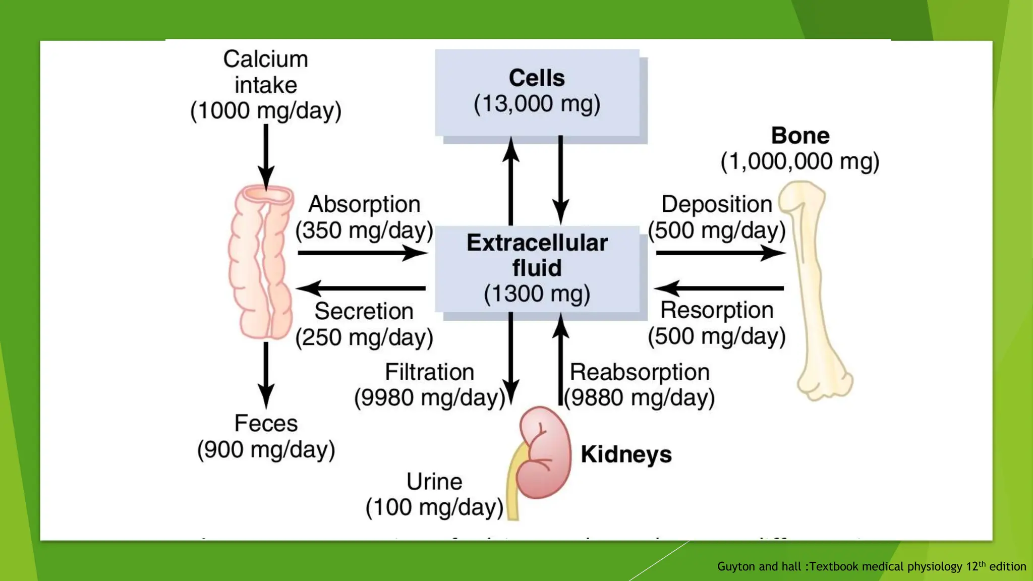
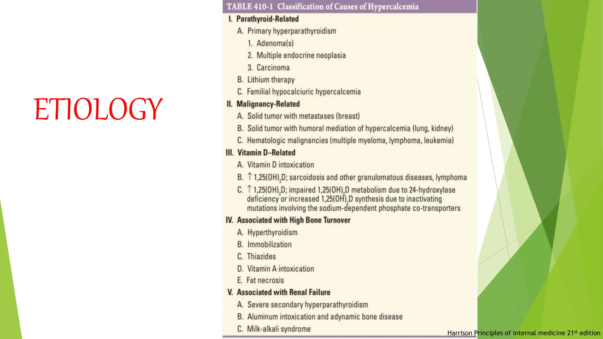
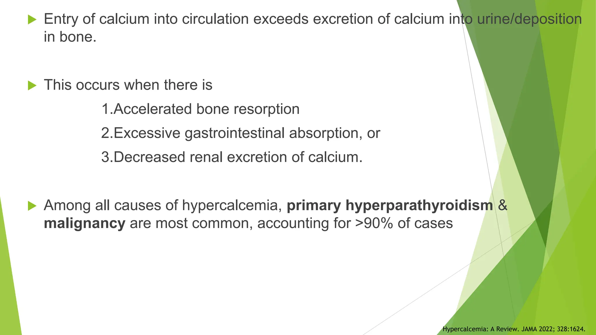
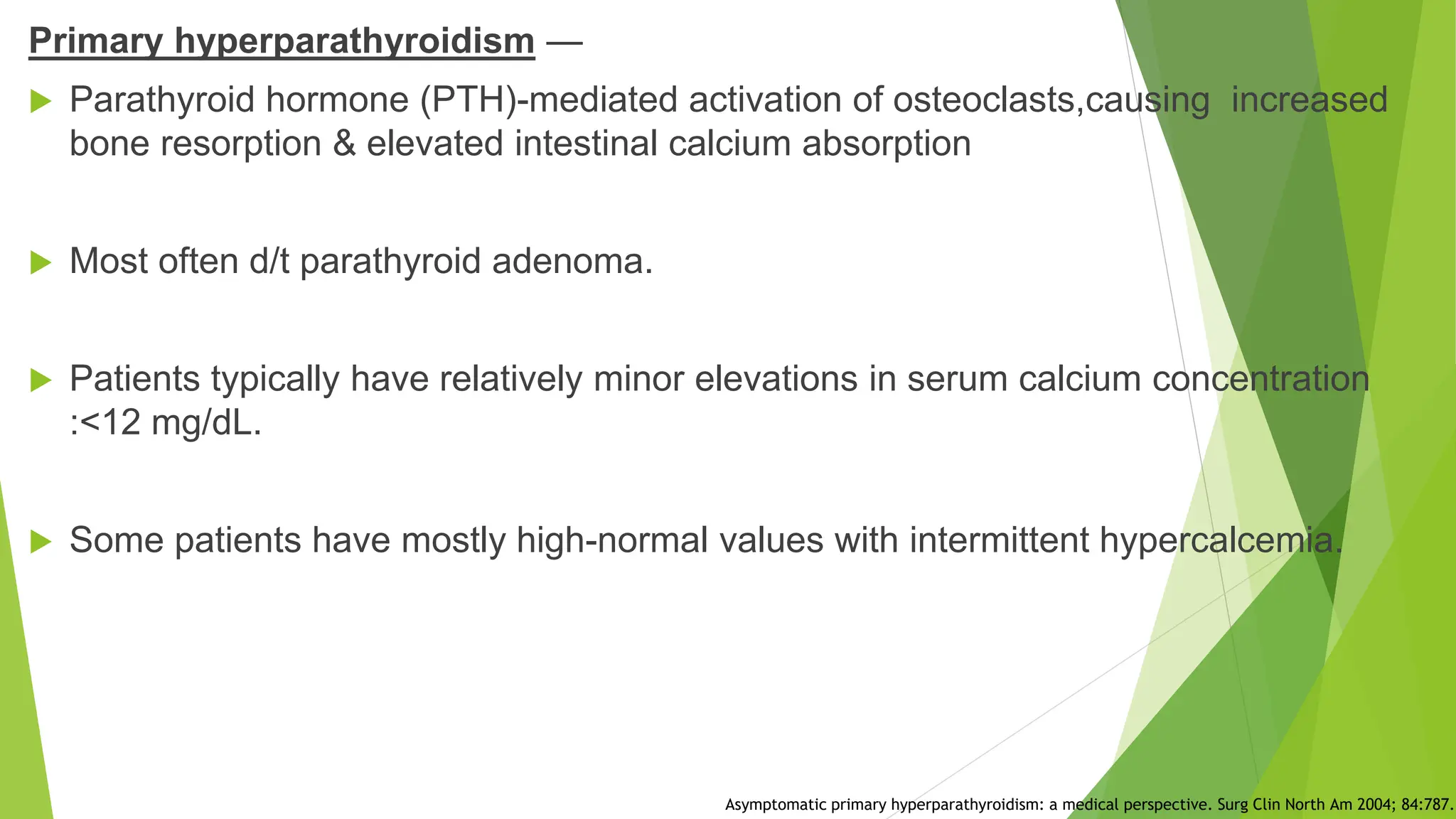
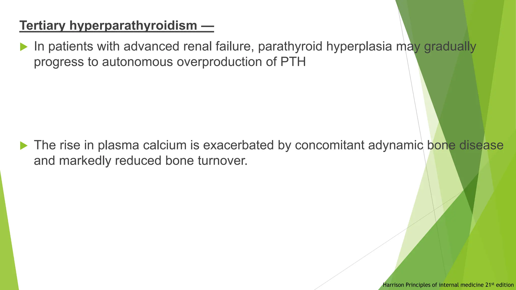
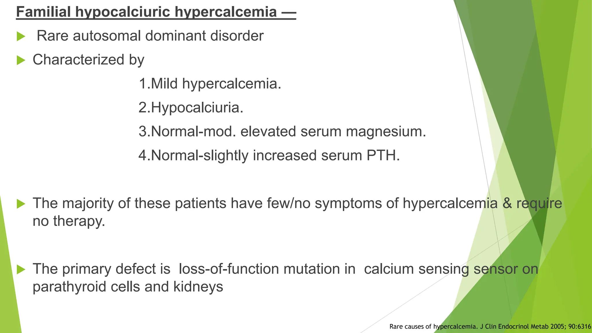
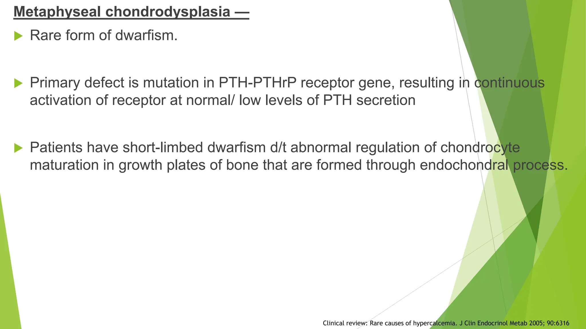
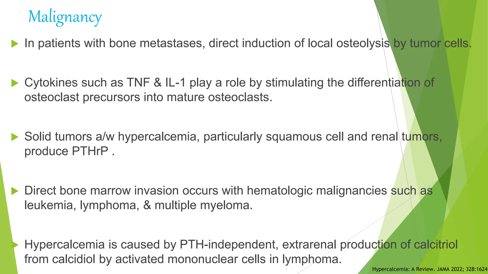
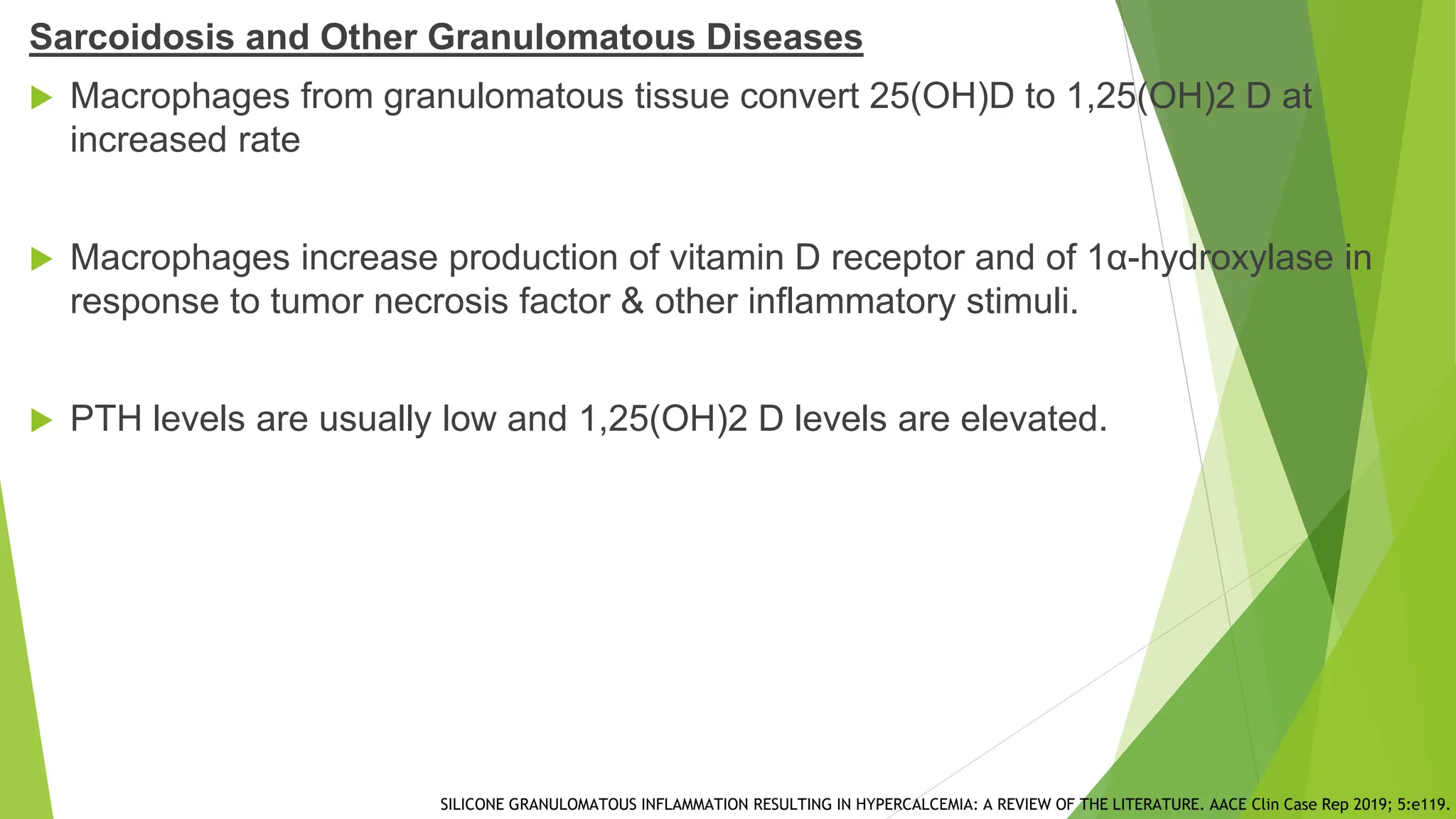
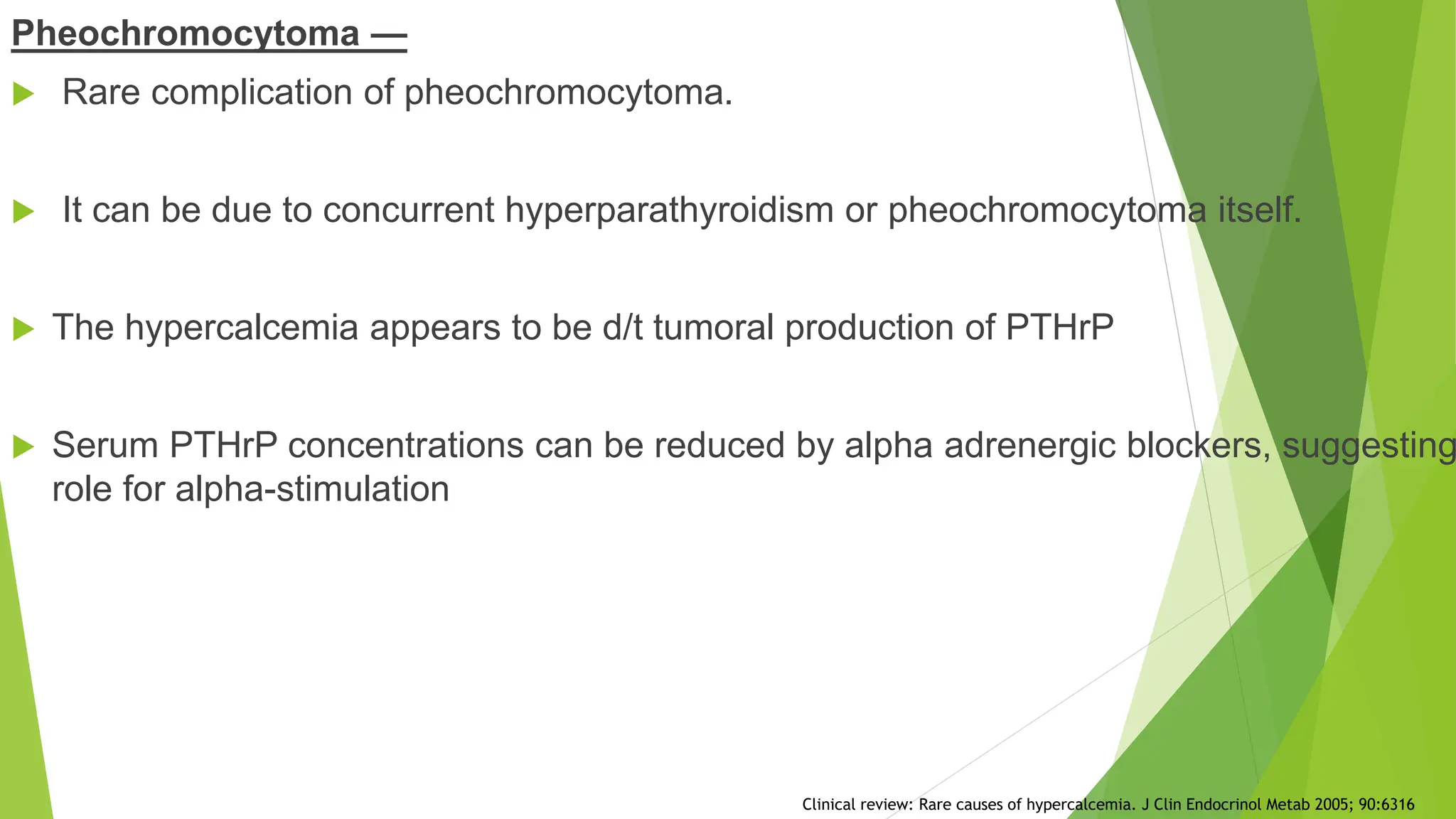
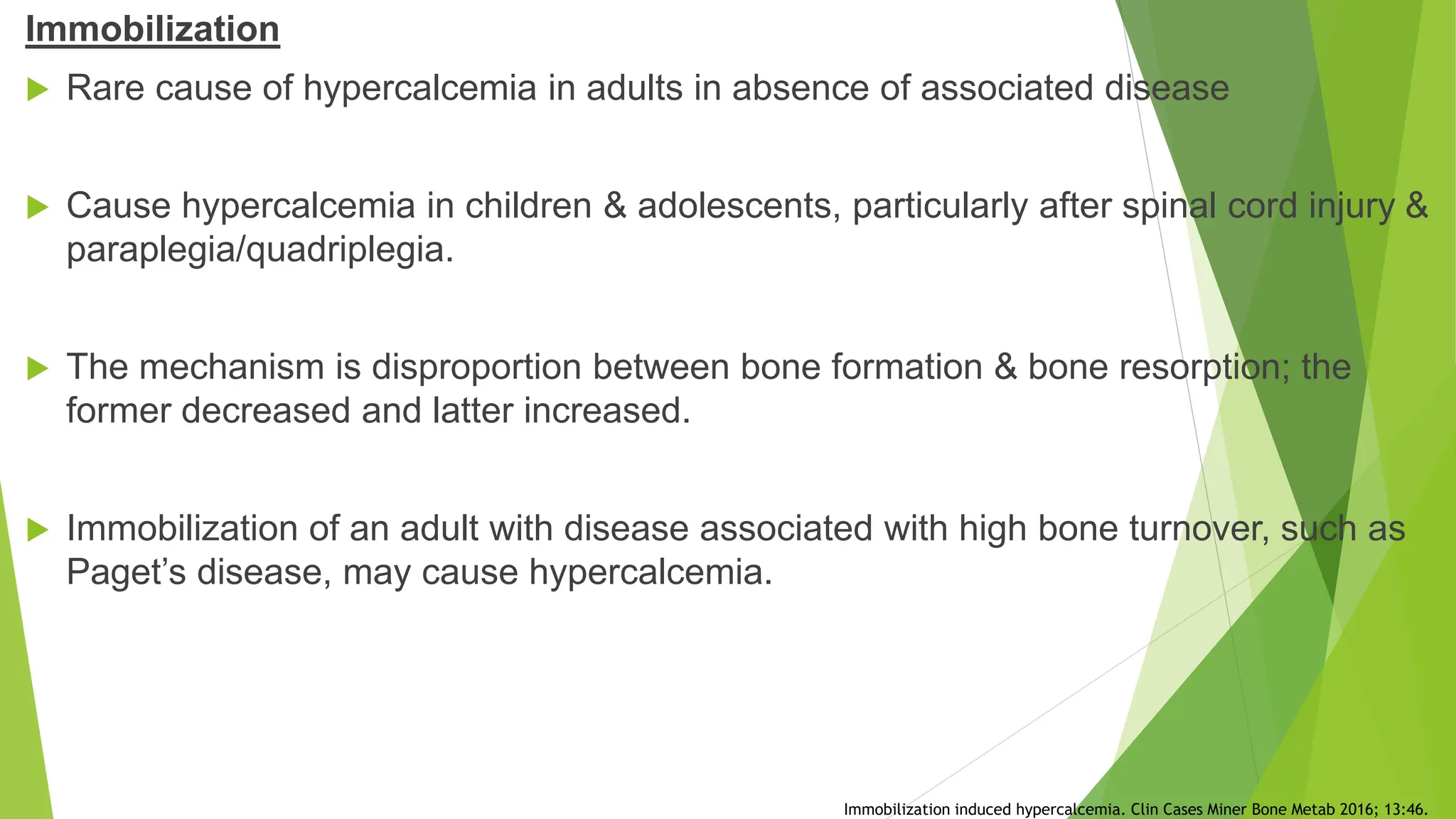
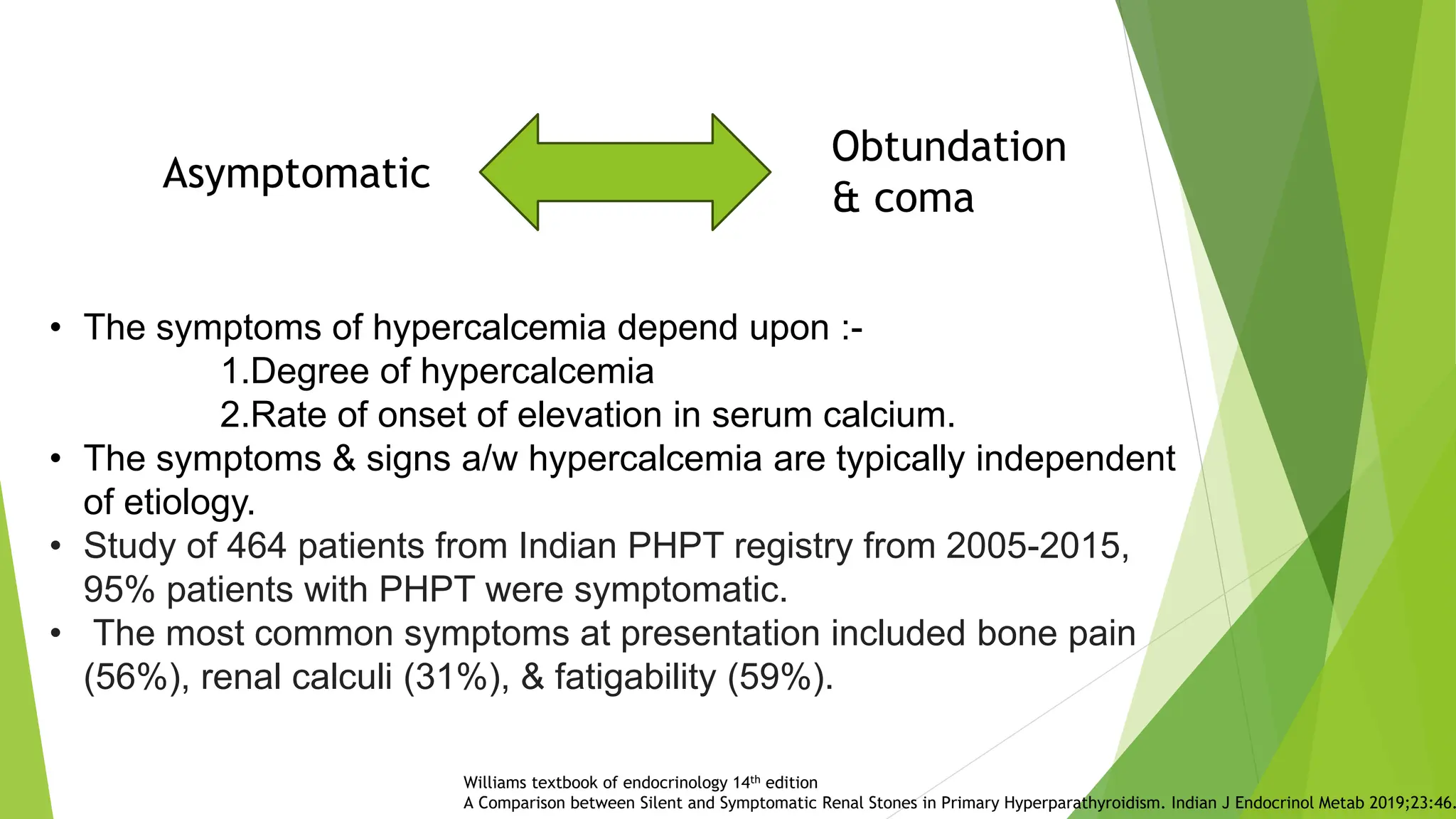
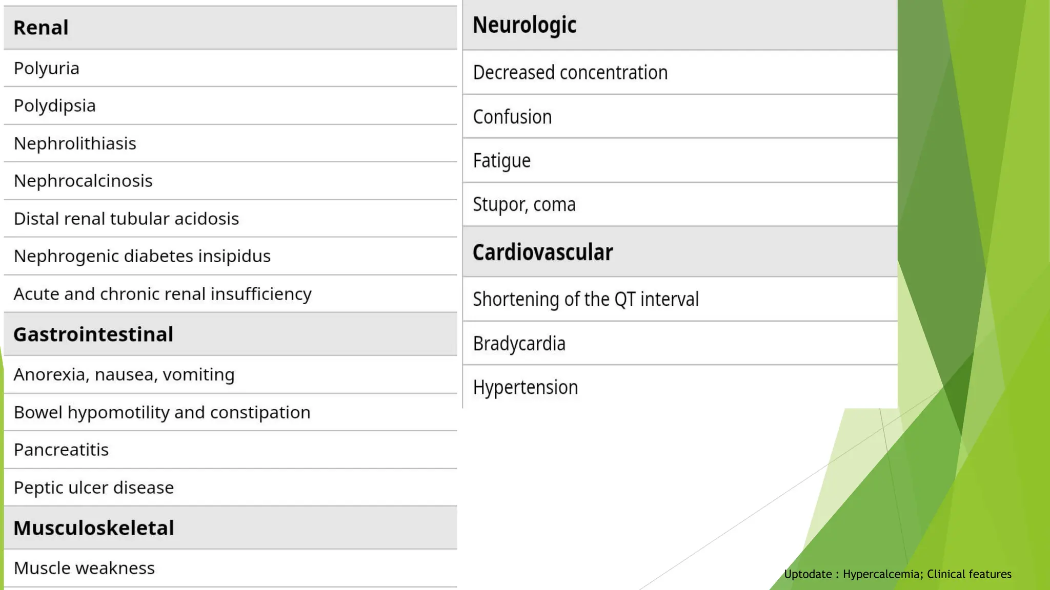
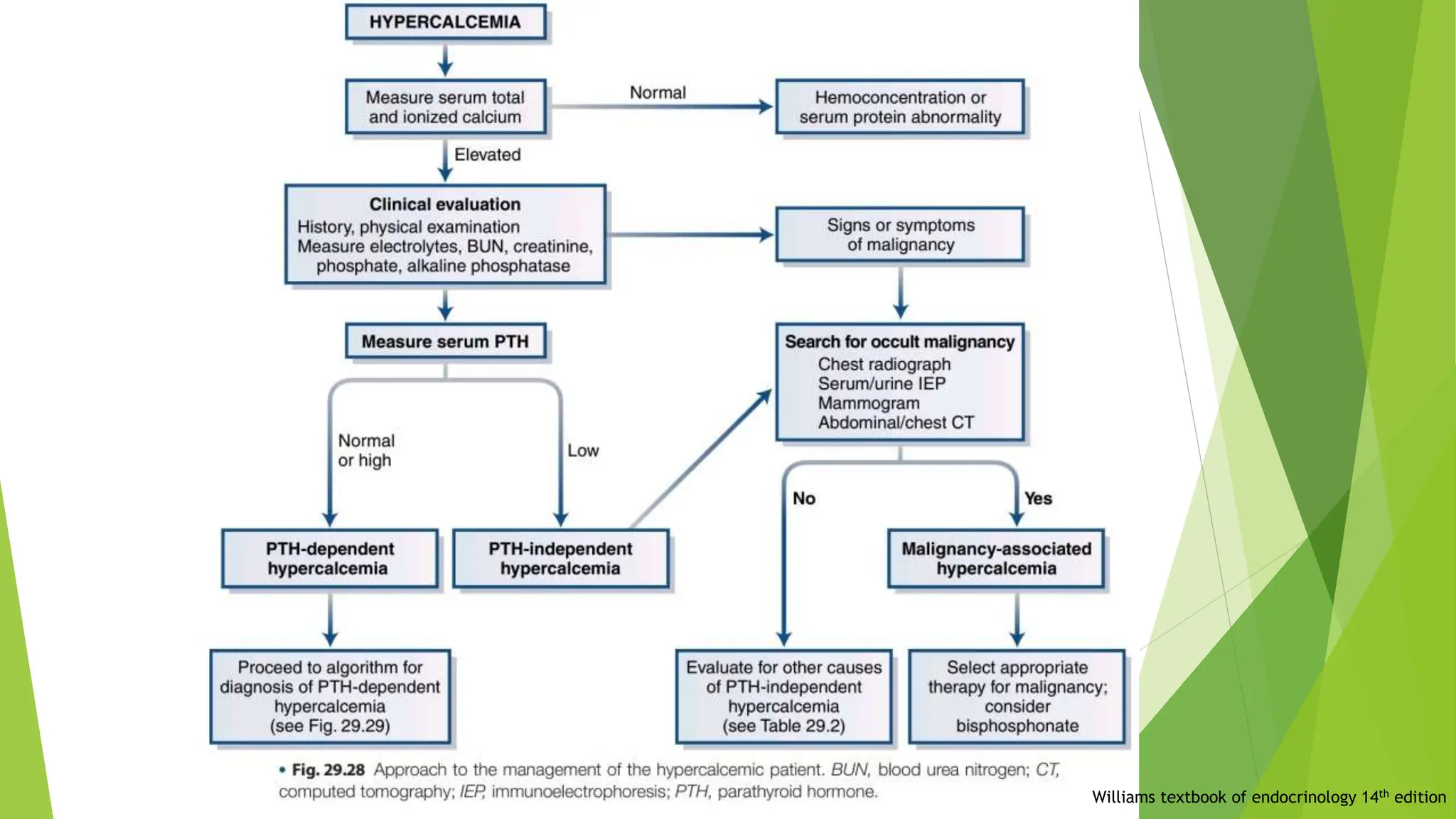
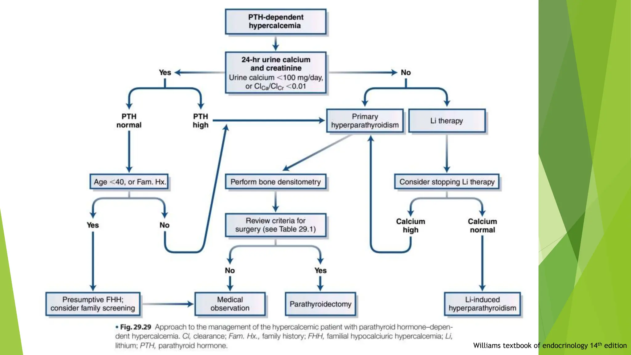
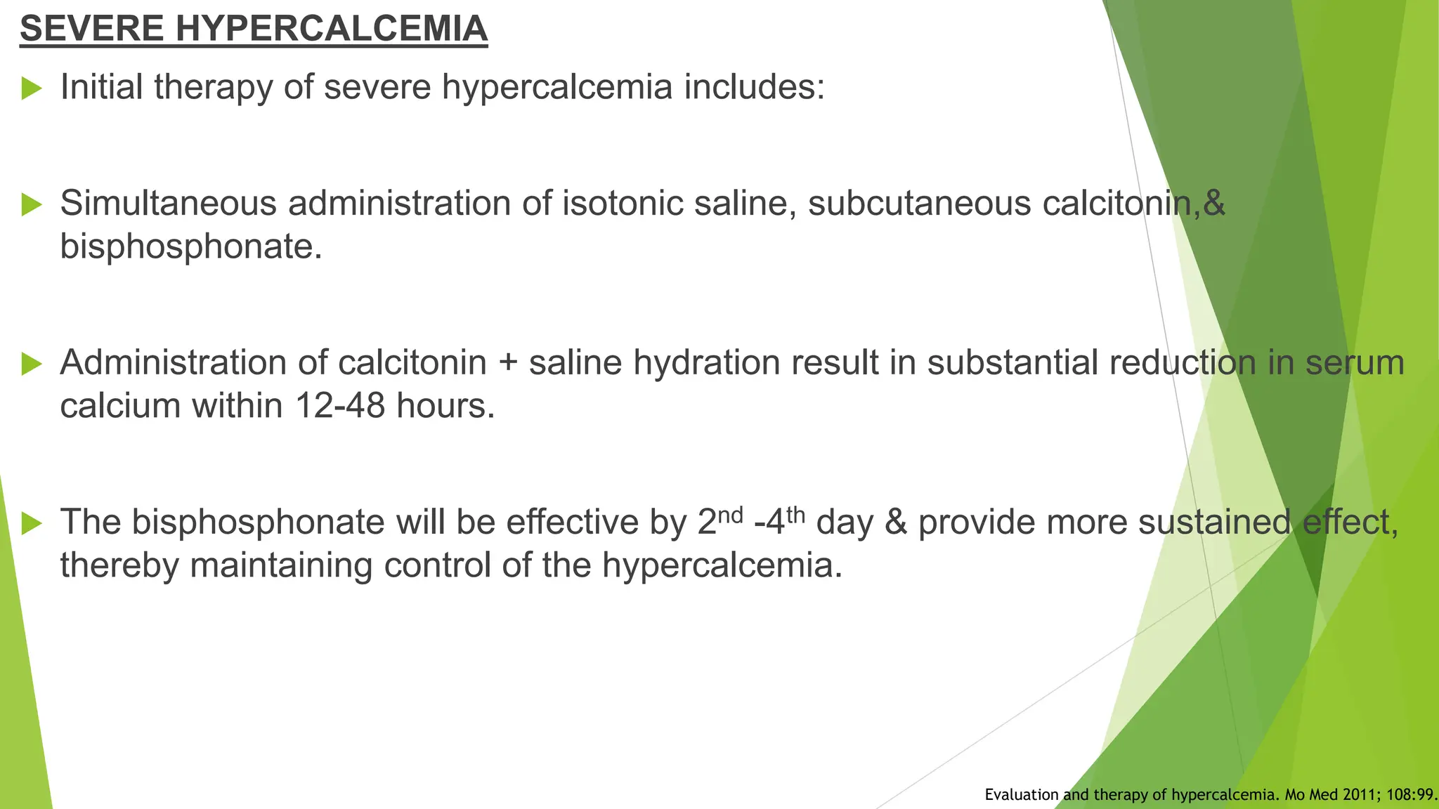
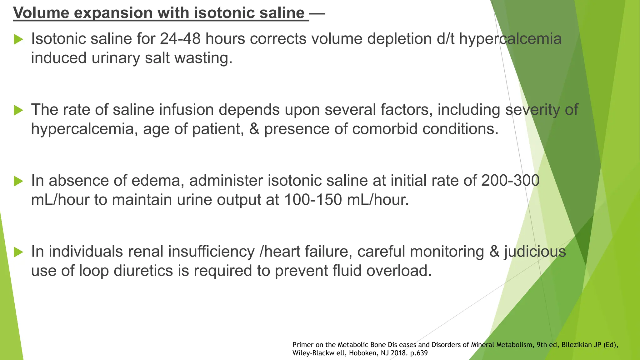
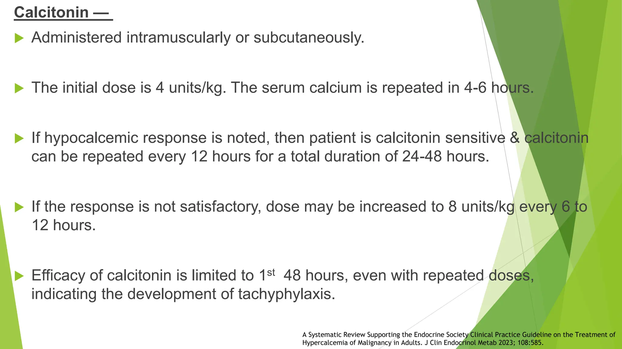
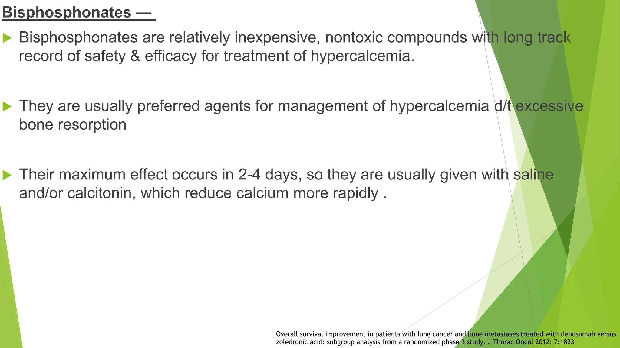
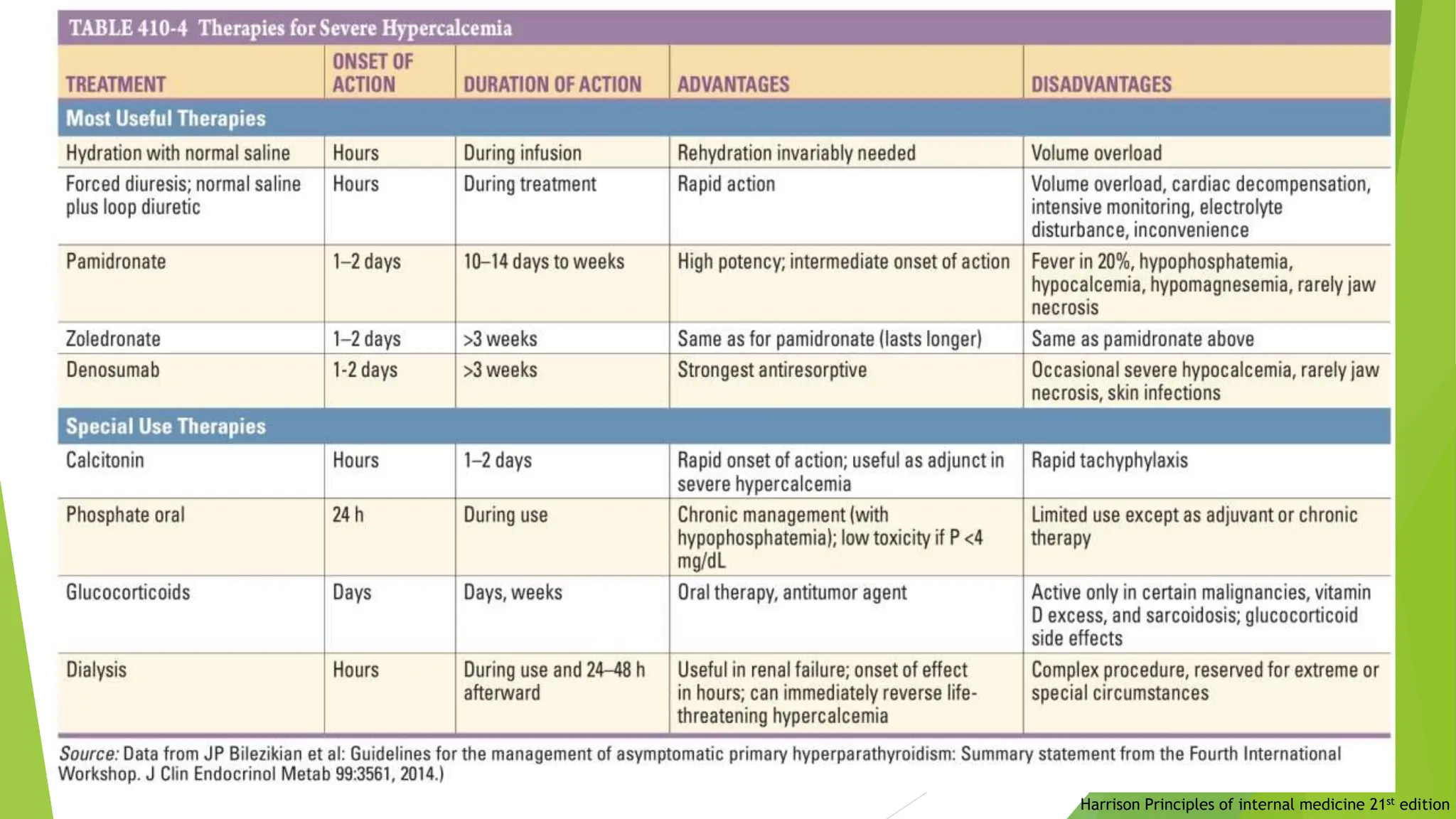
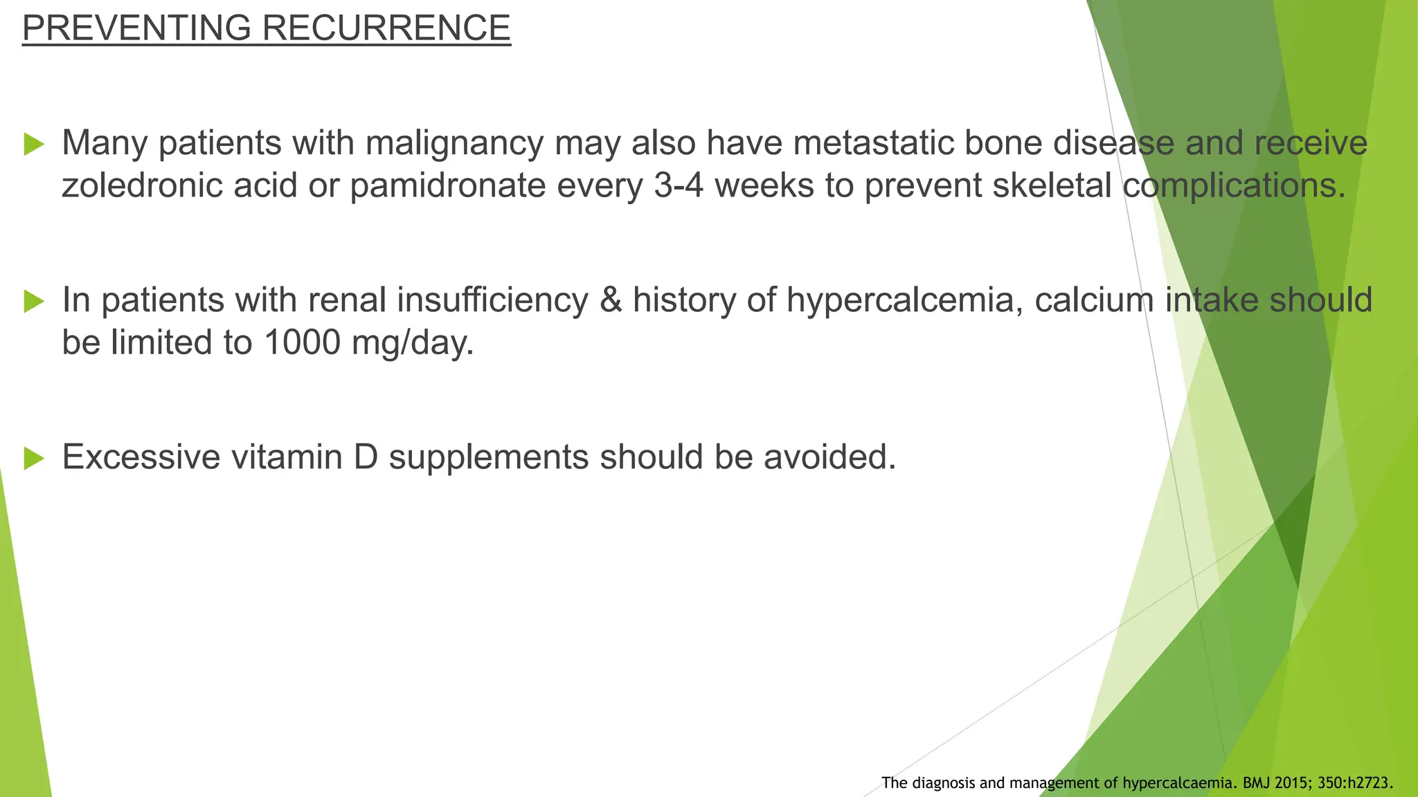
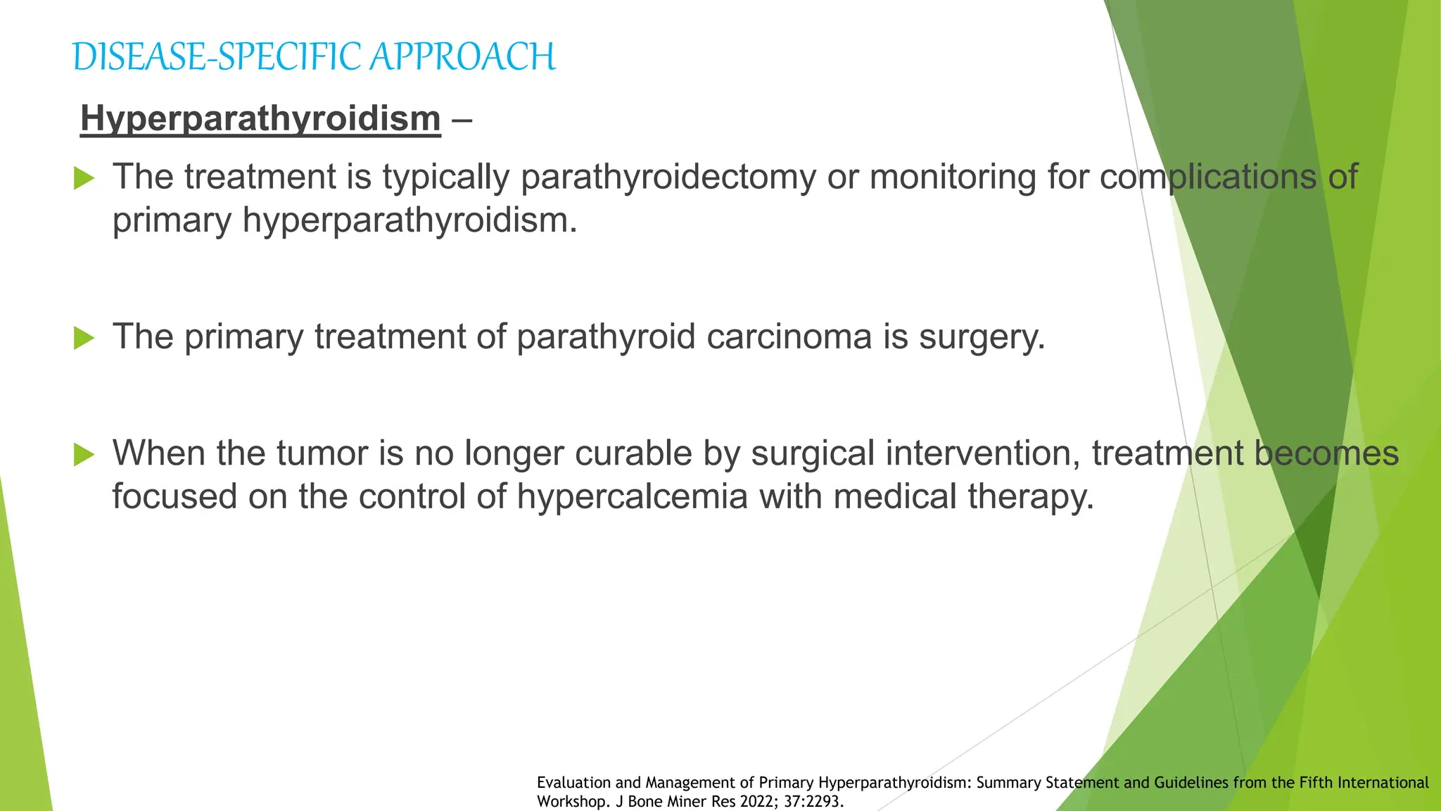
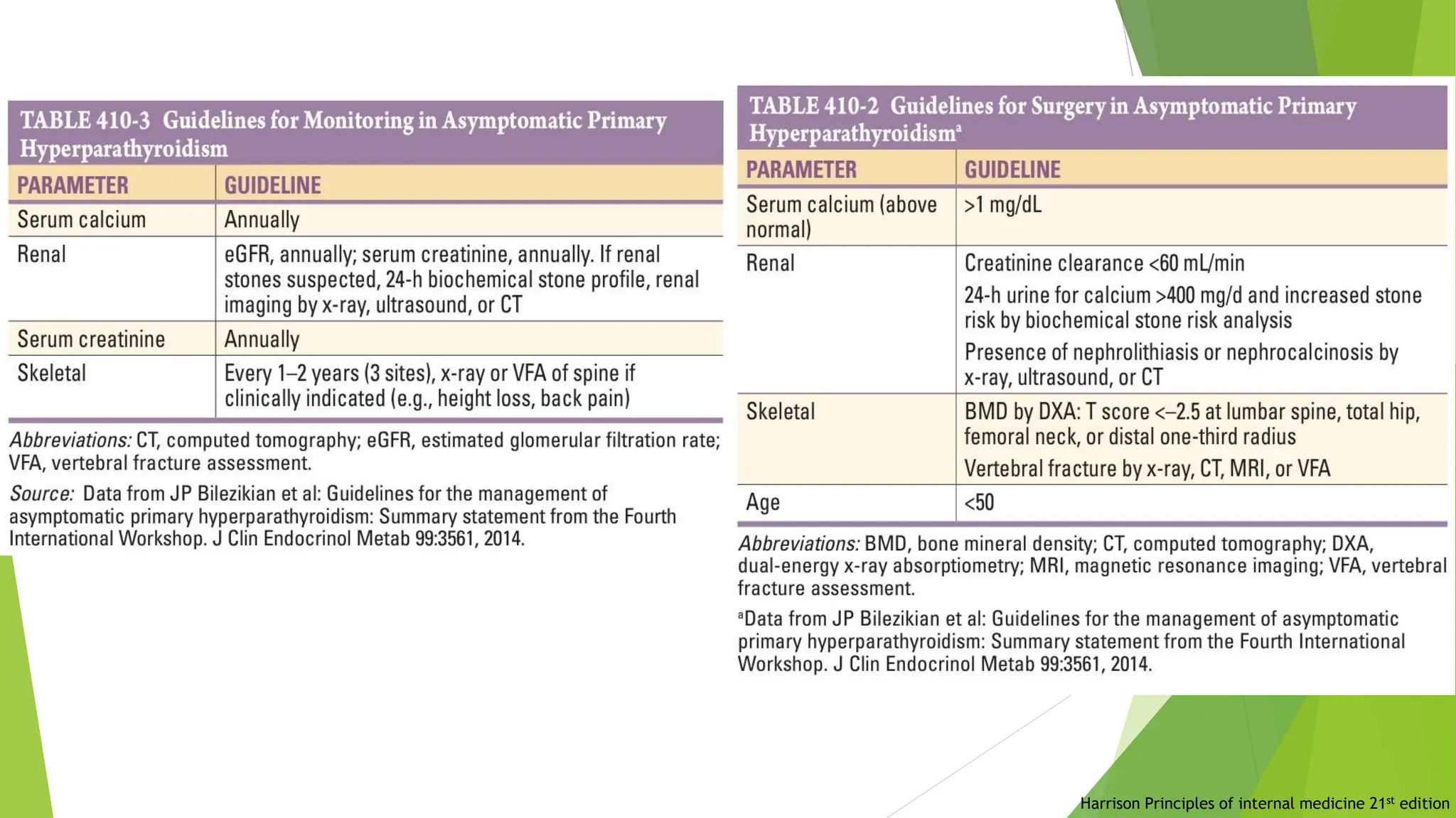
CALCIUM HOMEOSTASIS:
HYPERCALCEMIA
PRESENTER:SIDDHARTHA VATS
MODERATOR:PROF. NILESH KUMAR
 Calcium plays key role in:-
1. Contraction of skeletal, cardiac, & smooth muscles
2. Blood clotting
3. Transmission of nerve impulses
 Normal calcium levels :9-11mg/dl
 0.1% of total body calcium is in extracellular fluid
 About 1 % in cells & organelles.
 Rest is stored in bones.
Guyton and hall :Textbook medical physiology 12th edition
CALCIUM REGULATION
Guyton and hall :Textbook medical physiology 12th edition
Williams textbook of endocrinology 14th edition
Increases Ca++
and PO4
absorbtion
Williams textbook of endocrinology 14th edition
Guyton and hall :Textbook medical physiology 12th edition
HYPERCALCEMIA
Harrison Principles of internal medicine 21st edition
ETIOLOGY
 Entry of calcium into circulation exceeds excretion of calcium into urine/deposition
in bone.
 This occurs when there is
1.Accelerated bone resorption
2.Excessive gastrointestinal absorption, or
3.Decreased renal excretion of calcium.
 Among all causes of hypercalcemia, primary hyperparathyroidism &
malignancy are most common, accounting for >90% of cases
Hypercalcemia: A Review. JAMA 2022; 328:1624.
Primary hyperparathyroidism —
 Parathyroid hormone (PTH)-mediated activation of osteoclasts,causing increased
bone resorption & elevated intestinal calcium absorption
 Most often d/t parathyroid adenoma.
 Patients typically have relatively minor elevations in serum calcium concentration
:<12 mg/dL.
 Some patients have mostly high-normal values with intermittent hypercalcemia.
Asymptomatic primary hyperparathyroidism: a medical perspective. Surg Clin North Am 2004; 84:787.
Tertiary hyperparathyroidism —
 In patients with advanced renal failure, parathyroid hyperplasia may gradually
progress to autonomous overproduction of PTH
 The rise in plasma calcium is exacerbated by concomitant adynamic bone disease
and markedly reduced bone turnover.
Harrison Principles of internal medicine 21st edition
Familial hypocalciuric hypercalcemia —
 Rare autosomal dominant disorder
 Characterized by
1.Mild hypercalcemia.
2.Hypocalciuria.
3.Normal-mod. elevated serum magnesium.
4.Normal-slightly increased serum PTH.
 The majority of these patients have few/no symptoms of hypercalcemia & require
no therapy.
 The primary defect is loss-of-function mutation in calcium sensing sensor on
parathyroid cells and kidneys
Rare causes of hypercalcemia. J Clin Endocrinol Metab 2005; 90:6316.
Metaphyseal chondrodysplasia —
 Rare form of dwarfism.
 Primary defect is mutation in PTH-PTHrP receptor gene, resulting in continuous
activation of receptor at normal/ low levels of PTH secretion
 Patients have short-limbed dwarfism d/t abnormal regulation of chondrocyte
maturation in growth plates of bone that are formed through endochondral process.
Clinical review: Rare causes of hypercalcemia. J Clin Endocrinol Metab 2005; 90:6316
 In patients with bone metastases, direct induction of local osteolysis by tumor cells.
 Cytokines such as TNF & IL-1 play a role by stimulating the differentiation of
osteoclast precursors into mature osteoclasts.
 Solid tumors a/w hypercalcemia, particularly squamous cell and renal tumors,
produce PTHrP .
 Direct bone marrow invasion occurs with hematologic malignancies such as
leukemia, lymphoma, & multiple myeloma.
 Hypercalcemia is caused by PTH-independent, extrarenal production of calcitriol
from calcidiol by activated mononuclear cells in lymphoma.
Hypercalcemia: A Review. JAMA 2022; 328:1624.
Malignancy
Sarcoidosis and Other Granulomatous Diseases
 Macrophages from granulomatous tissue convert 25(OH)D to 1,25(OH)2 D at
increased rate
 Macrophages increase production of vitamin D receptor and of 1α-hydroxylase in
response to tumor necrosis factor & other inflammatory stimuli.
 PTH levels are usually low and 1,25(OH)2 D levels are elevated.
SILICONE GRANULOMATOUS INFLAMMATION RESULTING IN HYPERCALCEMIA: A REVIEW OF THE LITERATURE. AACE Clin Case Rep 2019; 5:e119.
MEDICATIONS
Lithium —
 Most likely d/t increased secretion of PTH due to increase in set point at which
calcium suppresses PTH release.
 The hypercalcemia usually, but not always, subsides when the lithium is
stopped.
 Lithium can also unmask previously unrecognized mild hyperparathyroidism.
Williams textbook of endocrinology 14th edition
Thiazide diuretics:
 Thiazide diuretics lower urinary calcium excretion, an effect that is useful in treatment
of patients with hypercalciuria & recurrent calcium nephrolithiasis
 Hypocalciuric effect appears to reflect enhancement of proximal tubular resorption of
sodium and calcium in response to sodium depletion.
 If hormonal function & calcium and bone metabolism are normal, homeostatic controls
counteract the calcium-elevating effect of the thiazides.
 Can lead to hypercalcemia in patients with underlying hyperparathyroidism, since
homeostatic mechanisms are ineffective.
Williams textbook of endocrinology 14th edition
Milk alkali syndrome –
 High intake of milk/calcium carbonate, leading to hypercalcemia, metabolic alkalosis,
& renal insufficiency.
 Calcium is absorbed in small intestine via both active & passive transport. The former
is more important physiologically & stimulated by vitamin D metabolites.
 But when calcium intake is >2g/d, substantial amounts of calcium is absorbed
passively.
Williams textbook of endocrinology 14th edition
ENDOCRINE DISORDERS
Thyrotoxicosis —
 Mild hypercalcemia occurs in ~15 -20% of thyrotoxic patients, d/t thyroid
hormone-mediated increase in bone resorption.
 It typically resolves following correction of hyperthyroidism.
 If the hypercalcemia persists after restoration of euthyroidism, serum PTH
should be measured to assess for concomitant hyperparathyroidism.
Williams textbook of endocrinology 14th edition
Pheochromocytoma —
 Rare complication of pheochromocytoma.
 It can be due to concurrent hyperparathyroidism or pheochromocytoma itself.
 The hypercalcemia appears to be d/t tumoral production of PTHrP
 Serum PTHrP concentrations can be reduced by alpha adrenergic blockers, suggesting
role for alpha-stimulation
Clinical review: Rare causes of hypercalcemia. J Clin Endocrinol Metab 2005; 90:6316
Immobilization
 Rare cause of hypercalcemia in adults in absence of associated disease
 Cause hypercalcemia in children & adolescents, particularly after spinal cord injury &
paraplegia/quadriplegia.
 The mechanism is disproportion between bone formation & bone resorption; the
former decreased and latter increased.
 Immobilization of an adult with disease associated with high bone turnover, such as
Paget’s disease, may cause hypercalcemia.
Immobilization induced hypercalcemia. Clin Cases Miner Bone Metab 2016; 13:46.
CLINICAL FEATURES
Asymptomatic
Obtundation
& coma
• The symptoms of hypercalcemia depend upon :-
1.Degree of hypercalcemia
2.Rate of onset of elevation in serum calcium.
• The symptoms & signs a/w hypercalcemia are typically independent
of etiology.
• Study of 464 patients from Indian PHPT registry from 2005-2015,
95% patients with PHPT were symptomatic.
• The most common symptoms at presentation included bone pain
(56%), renal calculi (31%), & fatigability (59%).
Williams textbook of endocrinology 14th edition
A Comparison between Silent and Symptomatic Renal Stones in Primary Hyperparathyroidism. Indian J Endocrinol Metab 2019;23:46.
Uptodate : Hypercalcemia; Clinical features
DIAGNOSIS
Williams textbook of endocrinology 14th edition
TREATMENT
 The degree of hypercalcemia, along with rate of rise of serum calcium, determines
symptoms & urgency of therapy.
 Patients with asymptomatic /mildly symptomatic hypercalcemia don’t require
immediate treatment.
 An acute rise may cause marked changes in sensorium, which requires more
aggressive measures.
 Patients with serum calcium >14 mg/dL require more aggressive treatment,
regardless of symptoms.
A practical approach to hypercalcemia. Am Fam Physician 2003; 67:1959.
MILD-MODERATE HYPERCALCEMIA.
 Advised to avoid factors that aggravate hypercalcemia:
1. Thiazide diuretics
2. Lithium
3. Volume depletion
4. Prolonged bed rest.
5. A high-calcium diet (>1000 mg/day)
6. Vitamin D supplements in excess of 800 international units/day.
 Adequate hydration (at least 6-8 glasses of water/day) is recommended to minimize
risk of nephrolithiasis.
 Acute rise cause marked changes in sensorium, treated same for severe
hypercalcemia.
Evaluation and therapy of hypercalcemia. Mo Med 2011; 108:99.
SEVERE HYPERCALCEMIA
 Initial therapy of severe hypercalcemia includes:
 Simultaneous administration of isotonic saline, subcutaneous calcitonin,&
bisphosphonate.
 Administration of calcitonin + saline hydration result in substantial reduction in serum
calcium within 12-48 hours.
 The bisphosphonate will be effective by 2nd -4th day & provide more sustained effect,
thereby maintaining control of the hypercalcemia.
Evaluation and therapy of hypercalcemia. Mo Med 2011; 108:99.
Volume expansion with isotonic saline —
 Isotonic saline for 24-48 hours corrects volume depletion d/t hypercalcemia
induced urinary salt wasting.
 The rate of saline infusion depends upon several factors, including severity of
hypercalcemia, age of patient, & presence of comorbid conditions.
 In absence of edema, administer isotonic saline at initial rate of 200-300
mL/hour to maintain urine output at 100-150 mL/hour.
 In individuals renal insufficiency /heart failure, careful monitoring & judicious
use of loop diuretics is required to prevent fluid overload.
Primer on the Metabolic Bone Dis eases and Disorders of Mineral Metabolism, 9th ed, Bilezikian JP (Ed),
Wiley-Blackw ell, Hoboken, NJ 2018. p.639
Calcitonin —
 Administered intramuscularly or subcutaneously.
 The initial dose is 4 units/kg. The serum calcium is repeated in 4-6 hours.
 If hypocalcemic response is noted, then patient is calcitonin sensitive & calcitonin
can be repeated every 12 hours for a total duration of 24-48 hours.
 If the response is not satisfactory, dose may be increased to 8 units/kg every 6 to
12 hours.
 Efficacy of calcitonin is limited to 1st 48 hours, even with repeated doses,
indicating the development of tachyphylaxis.
A Systematic Review Supporting the Endocrine Society Clinical Practice Guideline on the Treatment of
Hypercalcemia of Malignancy in Adults. J Clin Endocrinol Metab 2023; 108:585.
Bisphosphonates —
 Bisphosphonates are relatively inexpensive, nontoxic compounds with long track
record of safety & efficacy for treatment of hypercalcemia.
 They are usually preferred agents for management of hypercalcemia d/t excessive
bone resorption
 Their maximum effect occurs in 2-4 days, so they are usually given with saline
and/or calcitonin, which reduce calcium more rapidly .
Overall survival improvement in patients with lung cancer and bone metastases treated with denosumab versus
zoledronic acid: subgroup analysis from a randomized phase 3 study. J Thorac Oncol 2012; 7:1823
Denosumab-
 For patients in whom bisphosphonates are contraindicated /patients with
hypercalcemia refractory to zoledronic acid, denosumab can be administered
concurrently with calcitonin & saline hydration
 The risk of subsequent hypocalcemia is lower with bisphosphonates than
denosumab.
Denosumab versus intravenous bisphosphonate use for hypercalcemia in multiple myeloma. Leuk
Lymphoma 2022; 63:3249.
 Pretreatment considerations
1.Vitamin D – Some patients have hypercalcemia and concomitant vitamin D
deficiency.
Such patients are more likely to develop hypocalcemia after treatment with zoledronic
acid /denosumab.
If the 25hydroxyvitamin D level is < 20 ng/dL, vitamin D should be cautiously replaced
(eg, 400-800IU/day)
2.Creatinine – If renal function is impaired, the bisphosphonate should be infused at a
slower rate.
Patient and Physician Decisional Factors Regarding Hypercalcemia of Malignancy Treatment: A Novel
Mixed-Methods Study. J Clin Endocrinol Metab 2023; 108:563.
Choice of drug and dosing —
 Zoledronic acid :4 mg IV over 15 minutes repeated every 7 days.
 Pamidronate :60 to 90 mg over 2-24 hours repeated every 7 days.
 Ibandronate : 2-6mg IV over 2 hours.
 Denosumab :120 mg once weekly for up to 3 doses ; if the cause of hypercalcemia
persists, may continue at 120 mg every 4 weeks starting 2 weeks after initial 3
weekly doses
Side effects and precautions —
1. Flu-like symptoms (fever, arthralgias, myalgia, fatigue, bone pain),
2. Ocular inflammation (uveitis),
3. Hypocalcemia
4. Hypophosphatemia A practical approach to hypercalcemia. Am Fam Physician 2003; 67:1959.
Patient and Physician Decisional Factors Regarding Hypercalcemia of Malignancy Treatment: A Novel
Mixed-Methods Study. J Clin Endocrinol Metab 2023; 108:563.
Dialysis –
 Hemodialysis with little or no calcium in dialysis fluid & peritoneal dialysis are
both effective therapies for hypercalcemia.
 Dialysis may be indicated in patients with severe malignancy-associated
hypercalcemia and renal insufficiency/ heart failure, in whom hydration cannot be
safely administered
A Systematic Review Supporting the Endocrine Society Clinical Practice Guideline on the Treatment of
Hypercalcemia of Malignancy in Adults. J Clin Endocrinol Metab 2023; 108:585.
Harrison Principles of internal medicine 21st edition
PREVENTING RECURRENCE
 Many patients with malignancy may also have metastatic bone disease and receive
zoledronic acid or pamidronate every 3-4 weeks to prevent skeletal complications.
 In patients with renal insufficiency & history of hypercalcemia, calcium intake should
be limited to 1000 mg/day.
 Excessive vitamin D supplements should be avoided.
The diagnosis and management of hypercalcaemia. BMJ 2015; 350:h2723.
DISEASE-SPECIFIC APPROACH
Hyperparathyroidism –
 The treatment is typically parathyroidectomy or monitoring for complications of
primary hyperparathyroidism.
 The primary treatment of parathyroid carcinoma is surgery.
 When the tumor is no longer curable by surgical intervention, treatment becomes
focused on the control of hypercalcemia with medical therapy.
Evaluation and Management of Primary Hyperparathyroidism: Summary Statement and Guidelines from the Fifth International
Workshop. J Bone Miner Res 2022; 37:2293.
Harrison Principles of internal medicine 21st edition
 Granulomatous diseases –The major modalities of therapy are a low-calcium
diet, glucocorticoids, and treatment of underlying disease.
 Glucocorticoids (prednisone: 20-40 mg/d) will reduce serum calcium
concentrations within 2-5 days.
 Hypervitaminosis D –
 Calcitriol –Usually lasts ~ 1-2 days. Stopping calcitriol, increasing salt & fluid
intake, or hydrating with IV saline may be only therapy that is needed.
 Vitamin D – Hypercalcemia caused by calcidiol lasts longer, so more aggressive
therapy such as glucocorticoids (eg, prednisone- 20-40 mg/d) & zoledronic acid or
pamidronate may be necessary.
SILICONE GRANULOMATOUS INFLAMMATION RESULTING IN HYPERCALCEMIA: A REVIEW OF THE
LITERATURE. AACE Clin Case Rep 2019; 5:e119.
Vitamin D intoxication causes hypercalcaemia by increased bone resorption which responds to
pamidronate. Clin Endocrinol (Oxf) 1995; 43:531.
THANKYOU

HYPERCALCEMIA : :::: CALCIUM HOMEOSTASIS

  • 1.
  • 2.
     Calcium playskey role in:- 1. Contraction of skeletal, cardiac, & smooth muscles 2. Blood clotting 3. Transmission of nerve impulses  Normal calcium levels :9-11mg/dl  0.1% of total body calcium is in extracellular fluid  About 1 % in cells & organelles.  Rest is stored in bones. Guyton and hall :Textbook medical physiology 12th edition CALCIUM REGULATION
  • 3.
    Guyton and hall:Textbook medical physiology 12th edition
  • 4.
    Guyton and hall:Textbook medical physiology 12th edition
  • 5.
    Williams textbook ofendocrinology 14th edition Increases Ca++ and PO4 absorbtion
  • 6.
    Williams textbook ofendocrinology 14th edition
  • 7.
    Guyton and hall:Textbook medical physiology 12th edition
  • 8.
  • 9.
    Harrison Principles ofinternal medicine 21st edition ETIOLOGY
  • 10.
     Entry ofcalcium into circulation exceeds excretion of calcium into urine/deposition in bone.  This occurs when there is 1.Accelerated bone resorption 2.Excessive gastrointestinal absorption, or 3.Decreased renal excretion of calcium.  Among all causes of hypercalcemia, primary hyperparathyroidism & malignancy are most common, accounting for >90% of cases Hypercalcemia: A Review. JAMA 2022; 328:1624.
  • 11.
    Primary hyperparathyroidism — Parathyroid hormone (PTH)-mediated activation of osteoclasts,causing increased bone resorption & elevated intestinal calcium absorption  Most often d/t parathyroid adenoma.  Patients typically have relatively minor elevations in serum calcium concentration :<12 mg/dL.  Some patients have mostly high-normal values with intermittent hypercalcemia. Asymptomatic primary hyperparathyroidism: a medical perspective. Surg Clin North Am 2004; 84:787.
  • 12.
    Tertiary hyperparathyroidism — In patients with advanced renal failure, parathyroid hyperplasia may gradually progress to autonomous overproduction of PTH  The rise in plasma calcium is exacerbated by concomitant adynamic bone disease and markedly reduced bone turnover. Harrison Principles of internal medicine 21st edition
  • 13.
    Familial hypocalciuric hypercalcemia—  Rare autosomal dominant disorder  Characterized by 1.Mild hypercalcemia. 2.Hypocalciuria. 3.Normal-mod. elevated serum magnesium. 4.Normal-slightly increased serum PTH.  The majority of these patients have few/no symptoms of hypercalcemia & require no therapy.  The primary defect is loss-of-function mutation in calcium sensing sensor on parathyroid cells and kidneys Rare causes of hypercalcemia. J Clin Endocrinol Metab 2005; 90:6316.
  • 14.
    Metaphyseal chondrodysplasia — Rare form of dwarfism.  Primary defect is mutation in PTH-PTHrP receptor gene, resulting in continuous activation of receptor at normal/ low levels of PTH secretion  Patients have short-limbed dwarfism d/t abnormal regulation of chondrocyte maturation in growth plates of bone that are formed through endochondral process. Clinical review: Rare causes of hypercalcemia. J Clin Endocrinol Metab 2005; 90:6316
  • 15.
     In patientswith bone metastases, direct induction of local osteolysis by tumor cells.  Cytokines such as TNF & IL-1 play a role by stimulating the differentiation of osteoclast precursors into mature osteoclasts.  Solid tumors a/w hypercalcemia, particularly squamous cell and renal tumors, produce PTHrP .  Direct bone marrow invasion occurs with hematologic malignancies such as leukemia, lymphoma, & multiple myeloma.  Hypercalcemia is caused by PTH-independent, extrarenal production of calcitriol from calcidiol by activated mononuclear cells in lymphoma. Hypercalcemia: A Review. JAMA 2022; 328:1624. Malignancy
  • 16.
    Sarcoidosis and OtherGranulomatous Diseases  Macrophages from granulomatous tissue convert 25(OH)D to 1,25(OH)2 D at increased rate  Macrophages increase production of vitamin D receptor and of 1α-hydroxylase in response to tumor necrosis factor & other inflammatory stimuli.  PTH levels are usually low and 1,25(OH)2 D levels are elevated. SILICONE GRANULOMATOUS INFLAMMATION RESULTING IN HYPERCALCEMIA: A REVIEW OF THE LITERATURE. AACE Clin Case Rep 2019; 5:e119.
  • 17.
    MEDICATIONS Lithium —  Mostlikely d/t increased secretion of PTH due to increase in set point at which calcium suppresses PTH release.  The hypercalcemia usually, but not always, subsides when the lithium is stopped.  Lithium can also unmask previously unrecognized mild hyperparathyroidism. Williams textbook of endocrinology 14th edition
  • 18.
    Thiazide diuretics:  Thiazidediuretics lower urinary calcium excretion, an effect that is useful in treatment of patients with hypercalciuria & recurrent calcium nephrolithiasis  Hypocalciuric effect appears to reflect enhancement of proximal tubular resorption of sodium and calcium in response to sodium depletion.  If hormonal function & calcium and bone metabolism are normal, homeostatic controls counteract the calcium-elevating effect of the thiazides.  Can lead to hypercalcemia in patients with underlying hyperparathyroidism, since homeostatic mechanisms are ineffective. Williams textbook of endocrinology 14th edition
  • 19.
    Milk alkali syndrome–  High intake of milk/calcium carbonate, leading to hypercalcemia, metabolic alkalosis, & renal insufficiency.  Calcium is absorbed in small intestine via both active & passive transport. The former is more important physiologically & stimulated by vitamin D metabolites.  But when calcium intake is >2g/d, substantial amounts of calcium is absorbed passively. Williams textbook of endocrinology 14th edition
  • 20.
    ENDOCRINE DISORDERS Thyrotoxicosis — Mild hypercalcemia occurs in ~15 -20% of thyrotoxic patients, d/t thyroid hormone-mediated increase in bone resorption.  It typically resolves following correction of hyperthyroidism.  If the hypercalcemia persists after restoration of euthyroidism, serum PTH should be measured to assess for concomitant hyperparathyroidism. Williams textbook of endocrinology 14th edition
  • 21.
    Pheochromocytoma —  Rarecomplication of pheochromocytoma.  It can be due to concurrent hyperparathyroidism or pheochromocytoma itself.  The hypercalcemia appears to be d/t tumoral production of PTHrP  Serum PTHrP concentrations can be reduced by alpha adrenergic blockers, suggesting role for alpha-stimulation Clinical review: Rare causes of hypercalcemia. J Clin Endocrinol Metab 2005; 90:6316
  • 22.
    Immobilization  Rare causeof hypercalcemia in adults in absence of associated disease  Cause hypercalcemia in children & adolescents, particularly after spinal cord injury & paraplegia/quadriplegia.  The mechanism is disproportion between bone formation & bone resorption; the former decreased and latter increased.  Immobilization of an adult with disease associated with high bone turnover, such as Paget’s disease, may cause hypercalcemia. Immobilization induced hypercalcemia. Clin Cases Miner Bone Metab 2016; 13:46.
  • 23.
  • 24.
    Asymptomatic Obtundation & coma • Thesymptoms of hypercalcemia depend upon :- 1.Degree of hypercalcemia 2.Rate of onset of elevation in serum calcium. • The symptoms & signs a/w hypercalcemia are typically independent of etiology. • Study of 464 patients from Indian PHPT registry from 2005-2015, 95% patients with PHPT were symptomatic. • The most common symptoms at presentation included bone pain (56%), renal calculi (31%), & fatigability (59%). Williams textbook of endocrinology 14th edition A Comparison between Silent and Symptomatic Renal Stones in Primary Hyperparathyroidism. Indian J Endocrinol Metab 2019;23:46.
  • 25.
    Uptodate : Hypercalcemia;Clinical features
  • 26.
  • 27.
    Williams textbook ofendocrinology 14th edition
  • 28.
    Williams textbook ofendocrinology 14th edition
  • 29.
  • 30.
     The degreeof hypercalcemia, along with rate of rise of serum calcium, determines symptoms & urgency of therapy.  Patients with asymptomatic /mildly symptomatic hypercalcemia don’t require immediate treatment.  An acute rise may cause marked changes in sensorium, which requires more aggressive measures.  Patients with serum calcium >14 mg/dL require more aggressive treatment, regardless of symptoms. A practical approach to hypercalcemia. Am Fam Physician 2003; 67:1959.
  • 31.
    MILD-MODERATE HYPERCALCEMIA.  Advisedto avoid factors that aggravate hypercalcemia: 1. Thiazide diuretics 2. Lithium 3. Volume depletion 4. Prolonged bed rest. 5. A high-calcium diet (>1000 mg/day) 6. Vitamin D supplements in excess of 800 international units/day.  Adequate hydration (at least 6-8 glasses of water/day) is recommended to minimize risk of nephrolithiasis.  Acute rise cause marked changes in sensorium, treated same for severe hypercalcemia. Evaluation and therapy of hypercalcemia. Mo Med 2011; 108:99.
  • 32.
    SEVERE HYPERCALCEMIA  Initialtherapy of severe hypercalcemia includes:  Simultaneous administration of isotonic saline, subcutaneous calcitonin,& bisphosphonate.  Administration of calcitonin + saline hydration result in substantial reduction in serum calcium within 12-48 hours.  The bisphosphonate will be effective by 2nd -4th day & provide more sustained effect, thereby maintaining control of the hypercalcemia. Evaluation and therapy of hypercalcemia. Mo Med 2011; 108:99.
  • 33.
    Volume expansion withisotonic saline —  Isotonic saline for 24-48 hours corrects volume depletion d/t hypercalcemia induced urinary salt wasting.  The rate of saline infusion depends upon several factors, including severity of hypercalcemia, age of patient, & presence of comorbid conditions.  In absence of edema, administer isotonic saline at initial rate of 200-300 mL/hour to maintain urine output at 100-150 mL/hour.  In individuals renal insufficiency /heart failure, careful monitoring & judicious use of loop diuretics is required to prevent fluid overload. Primer on the Metabolic Bone Dis eases and Disorders of Mineral Metabolism, 9th ed, Bilezikian JP (Ed), Wiley-Blackw ell, Hoboken, NJ 2018. p.639
  • 34.
    Calcitonin —  Administeredintramuscularly or subcutaneously.  The initial dose is 4 units/kg. The serum calcium is repeated in 4-6 hours.  If hypocalcemic response is noted, then patient is calcitonin sensitive & calcitonin can be repeated every 12 hours for a total duration of 24-48 hours.  If the response is not satisfactory, dose may be increased to 8 units/kg every 6 to 12 hours.  Efficacy of calcitonin is limited to 1st 48 hours, even with repeated doses, indicating the development of tachyphylaxis. A Systematic Review Supporting the Endocrine Society Clinical Practice Guideline on the Treatment of Hypercalcemia of Malignancy in Adults. J Clin Endocrinol Metab 2023; 108:585.
  • 35.
    Bisphosphonates —  Bisphosphonatesare relatively inexpensive, nontoxic compounds with long track record of safety & efficacy for treatment of hypercalcemia.  They are usually preferred agents for management of hypercalcemia d/t excessive bone resorption  Their maximum effect occurs in 2-4 days, so they are usually given with saline and/or calcitonin, which reduce calcium more rapidly . Overall survival improvement in patients with lung cancer and bone metastases treated with denosumab versus zoledronic acid: subgroup analysis from a randomized phase 3 study. J Thorac Oncol 2012; 7:1823
  • 36.
    Denosumab-  For patientsin whom bisphosphonates are contraindicated /patients with hypercalcemia refractory to zoledronic acid, denosumab can be administered concurrently with calcitonin & saline hydration  The risk of subsequent hypocalcemia is lower with bisphosphonates than denosumab. Denosumab versus intravenous bisphosphonate use for hypercalcemia in multiple myeloma. Leuk Lymphoma 2022; 63:3249.
  • 37.
     Pretreatment considerations 1.VitaminD – Some patients have hypercalcemia and concomitant vitamin D deficiency. Such patients are more likely to develop hypocalcemia after treatment with zoledronic acid /denosumab. If the 25hydroxyvitamin D level is < 20 ng/dL, vitamin D should be cautiously replaced (eg, 400-800IU/day) 2.Creatinine – If renal function is impaired, the bisphosphonate should be infused at a slower rate. Patient and Physician Decisional Factors Regarding Hypercalcemia of Malignancy Treatment: A Novel Mixed-Methods Study. J Clin Endocrinol Metab 2023; 108:563.
  • 38.
    Choice of drugand dosing —  Zoledronic acid :4 mg IV over 15 minutes repeated every 7 days.  Pamidronate :60 to 90 mg over 2-24 hours repeated every 7 days.  Ibandronate : 2-6mg IV over 2 hours.  Denosumab :120 mg once weekly for up to 3 doses ; if the cause of hypercalcemia persists, may continue at 120 mg every 4 weeks starting 2 weeks after initial 3 weekly doses Side effects and precautions — 1. Flu-like symptoms (fever, arthralgias, myalgia, fatigue, bone pain), 2. Ocular inflammation (uveitis), 3. Hypocalcemia 4. Hypophosphatemia A practical approach to hypercalcemia. Am Fam Physician 2003; 67:1959. Patient and Physician Decisional Factors Regarding Hypercalcemia of Malignancy Treatment: A Novel Mixed-Methods Study. J Clin Endocrinol Metab 2023; 108:563.
  • 39.
    Dialysis –  Hemodialysiswith little or no calcium in dialysis fluid & peritoneal dialysis are both effective therapies for hypercalcemia.  Dialysis may be indicated in patients with severe malignancy-associated hypercalcemia and renal insufficiency/ heart failure, in whom hydration cannot be safely administered A Systematic Review Supporting the Endocrine Society Clinical Practice Guideline on the Treatment of Hypercalcemia of Malignancy in Adults. J Clin Endocrinol Metab 2023; 108:585.
  • 40.
    Harrison Principles ofinternal medicine 21st edition
  • 41.
    PREVENTING RECURRENCE  Manypatients with malignancy may also have metastatic bone disease and receive zoledronic acid or pamidronate every 3-4 weeks to prevent skeletal complications.  In patients with renal insufficiency & history of hypercalcemia, calcium intake should be limited to 1000 mg/day.  Excessive vitamin D supplements should be avoided. The diagnosis and management of hypercalcaemia. BMJ 2015; 350:h2723.
  • 42.
    DISEASE-SPECIFIC APPROACH Hyperparathyroidism – The treatment is typically parathyroidectomy or monitoring for complications of primary hyperparathyroidism.  The primary treatment of parathyroid carcinoma is surgery.  When the tumor is no longer curable by surgical intervention, treatment becomes focused on the control of hypercalcemia with medical therapy. Evaluation and Management of Primary Hyperparathyroidism: Summary Statement and Guidelines from the Fifth International Workshop. J Bone Miner Res 2022; 37:2293.
  • 43.
    Harrison Principles ofinternal medicine 21st edition
  • 44.
     Granulomatous diseases–The major modalities of therapy are a low-calcium diet, glucocorticoids, and treatment of underlying disease.  Glucocorticoids (prednisone: 20-40 mg/d) will reduce serum calcium concentrations within 2-5 days.  Hypervitaminosis D –  Calcitriol –Usually lasts ~ 1-2 days. Stopping calcitriol, increasing salt & fluid intake, or hydrating with IV saline may be only therapy that is needed.  Vitamin D – Hypercalcemia caused by calcidiol lasts longer, so more aggressive therapy such as glucocorticoids (eg, prednisone- 20-40 mg/d) & zoledronic acid or pamidronate may be necessary. SILICONE GRANULOMATOUS INFLAMMATION RESULTING IN HYPERCALCEMIA: A REVIEW OF THE LITERATURE. AACE Clin Case Rep 2019; 5:e119. Vitamin D intoxication causes hypercalcaemia by increased bone resorption which responds to pamidronate. Clin Endocrinol (Oxf) 1995; 43:531.
  • 45.

Editor's Notes

  • #12 Occasionally, patients have more severe hypercalcemia with levels >12 mg/dL. When primary hyperparathyroidism is supected, and serum calcium is high-normal, it is necessary to obtain serial measurements of serum calcium to detect hypercalcemia.
  • #13 that is not suppressible by elevated serum calcium concentrations. Which causes reduction in the skeletal uptake of calcium after a calcium load, eg. when calcium carbonate used as a phosphate binder to treat hyperphosphatemia.
  • #14 PTH may be elevated in the different forms of FHH, but the values are usually normal or lower for the same degree of calcium elevation than is observed in patients with primary hyperparathyroidism. Higher than normal serum calcium concentrations are needed to suppress PTH release. Subtotal parathyroidectomy does not correct the hypercalcemia.
  • #15 Asymptomatic but significant hypercalcemia & hypophosphatemia. In adult life, abnormalities in bone, includes multiple cystic resorptive areas resembling those in severe hyperparathyroidism. Parathyroid glands are normal, & serum PTH and PTH-related protein (PTHrP) concentrations are normal or low.
  • #16 Lymphokines and cytokines (including PTHrP) produced by cells involved in the marrow response to the tumors promote resorption of bone through local destruction. The mechanism of increased bone resorption with malignancy depends upon cancer. PTHrP .that causes increased bone resorption & mediates hypercalcemia via systemic actions on skeleton. Hypercalcemia occurs in patients with many malignancies, both solid tumors & leukemias. In general, serum calcium levels are higher in patients with malignancy than those with primary hyperparathyroidism. Values above 13 mg/dL are less commonly seen in primary hyperparathyroidism and are more likely d/t malignancy.
  • #17 positive correlation in patients with sarcoidosis between 25(OH)D levels (reflecting vitamin D intake) and the circulating concentrations of 1,25(OH)2 D, whereas normally, there is no increase in 1,25(OH)2 D with increasing 25(OH)D levels due to multiple feedback controls on renal 1α-hydroxylase Clearance of 1,25(OH)2 D from blood may be decreased in sarcoidosis as well. Alternatively, glucocorticoids in the equivalent of 100 mg/d of hydrocortisone or equivalent doses of glucocorticoids may help control hypercalcemia. Glucocorticoids appear to act by blocking excessive production of 1,25(OH)2 D, as well as response to it in target organs. Management of hypercalcemia can be accomplished by limiting vitamin D & calcium intake. The abnormal sensitivity to vitamin D & abnormal regulation of 1,25(OH)2 D synthesis will persist as long as disease is active. In patients with sarcoidosis & other granulomatous diseases, such as TB & fungal infections, excess 1,25(OH)2 D is synthesized in macrophages & other cells in granulomas.
  • #20 The metabolic alkalosis augments the hypercalcemia by directly stimulating calcium reabsorption in the distal tubule, thereby diminishing calcium excretion. Several clinical presentations—acute, subacute, and chronic—have been described, all of which feature hypercalcemia, alkalosis, and renal failure The chronic form of disease “Burnett’s syndrome”, a/w irreversible renal damage. The acute syndromes reverse if the excess calcium and absorbable alkali are stopped. The milk-alkali syndrome typically occurs in excess calcium carbonate supplementation to treat osteoporosis or dyspepsia. A calcium-induced decline in renal function, due to renal vasoconstriction,& with chronic hypercalcemia- structural injury, cause inability to excrete excess calcium. Renal function returns to baseline after cessation of milk or calcium carbonate intake, but irreversible injury can occur with prolonged hypercalcemia.
  • #23 With resumption of ambulation, hypercalcemia in children usually returns to normal.
  • #25 Parathyroid crisis is characterized by severe hypercalcemia, with the serum calcium concentration usually above 15 mg/dL (3.8 mmol/L) and marked symptoms of hypercalcemia: in particular, central nervous system dysfunction. (mean age 41 years
  • #26 Downregulation of aquaporin-2 water channels & calcium deposition in medulla with secondary tubulointerstitial injury & impaired generation of interstitial osmotic gradient play important roles.
  • #31 Patients with serum calcium of 12 -14 mg/dL may not require immediate treatment, because it is tolerated well chronically.
  • #32 Patients with asymptomatic/ mildly symptomatic hypercalcemia don’t require immediate treatment
  • #34 Most patients presenting with severe hypercalcemia have marked intravascular volume depletion. Hypovolemia exacerbates hypercalcemia by impairing renal clearance of calcium. Saline therapy rarely normalizes serum calcium in patients with more than mild hypercalcemia.
  • #35 Because of its limited duration of effect, calcitonin is most useful in symptomatic patients with calcium >14 mg/dL (3.5 mmol/L), when combined with hydration and bisphosphonates (or denosumab, in bisphosphonate-refractory patients). (other than mild nausea and the rare hypersensitivity reaction). Calcitonin is safe and relatively nontoxic Although a relatively weak agent, it works rapidly, lowering the serum calcium concentration by a maximum of 1 to 2 mg/dL (0.3 to 0.5 mmol/L) beginning within four to six hours [1,5,15,16]. Thus, it is useful in combination with hydration for the initial management of severe hypercalcemia. perhaps due to receptor downregulation [1,11-13] Nasal administration of calcitonin is not efficacious for treatment of hypercalcemia Pharmacologic doses of calcitonin reduce serum calcium by increasing renal calcium excretion & by decreasing bone resorption via interference with osteoclast function
  • #36 For longer-term control of hypercalcemia in patients with severe/symptomatic hypercalcemia Randomized trials show evidence of bisphosphonate efficacy compared with saline alone for the treatment hypercalcemia of malignancy [18-22]. (eg, due to severe kidney impairment <35ml/min, bisphosphonate allergy), For patients in whom bisphosphonates are contraindicated (eg, due to severe renal impairment, bisphosphonate allergy), or in patients with hypercalcemia refractory to zoledronic acid, denosumab is an option and can be administered concurrently with calcitonin and saline hydration
  • #37 There are no randomized trials specifically evaluating denosumab for hypercalcemia of malignancy. In an observational study in patients with hypercalcemia of malignancy, there was no difference between denosumab & IV bisphosphonates in response rate by day 7 (89 & 86 %, respectively).
  • #38 An elevated serum concentration of 25-hydroxyvitamin D is indicative of vitamin D intoxication due to the ingestion of either vitamin D or calcidiol itself. However, some patients with hypercalcemia due to another etiology (eg, hypercalcemia of malignancy)
  • #39 Repetitive use of bisphosphonates has been associated with risk of developing osteonecrosis of the jaw and atypical femur fractures (in patients who require long-term therapy) [48,52]. These side effects associated with long-term therapy may be of limited relevance in the management of acute hypercalcemia
  • #40 The use of hemodialysis for patients with hypercalcemia but without renal failure may require alterations in the composition of conventional dialysis solutions in order to avoid an exacerbation or induction of other metabolic abnormalities, particularly hypophosphatemia. As an example, hemodialysis with a dialysis solution enriched with phosphorus (final phosphorous concentration of 4 mg/dL) resulted in rapid correction of all abnormalities in one patient in whom medical therapy had failed to reverse hypercalcemia, mental status changes, and hypophosphatemia due to primary hyperparathyroidism [65].
  • #42 In patients with hypercalcemia of malignancy, progressive hypercalcemia will accompany tumor progression therefore, underlying disease causing hypercalcemia should be treated, if possible.
  • #43 which can include bisphosphonates, calcimimetic agents, or denosumab. (See "Parathyroid carcinoma", section on 'Treatment'.) The calcimimetic agent cinacalcet reduces the serum calcium concentration in patients with severe hypercalcemia due to parathyroid carcinoma and in hemodialysis patients with an elevated calcium-phosphorous product and secondary hyperparathyroidism. Calcimimetics have also been evaluated in patients with primary hyperparathyroidism but are not standard therapy. intraoperative sampling of PTH before and at 5-min intervals after removal of a suspected adenoma to confirm a rapid fall (>50%) to normal levels of PTH
  • #45 Glucocorticoids decreases calcitriol production by activated mononuclear cells in lung & lymph nodes and reducing intestinal calcium absorption.