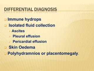
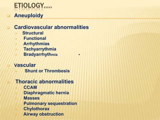
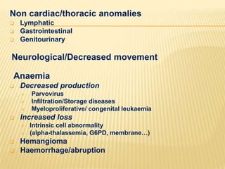
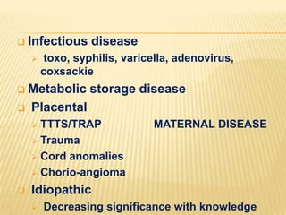
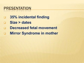
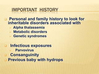
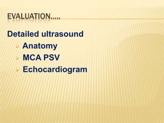
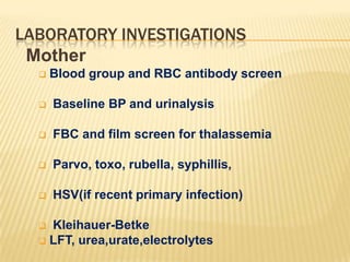
NONIMMUNE HYDROPS FETALIS

Dr Bibek Mohan Rakshit
MD; MRCOG; DNB; MNAMS
Associate Professor (Gynaecology & Obstetrics)
Burdwan Medical college and Hospital
BACKGROUND
Nonimmune Hydrops Fetalis (NIHF) is now
responsible for 90% of all hydrops.
 It has an estimated incidence of 1/1500 to
1/3800 births.
 The pathogenesis of NIHF is not clear, but it
is associated with numerous potential
mechanisms and underlying disorders.
 NIHF has an obvious poor prognosis for the
fetus, but can also have maternal
consequences as well with a 10% incidence
of Mirror syndrome.


Newzealand maternal fetal medicine network
DEFINITION.......
NIHF is established by the ultrasound
findings of at least two of the following
 Ascites
 Pleural effusion: ANY fluid abnormal
 Pericardial effusion: > than 2mm
 Skin edema: > than 5mm on chest and
scalp
 Polyhydramnios: Max pocket > 8cm, or
AFI >
24cm
DIFFERENTIAL DIAGNOSIS
Immune hydrops
 Isolated fluid collection


Ascites
 Pleural effusion
 Pericardial effusion


Skin Oedema
 Polyhydramnios or placentomegaly.

ETIOLOGY.....


Aneuploidy



Cardiovascular abnormalities










Structural
Functional
Arrhythmias
Tachyarrythmia
Bradyarrhythmia

Vascular

Shunt or Thrombosis

Thoracic abnormalities








CCAM
Diaphragmatic hernia
Masses
Pulmonary sequestration
Chylothorax
Airway obstruction

•
Non cardiac/thoracic anomalies




Lymphatic
Gastrointestinal
Genitourinary

Neurological/Decreased movement
Anaemia


Decreased production







Increased loss







Parvovirus
Infiltration/Storage diseases
Myeloproliferative/ congenital leukaemia
Intrinsic cell abnormality
(alpha-thalassemia, G6PD, membrane…)

Hemangioma
Haemorrhage/abruption


Infectious disease


toxo, syphilis, varicella, adenovirus,
coxsackie

Metabolic storage disease
 Placental


TTTS/TRAP
 Trauma
 Cord anomalies
 Chorio-angioma




MATERNAL DISEASE

Idiopathic


Decreasing significance with knowledge
PRESENTATION





35% incidental finding
Size > dates
Decreased fetal movement
Mirror Syndrome in mother
IMPORTANT HISTORY


Personal and family history to look for
inheritable disorders associated with






Alpha thalassemia
Metabolic disorders
Genetic syndromes

Infectious exposures


Parvovirus

Consanguinity
 Previous baby with hydrops

EVALUATION.....
Detailed ultrasound
 Anatomy
 MCA PSV
 Echocardiogram
LABORATORY INVESTIGATIONS
Mother


Blood group and RBC antibody screen



Baseline BP and urinalysis



FBC and film screen for thalassemia



Parvo, toxo, rubella, syphillis,



HSV(if recent primary infection)

Kleihauer-Betke
 LFT, urea,urate,electrolytes

Infant





Amniocentesis
Karyotype
PCR for TORCH pathogens
Storage for further testing

Cordocentesis
RBC for anaemia
Metabolic/genetic tests
UTERINE VENOUS PRESSURE
PROGNOSIS
The overall perinatal mortality rate is 50 –
98%.
 There has been no significant change over
past 15 years.
 Mortality rates will vary according to






Gestation (earlier has worse prognosis)
Pleural effusions (worse prognosis)
Underlying etiology
MANAGEMENT OPTIONS
Termination

Selective therapeutic intervention if
possible
Ongoing pregnancy with
hydrops

Monitor for PET/Mirror syndrome

Consider neonatal palliation

INTERVENTION
If for active intervention
Monitor with CTG’s or BPP’s
2. Delivery at tertiary care center
3. Consider risks of:







birth trauma
PPH
non reassuring fetal heart
dystocia
caesarean

Low recurrence if no inheritable disorder
identified
SOGC CLINICAL PRACTICE GUIDELINES
Recommendations ..
1. All patients with fetal hydrops should be
referred promptly to a tertiary care centre for
evaluation. Some conditions amenable to
prenatal treatment represent a therapeutic
emergency after 18 weeks. (II-2A)
2. Fetal chromosome analysis and genetic
microarray molecular testing should be offered
where available in all cases of non-immune fetal
hydrops. (II-2A)

3. Imaging studies should include
comprehensive obstetrical ultrasound (including
4.Investigation for maternal–fetal infections, and alphathalassemia in women at risk because of their
ethnicity, should be performed in all cases of
unexplained fetal hydrops. (II-2A)
5. To evaluate the risk of fetal anemia, Doppler
measurement of the middle cerebral artery peak
systolic velocity should be performed in all hydropic
fetuses after 16 weeks of gestation. In case of
suspected fetal anemia, fetal blood sampling and
intrauterine transfusion should be offered rapidly. (II2A)
6. All cases of unexplained fetal hydrops should be
referred to a medical genetics service where available.
Detailed postnatal evaluation by a medical geneticist
should be performed on all cases of newborns with
unexplained non-immune hydrops. (II-2A)
7.

Autopsy should be recommended in all
cases of fetal or neonatal death or
pregnancy termination. (II-2A) Amniotic
fluid and/or fetal cells should be stored
for future genetic testing. (II-2B)

8.

SOGC GUIDELINES.....2013
Nonimmune  hydrops  fetalis .  Dr B M Rakshit

Nonimmune hydrops fetalis . Dr B M Rakshit

  • 1.
    NONIMMUNE HYDROPS FETALIS DrBibek Mohan Rakshit MD; MRCOG; DNB; MNAMS Associate Professor (Gynaecology & Obstetrics) Burdwan Medical college and Hospital
  • 2.
    BACKGROUND Nonimmune Hydrops Fetalis(NIHF) is now responsible for 90% of all hydrops.  It has an estimated incidence of 1/1500 to 1/3800 births.  The pathogenesis of NIHF is not clear, but it is associated with numerous potential mechanisms and underlying disorders.  NIHF has an obvious poor prognosis for the fetus, but can also have maternal consequences as well with a 10% incidence of Mirror syndrome.  Newzealand maternal fetal medicine network
  • 3.
    DEFINITION....... NIHF is establishedby the ultrasound findings of at least two of the following  Ascites  Pleural effusion: ANY fluid abnormal  Pericardial effusion: > than 2mm  Skin edema: > than 5mm on chest and scalp  Polyhydramnios: Max pocket > 8cm, or AFI > 24cm
  • 5.
    DIFFERENTIAL DIAGNOSIS Immune hydrops Isolated fluid collection  Ascites  Pleural effusion  Pericardial effusion  Skin Oedema  Polyhydramnios or placentomegaly. 
  • 6.
    ETIOLOGY.....  Aneuploidy  Cardiovascular abnormalities        Structural Functional Arrhythmias Tachyarrythmia Bradyarrhythmia Vascular  Shunt orThrombosis Thoracic abnormalities       CCAM Diaphragmatic hernia Masses Pulmonary sequestration Chylothorax Airway obstruction •
  • 7.
    Non cardiac/thoracic anomalies    Lymphatic Gastrointestinal Genitourinary Neurological/Decreasedmovement Anaemia  Decreased production     Increased loss     Parvovirus Infiltration/Storage diseases Myeloproliferative/ congenital leukaemia Intrinsic cell abnormality (alpha-thalassemia, G6PD, membrane…) Hemangioma Haemorrhage/abruption
  • 8.
     Infectious disease  toxo, syphilis,varicella, adenovirus, coxsackie Metabolic storage disease  Placental  TTTS/TRAP  Trauma  Cord anomalies  Chorio-angioma   MATERNAL DISEASE Idiopathic  Decreasing significance with knowledge
  • 9.
    PRESENTATION     35% incidental finding Size> dates Decreased fetal movement Mirror Syndrome in mother
  • 10.
    IMPORTANT HISTORY  Personal andfamily history to look for inheritable disorders associated with     Alpha thalassemia Metabolic disorders Genetic syndromes Infectious exposures  Parvovirus Consanguinity  Previous baby with hydrops 
  • 11.
  • 12.
    LABORATORY INVESTIGATIONS Mother  Blood groupand RBC antibody screen  Baseline BP and urinalysis  FBC and film screen for thalassemia  Parvo, toxo, rubella, syphillis,  HSV(if recent primary infection) Kleihauer-Betke  LFT, urea,urate,electrolytes 
  • 13.
    Infant     Amniocentesis Karyotype PCR for TORCHpathogens Storage for further testing Cordocentesis RBC for anaemia Metabolic/genetic tests UTERINE VENOUS PRESSURE
  • 14.
    PROGNOSIS The overall perinatalmortality rate is 50 – 98%.  There has been no significant change over past 15 years.  Mortality rates will vary according to     Gestation (earlier has worse prognosis) Pleural effusions (worse prognosis) Underlying etiology
  • 15.
    MANAGEMENT OPTIONS Termination  Selective therapeuticintervention if possible Ongoing pregnancy with hydrops  Monitor for PET/Mirror syndrome  Consider neonatal palliation 
  • 16.
    INTERVENTION If for activeintervention Monitor with CTG’s or BPP’s 2. Delivery at tertiary care center 3. Consider risks of:      birth trauma PPH non reassuring fetal heart dystocia caesarean Low recurrence if no inheritable disorder identified
  • 17.
    SOGC CLINICAL PRACTICEGUIDELINES Recommendations .. 1. All patients with fetal hydrops should be referred promptly to a tertiary care centre for evaluation. Some conditions amenable to prenatal treatment represent a therapeutic emergency after 18 weeks. (II-2A) 2. Fetal chromosome analysis and genetic microarray molecular testing should be offered where available in all cases of non-immune fetal hydrops. (II-2A) 3. Imaging studies should include comprehensive obstetrical ultrasound (including
  • 18.
    4.Investigation for maternal–fetalinfections, and alphathalassemia in women at risk because of their ethnicity, should be performed in all cases of unexplained fetal hydrops. (II-2A) 5. To evaluate the risk of fetal anemia, Doppler measurement of the middle cerebral artery peak systolic velocity should be performed in all hydropic fetuses after 16 weeks of gestation. In case of suspected fetal anemia, fetal blood sampling and intrauterine transfusion should be offered rapidly. (II2A) 6. All cases of unexplained fetal hydrops should be referred to a medical genetics service where available. Detailed postnatal evaluation by a medical geneticist should be performed on all cases of newborns with unexplained non-immune hydrops. (II-2A)
  • 19.
    7. Autopsy should berecommended in all cases of fetal or neonatal death or pregnancy termination. (II-2A) Amniotic fluid and/or fetal cells should be stored for future genetic testing. (II-2B) 8. SOGC GUIDELINES.....2013