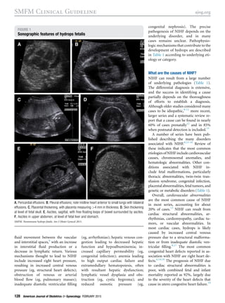
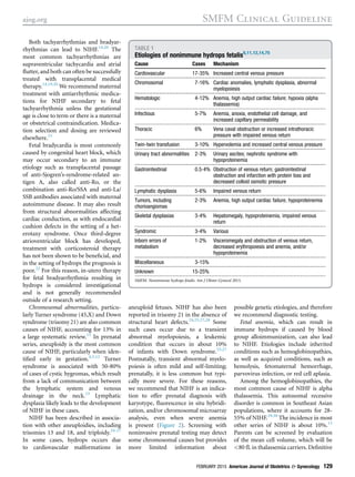
Society for Maternal-Fetal Medicine
(SMFM) Clinical Guideline #7:
nonimmune hydrops fetalis
Society for Maternal-Fetal Medicine (SMFM); Mary E. Norton, MD;
Suneet P. Chauhan, MD; and Jodi S. Dashe, MD
Hydrops fetalis is a Greek term
that describes pathological fluid
(“ὕdur,” Greek for water) accumulation
in fetal soft tissues and serous cavities.
The features are detected by ultrasound,
and are defined as the presence of !2
abnormal fluid collections in the fetus.
These include ascites, pleural effusions,
pericardial effusion, and generalized
skin edema (defined as skin thickness
>5 mm).1
Other frequent sonographic
findings include placental thickening
(typically defined as a placental thickness
!4 cm in the second trimester or !6
cm in the third trimester)2,3
and poly-
hydramnios (Figure 1). Nonimmune
hydrops fetalis (NIHF) refers specifically
to cases not caused by red cell alloim-
munization. With the development
and widespread use of Rh(D) immune
globulin, the prevalence of Rh(D)
alloimmunization and associated
hydrops has dramatically decreased. As
a result, NIHF now accounts for almost
90% of cases of hydrops,4
with the
prevalence in published series reported
as 1 in 1700-3000 pregnancies.5-7
However, many of these reports pre-
date routine sonography and limited
information is available on contempo-
rary incidence of NIHF in a prenatal
population.
What is the underlying pathogenesis
of NIHF?
The common pathophysiology underly-
ing the many etiologies of hydrops fetalis
is an imbalance in the regulation of
From the Society for Maternal-Fetal Medicine
Publications Committee, Washington, DC; the
Division of Maternal-Fetal Medicine, University of
California, San Francisco, San Francisco, CA
(Dr Norton); the Department of Obstetrics,
Gynecology and Reproductive Sciences, The
University of Texas Health Science Center at
Houston, Houston, TX (Dr Chauhan); and the
Department of Obstetrics and Gynecology,
University of Texas Southwestern Medical
Center, Dallas, TX (Dr Dashe).
Received Nov. 5, 2014; accepted Dec. 12,
2014.
The authors report no conflict of interest.
Corresponding author: Society for Maternal-
Fetal Medicine: Publications Committee. pubs@
smfm.org
0002-9378/free
ª 2015 Elsevier Inc. All rights reserved.
http://dx.doi.org/10.1016/j.ajog.2014.12.018
OBJECTIVE: Nonimmune hydrops is the presence of !2 abnormal fetal fluid collections
in the absence of red cell alloimmunization. The most common etiologies include car-
diovascular, chromosomal, and hematologic abnormalities, followed by structural fetal
anomalies, complications of monochorionic twinning, infection, and placental abnor-
malities. We sought to provide evidence-based guidelines for the evaluation and man-
agement of nonimmune hydrops fetalis.
METHODS: A systematic literature review was performed using MEDLINE, PubMed,
EMBASE, and Cochrane Library. The search was restricted to English-language articles
published from 1966 through June 2014. Priority was given to articles reporting original
research, although review articles and commentaries also were consulted. Abstracts of
research presented at symposia and scientific conferences were not considered
adequate for inclusion in this document. Evidence reports and guidelines published by
organizations or institutions such as the National Institutes of Health, Agency for Health
Research and Quality, American Congress of Obstetricians and Gynecologists, and
Society for Maternal-Fetal Medicine were also reviewed, and additional studies were
located by reviewing bibliographies of identified articles. Grading of Recommendations
Assessment, Development, and Evaluation methodology was employed for defining
strength of recommendations and rating quality of evidence. Consistent with US Pre-
ventive Task Force guidelines, references were evaluated for quality based on the highest
level of evidence.
RESULTS AND RECOMMENDATIONS: Evaluation of hydrops begins with an antibody
screen (indirect Coombs test) to determine if it is nonimmune, detailed sonography of the
fetus(es) and placenta, including echocardiography and assessment for fetal arrhythmia,
and middle cerebral artery Doppler evaluation for anemia, as well as fetal karyotype and/
or chromosomal microarray analysis, regardless of whether a structural fetal anomaly is
identified. Recommended treatment depends on the underlying etiology and gestational
age; preterm delivery is recommended only for obstetric indications including devel-
opment of mirror syndrome. Candidates for corticosteroids and antepartum surveillance
include those with an idiopathic etiology, an etiology amenable to prenatal or postnatal
treatment, and those in whom intervention is planned if fetal deterioration occurs. Such
pregnancies should be delivered at a facility with the capability to stabilize and treat
critically ill newborns. The prognosis depends on etiology, response to therapy if
treatable, and the gestational age at detection and delivery. Aneuploidy confers a poor
prognosis, and even in the absence of aneuploidy, neonatal survival is often <50%.
Mirror syndrome is a form of severe preeclampsia that may develop in association with
fetal hydrops and in most cases necessitates delivery.
Key words: fetal complications, hydrops, nonimmune hydrops fetalis
FEBRUARY 2015 American Journal of Obstetrics & Gynecology 127
SMFM Clinical Guideline ajog.org
fluid movement between the vascular
and interstitial spaces,8
with an increase
in interstitial fluid production or a
decrease in lymphatic return. Various
mechanisms thought to lead to NIHF
include increased right heart pressure,
resulting in increased central venous
pressure (eg, structural heart defects);
obstruction of venous or arterial
blood flow (eg, pulmonary masses);
inadequate diastolic ventricular filling
(eg, arrhythmias); hepatic venous con-
gestion leading to decreased hepatic
function and hypoalbuminemia; in-
creased capillary permeability (eg,
congenital infection); anemia leading
to high output cardiac failure and
extramedullary hematopoiesis, often
with resultant hepatic dysfunction;
lymphatic vessel dysplasia and obs-
truction (eg, cystic hygroma); and
reduced osmotic pressure (eg,
congenital nephrosis). The precise
pathogenesis of NIHF depends on the
underlying disorder, and in many
cases remains unclear. Pathophysio-
logic mechanisms that contribute to the
development of hydrops are described
in Table 1 according to underlying eti-
ology or category.
What are the causes of NIHF?
NIHF can result from a large number
of underlying pathologies (Table 1).
The differential diagnosis is extensive,
and the success in identifying a cause
partially depends on the thoroughness
of efforts to establish a diagnosis.
Although older studies considered many
cases to be idiopathic,9-11
more recent,
larger series and a systematic review re-
port that a cause can be found in nearly
60% of cases prenatally12
and in 85%
when postnatal detection is included.13
A number of series have been pub-
lished describing the many disorders
associated with NIHF.8,13-16
Review of
these indicates that the most common
etiologiesofNIHF includecardiovascular
causes, chromosomal anomalies, and
hematologic abnormalities. Other con-
ditions associated with NIHF in-
clude fetal malformations, particularly
thoracic abnormalities, twin-twin tran-
sfusion syndrome, congenital infection,
placentalabnormalities,fetaltumors,and
genetic or metabolic disorders (Table 1).
Overall, cardiovascular abnormalities
are the most common cause of NIHF
in most series, accounting for about
20% of cases.13
NIHF can result from
cardiac structural abnormalities, ar-
rhythmias, cardiomyopathy, cardiac tu-
mors, or vascular abnormalities. In
most cardiac cases, hydrops is likely
caused by increased central venous
pressure due to a structural malforma-
tion or from inadequate diastolic ven-
tricular filling.8,17
The most common
congenital heart defects reported in as-
sociation with NIHF are right heart de-
fects.6,14,18
The prognosis of NIHF due
to cardiac structural abnormalities is
poor, with combined fetal and infant
mortality reported as 92%, largely due
to the severity of the heart defects that
cause in utero congestive heart failure.19
FIGURE 1
Sonographic features of hydrops fetalis
A, Pericardial effusions. B, Pleural effusions; note midline heart anterior to small lungs with bilateral
effusions. C, Placental thickening, with placenta measuring >4 mm in thickness. D, Skin thickening
at level of fetal skull. E, Ascites, sagittal, with free-floating loops of bowel surrounded by ascites.
F, Ascites in upper abdomen, at level of fetal liver and stomach.
SMFM. Nonimmune hydrops fetalis. Am J Obstet Gynecol 2015.
SMFM Clinical Guideline ajog.org
128 American Journal of Obstetrics & Gynecology FEBRUARY 2015
Both tachyarrhythmias and bradyar-
rhythmias can lead to NIHF.14,20
The
most common tachyarrhythmias are
supraventricular tachycardia and atrial
flutter, and both can often be successfully
treated with transplacental medical
therapy.14,19,20
We recommend maternal
treatment with antiarrhythmic medica-
tions for NIHF secondary to fetal
tachyarrhythmia unless the gestational
age is close to term or there is a maternal
or obstetrical contraindication. Medica-
tion selection and dosing are reviewed
elsewhere.21
Fetal bradycardia is most commonly
caused by congenital heart block, which
may occur secondary to an immune
etiology such as transplacental passage
of anti-Sjogren’s-syndrome-related an-
tigen A, also called anti-Ro, or the
combination anti-Ro/SSA and anti-La/
SSB antibodies associated with maternal
autoimmune disease. It may also result
from structural abnormalities affecting
cardiac conduction, as with endocardial
cushion defects in the setting of a het-
erotaxy syndrome. Once third-degree
atrioventricular block has developed,
treatment with corticosteroid therapy
has not been shown to be beneficial, and
in the setting of hydrops the prognosis is
poor.22
For this reason, in-utero therapy
for fetal bradyarrhythmia resulting in
hydrops is considered investigational
and is not generally recommended
outside of a research setting.
Chromosomal abnormalities, particu-
larly Turner syndrome (45,X) and Down
syndrome (trisomy 21) are also common
causes of NIHF, accounting for 13% in
a large systematic review.13
In prenatal
series, aneuploidy is the most common
cause of NIHF, particularly when iden-
tified early in gestation.4,5,12
Turner
syndrome is associated with 50-80%
of cases of cystic hygromas, which result
from a lack of communication between
the lymphatic system and venous
drainage in the neck.23
Lymphatic
dysplasia likely leads to the development
of NIHF in these cases.
NIHF has been described in associa-
tion with other aneuploidies, including
trisomies 13 and 18, and triploidy.24-27
In some cases, hydrops occurs due
to cardiovascular malformations in
aneuploid fetuses. NIHF has also been
reported in trisomy 21 in the absence of
structural heart defects.24,25,27,28
Some
such cases occur due to a transient
abnormal myelopoiesis, a leukemic
condition that occurs in about 10%
of infants with Down syndrome.25,27
Postnatally, transient abnormal myelo-
poiesis is often mild and self-limiting;
prenatally, it is less common but typi-
cally more severe. For these reasons,
we recommend that NIHF is an indica-
tion to offer prenatal diagnosis with
karyotype, fluorescence in situ hybridi-
zation, and/or chromosomal microarray
analysis, even when severe anemia
is present (Figure 2). Screening with
noninvasive prenatal testing may detect
some chromosomal causes but provides
more limited information about
possible genetic etiologies, and therefore
we recommend diagnostic testing.
Fetal anemia, which can result in
immune hydrops if caused by blood
group alloimmunization, can also lead
to NIHF. Etiologies include inherited
conditions such as hemoglobinopathies,
as well as acquired conditions, such as
hemolysis, fetomaternal hemorrhage,
parvovirus infection, or red cell aplasia.
Among the hemoglobinopathies, the
most common cause of NIHF is alpha
thalassemia. This autosomal recessive
disorder is common in Southeast Asian
populations, where it accounts for 28-
55% of NIHF.29,30
The incidence in most
other series of NIHF is about 10%.13
Parents can be screened by evaluation
of the mean cell volume, which will be
<80 fL in thalassemia carriers. Definitive
TABLE 1
Etiologies of nonimmune hydrops fetalis6,11,12,14,75
Cause Cases Mechanism
Cardiovascular 17-35% Increased central venous pressure
Chromosomal 7-16% Cardiac anomalies, lymphatic dysplasia, abnormal
myelopoiesis
Hematologic 4-12% Anemia, high output cardiac failure; hypoxia (alpha
thalassemia)
Infectious 5-7% Anemia, anoxia, endothelial cell damage, and
increased capillary permeability
Thoracic 6% Vena caval obstruction or increased intrathoracic
pressure with impaired venous return
Twin-twin transfusion 3-10% Hypervolemia and increased central venous pressure
Urinary tract abnormalities 2-3% Urinary ascites; nephrotic syndrome with
hypoproteinemia
Gastrointestinal 0.5-4% Obstruction of venous return; gastrointestinal
obstruction and infarction with protein loss and
decreased colloid osmotic pressure
Lymphatic dysplasia 5-6% Impaired venous return
Tumors, including
chorioangiomas
2-3% Anemia, high output cardiac failure, hypoproteinemia
Skeletal dysplasias 3-4% Hepatomegaly, hypoproteinemia, impaired venous
return
Syndromic 3-4% Various
Inborn errors of
metabolism
1-2% Visceromegaly and obstruction of venous return,
decreased erythropoiesis and anemia, and/or
hypoproteinemia
Miscellaneous 3-15%
Unknown 15-25%
SMFM. Nonimmune hydrops fetalis. Am J Obstet Gynecol 2015.
ajog.org SMFM Clinical Guideline
FEBRUARY 2015 American Journal of Obstetrics & Gynecology 129
diagnosis of an affected fetus can be
made by detection of one of the common
DNA deletions or point mutations that
account for most cases.31
Conversely, a
fetal blood sample can be evaluated for
the presence of the abnormal Bart’s he-
moglobin seen in this condition. Bart’s
hemoglobin is an ineffective oxygen
carrier, thus the fetus with alpha thalas-
semia will suffer severe intrauterine
hypoxia from an early gestational age.
The resultant NIHF will typically present
in the late second or early third trimester.
Fetal anemia may also occur due to
fetal hemorrhage. NIHF occurs only with
significant fetomaternal bleeding that
is not enough to lead to fetal hypo-
volemia and death. Fetomaternal hem-
orrhage leading to hydrops may occur
as either an isolated acute event, or as a
chronic, ongoing hemorrhage.32,33
With
either, a Kleihauer-Betke smear will
show the presence of fetal cells in the
maternal peripheral blood in most cases.
Flow cytometry can also be used to es-
timate the volume of fetal bleeding
into the mother. This is an important
diagnosis to make, because even with
a massive fetomaternal hemorrhage,
intravascular fetal transfusion can be
lifesaving.34-36
For this reason, we
recommend that NIHF due to anemia
from fetomaternal hemorrhage be
treated with transfusion, unless the
pregnancy is at an advanced gestational
age and risks associated with delivery
are considered to be less than those
associated with the procedure.
Other, less common causes of fetal
anemia and hydrops include G-6-PD
deficiency, erythrocyte enzymopathies
such as pyruvate kinase deficiency, and
maternal acquired red cell aplasia.37,38
NIHF has been reported in asso-
ciation with a number of viral, bacterial,
and parasitic infectious diseases, in-
cluding parvovirus, cytomegalovirus,
syphilis, and toxoplasmosis.39-42
In
most series, such infections account for
5-10% of NIHF.6,13,14,16
Although the
associations are less clear, NIHF has
also been reported to occur with Cox-
sackie virus, trypanosomiasis, varicella,
human herpesvirus 6 and 7, herpes
simplex type 1, respiratory syncytial vi-
rus, congenital lymphocytic choriome-
ningitis virus, and leptospirosis.6,43-47
Fetal infection can cause NIHF due to
anemia, anoxia, endothelial cell damage,
increased capillary permeability, and
myocarditis.
Parvovirus is the most commonly re-
ported infectious cause of NIHF. In
the fetus, the virus has a predilection
for erythroid progenitor cells, leading
to inhibition of erythropoiesis and sub-
sequent anemia.48,49
The risk of a poor
outcome for the fetus is greatest when
the congenital infection occurs in the
early second trimester (<20 weeks of
gestation).50-53
The risk of fetal death has
been reported to be 15% at 13-20 weeks
of gestation, and 6% after 20 weeks.54
In most cases, the anemia is transient
and fetal intravascular transfusion can
support a fetus through this aplastic
crisis.55,56
However, development of
NIHF is associated with high mortality,
and outcomes are reported to be signif-
icantly improved following fetal intra-
uterine transfusion.51,53
For this reason,
we recommend fetal intrauterine trans-
fusion for NIHF due to parvovirus
infection, unless the pregnancy is at an
advanced gestational age and risks asso-
ciated with delivery are considered to
be less than those associated with the
procedure.
Fetal thoracic abnormalities, including
masses as well as congenital hydrothorax,
can also be associated with NIHF. The
most frequent pulmonary lesion associ-
ated with NIHF is a congenital pulmo-
nary airway malformation (CPAM).
FIGURE 2
Workup of nonimmune hydrops fetalis*
AFAFP, amniotic fluid alpha fetoprotein; CBC, complete blood count; CMA, chromosomal microarray; CMV, cytomegalovirus; G6PD,
glucose-6-phosphate dehydrogenase deficiency; MCA PSV, middle cerebral artery peak systolic velocity; MCV, mean corpuscular
volumne; MoM, multiple of the median; PCR, polymerase chain reaction; RPR, rapid plasma reagin.
*Assuming negative indirect Coombs test, thereby excluding alloimmunization; **CMV/toxoplasmosis testing if fetal anomalies sug-
gestive of infection; ***Either amniocentesis or FBS; ****Available in some laboratories.67,76
SMFM. Nonimmune hydrops fetalis. Am J Obstet Gynecol 2015.
SMFM Clinical Guideline ajog.org
130 American Journal of Obstetrics & Gynecology FEBRUARY 2015
With a large lesion or effusion, medias-
tinal shift may impair venous return
and cardiac output, and the associated
esophageal compression may result in
polyhydramnios. Hydrops occurs in only
about 5% of fetuses with CPAM but
confers a poor prognosis without treat-
ment.57
If the lesion is macrocystic,
the cyst may be treated with needle
drainage or thoracoamniotic shunt
placement.58,59
If predominantly solid
(microcystic), both corticosteroid ther-
apy and in utero resection have been
advocated, and corticosteroid treatment
is currently recommended as a first-line
treatment.60
Large bronchopulmonary
sequestrations have also been treated
with a needle procedure involving
neodymium:yttrium-aluminium-garnet
laser of the feeding vessel.57
The most common etiology of an
isolated effusion leading to NIHF is
chylothorax, caused by lymphatic
obstruction. The fluid may be sampled at
the time of needle drainage or shunt
placement, and the diagnosis is con-
firmed by the finding of a fetal pleural
cell count with >80% lymphocytes in
the absence of infection. Reported sur-
vival exceeds 50% in hydropic fetuses
treated with thoracoamniotic shunt
placement.61
Twin-twin transfusion syndrome results
from an imbalance in blood flow caused
by anastomoses in the placentas of
monochorionic twin pregnancies. In se-
vere cases, one or both twins may develop
NIHF, although more commonly the
recipient twin is affected, likely due to
hypervolemia and increased central
venous pressure.62
Cases of twin-twin
transfusion sequence with hydrops
have a very poor prognosis without
treatment, and laser therapy is consid-
ered by most experts to be the best
available therapeutic approach to
improve the prognosis.63
Selective ter-
mination via umbilical cord coagulation
is also an option for pregnancies with
twin-twin transfusion sequence result-
ing in NIHF. Another complication of
monochorionic twinning that may
result in NIHF is twin-reversed arterial
perfusion sequence. Radiofrequency
ablation of the acardiac twin has been
advocated for severe cases, including
those with hydrops, with reported
overall survival of 80%.64
Structural urinary and gastrointestinal
abnormalities are less common causes of
NIHF. A ruptured bladder or renal col-
lecting system may cause urinary ascites
and mimic NIHF. Congenital nephrotic
syndromes have been reported to cause
NIHF due to hypoproteinemia.19,65,66
Surviving infants may have massive
proteinuria at birth and develop renal
failure in childhood.
Few primary abnormalities of the
gastrointestinal tract have been associ-
ated with NIHF. Those that have been
reported include diaphragmatic hernia,
midgut volvulus, gastrointestinal ob-
struction, jejunal atresia, malrotation of
the intestines, and meconium perito-
nitis.6,19
Intraabdominal masses may
cause NIHF due to obstruction of
venous return, while gastrointestinal
obstruction and infarction may lead to
decreased colloid osmotic pressure due
to protein loss.19
Hepatic disorders
such as cirrhosis, hepatic necrosis,
cholestasis, polycystic disease of the liver,
and biliary atresia have been reported in
association with NIHF, most likely due
to hypoproteinemia.7
Hemangioma
of the liver has also been reported as a
cause of NIHF, probably due to arterio-
venous shunting resulting in cardiac
failure.
Neoplastic diseases or fetal tumors can
occur in utero and have been associated
with NIHF. Relatively common in this
category are lymphangiomas, hemangi-
omas, sacrococcygeal, mediastinal, and
pharyngeal teratomas, and neuroblas-
tomas.19,67,68
Many of these are very
vascular and lead to NIHF due to high
output cardiac failure. Fetal therapy has
been offered for cases of solid sacro-
coccygeal teratoma resulting in NIHF,
and in a recent systematic review, open
fetal surgery resulted in survival in 6 of
11 cases (55%), and minimally invasive
therapy was associated with survival in
6 of 20 (30%).69
Tuberous sclerosis is
an autosomal dominant disorder char-
acterized by fibroangiomatous tumors
in multiple organs, most typically the
cortex of the brain, the skin, and the
kidneys. Cardiac rhabdomyomas and
liver fibrosis are also sometimes present.
NIHF has been reported in association
with tuberous sclerosis, probably either
as a result of cardiac failure due to
rhabdomyomas (resulting in obstruction
to filling or outflow), or hepatic failure
due to fibrosis.70
Placental and cord lesions that have
been associated with NIHF include cho-
rioangiomas, angiomyxoma of the cord,
aneurysm of the umbilical artery, cord
vein thrombosis, umbilical vein torsion,
true knots, and amniotic bands.7,19,68
Placental chorioangiomas are relatively
common, occurring in about 1% of
pregnancies. While small lesions are
usually not clinically significant, those
measuring >5 cm can act as high
volume arteriovenous shunts and lead
to hydrops due to high output cardiac
failure. Other vascular tumors and arte-
riovenous malformations can similarly
cause NIHF. Hemangiomas have been
reported to cause NIHF, likely due to
severe anemia, hypoproteinemia, and/or
extramedullary erythropoiesis.
A large number of skeletal dysplasias
have been associated with NIHF, including
achondroplasia, achondrogenesis, osteo-
genesis imperfecta, osteopetrosis, thana-
tophoric dysplasia, short-rib polydactyly
syndrome, and asphyxiating thoracic
dysplasia.14,19,71-73
In all of these, the
mechanism is unclear, although it has
been proposed that hepatic enlargement
occurs secondary to intrahepatic prolifer-
ation of blood cell precursors to
compensate for a small bone-marrow
volume. This may cause large vessel
compression and lead to anasarca in these
fetuses.
Inborn errors of metabolism and other
genetic conditions are historically asso-
ciated with 1-2% of cases of NIHF, which
may be transient or manifest as isolated
ascites. Inherited metabolic disorders
that have been implicated as a cause of
NIHF are most typically lysosomal
storage diseases such as various muco-
polysaccharidoses, Gaucher disease, and
Niemann-Pick disease.74,75
In a recent
review of the literature including 678
cases of NIHF, lysosomal storage diseases
occurred in 5.2% of all NIHF cases, and
in 29.6% of idiopathic NIHF cases if
a comprehensive workup for these con-
ditions is done.76
Proposed mechanisms
ajog.org SMFM Clinical Guideline
FEBRUARY 2015 American Journal of Obstetrics & Gynecology 131
involve visceromegaly and obstruction
of venous return, decreased erythropoi-
esis and anemia, and/or hypopro-
teinemia. Although such disorders are a
relatively uncommon cause of NIHF,
they are important because of the high
recurrence risk of these mainly auto-
somal recessive disorders. Careful his-
tology of the placenta, liver, spleen,
and bone marrow will often provide a
clue that a metabolic storage disorder
was present. For many such disorders,
testing is available to determine a diag-
nosis and for prenatal diagnosis in a
subsequent pregnancy. Panels of causa-
tive storage disorders can be tested for
in some laboratories, and this should
be considered for cases of NIHF in
a structurally normal fetus in which
another cause has not been identified,
or with cases of recurrence within a
family.76,77
A number of other syndromes have
been associated with NIHF. Many of
these are disorders associated with
lymphatic dysfunction, such as Noonan
and multiple pterygium syndrome, both
of which frequently present with cystic
hygroma; idiopathic chylothorax, in
which a local pleuromediastinal lymph
vessel disturbance occurs as the possible
pathogenic mechanism; yellow nail syn-
drome, a dominantly inherited congen-
ital lymphedema syndrome; and
congenital pulmonary lymphangiectasia.
Familial recurrence in some of these
cases suggests a hereditary maldevelop-
ment of lymphatic vessels.6,14,19,78
What is the appropriate evaluation
when fetal hydrops is detected?
Sonographic identification of the
hydropic fetus is not difficult. The diag-
nostic challenge is to establish the etiol-
ogy and determine the appropriate
therapy (if available) and timing of de-
livery. It has been reported that the cause
of hydrops can be determined in about
60-85% of cases, although this includes
postnatal evaluation.13
Figure 2 outlines the various steps in
the evaluation of the hydropic fetus. It is
especially important to rule out poten-
tially treatable conditions, as well as ge-
netic disorders with a risk of recurrence
in future pregnancies. Often, the etiology
of the hydrops can be determined at the
time of diagnosis, since several etiologies
are confirmed or excluded based upon
ultrasound findings (eg, twin-to-twin
transfusion, cardiac arrhythmias, and
structural anomalies associated with
NIHF).
Management is guided by the pres-
ence or absence of additional anomalies.
Sonographic evaluation should include
a detailed survey for anomalies of the
fetus, umbilical cord, and placenta, and
estimation of amniotic fluid volume. A
fetal echocardiogram should be in-
cluded, as fetal cardiac anomalies are
among the most common causes of
NIHF.
In a structurally normal fetus, the first
step is to rule out alloimmunization as a
cause. The maternal blood type and
Rh(D) antigen status are assessed as part
of routine prenatal care, along with an
indirect Coombs test (an antibody
screen) to evaluate for circulating red
blood cell antibodies. These results
should be reviewed, and if the indirect
Coombs test was previously normal, it
should be repeated. Maternal blood
studies should also include a complete
blood cell count with differential
and indices, Kleihauer-Betke stain for
fetal hemoglobin, and parvovirus B19
serology. Serologic test results for syph-
ilis should be reviewed or repeated, and
consideration should be given to acute
phase titers for cytomegalovirus and
toxoplasmosis.
It is particularly important to perform
middle cerebral artery Doppler studies
to assess for the presence of fetal anemia,
which may be treatable with intravas-
cular transfusion. The fetus with NIHF
due to severe anemia will have increased
velocity through the middle cerebral
artery.79
A fetal karyotype, fluorescence in situ
hybridization studies, and/or chromo-
somal microarray analysis should be
offered with or without identified so-
nographic anomalies.80
This can be
performed by amniocentesis or fetal
blood sampling; the latter allows direct
analysis of fetal hematocrit and hemo-
globin if anemia is suspected. Invasive
testing also allows testing for lysosomal
storage disorders, and polymerase chain
reaction studies for parvovirus, toxo-
plasmosis, and cytomegalovirus infec-
tion. Such testing should be performed
in a structurally normal fetus in which
no other cause has been identified.
An important step in the evalua-
tion of NIHF is to exclude a genetic
abnormality. Genetically transmitted
disorders account for about one
third of cases of NIHF, and include
chromosomal abnormalities, hemo-
globinopathies, skeletal dysplasias,
metabolic storage disorders, and eryth-
rocyte enzymopathies. A complete fam-
ily history is thus imperative to rule out a
known inherited disorder in the family
and to assess for consanguinity, which
will increase the likelihood of a recessive
disorder. Although idiopathic NIHF
has a low recurrence risk, the risk for
some cases of NIHF may be as high
as 25%, making genetic counseling an
integral part of the management of any
patient with NIHF.
What maternal risks are associated
with NIHF?
Women with NIHF may develop mirror
syndrome, an uncommon complication
in which the mother develops edema
that “mirrors” that of her hydropic fetus.
Mirror syndrome may represent a form
of preeclampsia, and is characterized by
edema in approximately 90%, hyper-
tension in 60%, and proteinuria in 40%
of cases.81
As it is uncommon and likely
underdiagnosed, the incidence is un-
clear. Additional associated findings with
the syndrome include headache, visual
disturbances, oliguria, elevated uric acid,
liver function tests, or creatinine levels,
low platelets, anemia, and hemodilu-
tion.82
A review of the literature (1956
through 2009) by Braun et al81
noted
that among 56 cases of mirror syndrome,
the major maternal morbidity was pul-
monary edema, which occurred in 21%.
Resolution occurs with either the treat-
ment of the hydrops or with delivery.81,82
There have been case reports in
which pregnancies with mirror syn-
drome and various treatable causes of
hydropsesecondary to fetal arrhythmia,
hydrothorax, parvovirus, and bladder
obstructionehave experienced resolu-
tion of both hydrops and mirror
SMFM Clinical Guideline ajog.org
132 American Journal of Obstetrics & Gynecology FEBRUARY 2015
syndrome following treatment.83-86
The
same imbalance of angiogenic and anti-
angiogenic factors described with severe
preeclampsia has also been observed in
cases of mirror syndrome, with correc-
tion following treatment and resolution
of the NIHF.83,84
However, there are
no data regarding the likelihood of res-
olution or long-term benefits. Given
risks of expectant management of
severe preeclampsia, it is recommended
that this approach be taken only with
caution, and that delivery not be delayed
if the maternal condition deteriorates.
Thus for most cases of NIHF, including
all cases without a treatable etiology,
development of mirror syndrome ne-
cessitates delivery.
What obstetric complications are
associated with NIHF?
Polyhydramnios and preterm birth
occur frequently with NIHF, with
reported incidences as high as 29%71
and
66%,87
respectively. If the poly-
hydramnios is associated with maternal
respiratory symptoms, reported man-
agement options have included a short
course of a prostaglandin inhibitor or
serial amnioreduction. Since both treat-
ment modalities lack evidence of bene-
fit and have potential complications,
including in utero constriction of the
ductus arteriosus, abruption, premature
rupture of the membranes, and neo-
natal complications such as necrotizing
enterocolitis and patent ductus arterio-
sus, they should be used judiciously.88,89
Tocolytic agents are a consideration <24
weeks if contractions occur secondary to
a known inciting event, such as an
invasive procedure performed for the
diagnosis or management of NIHF.88
Although in the past preterm delivery
has been advocated by some to poten-
tially improve the outcome of NIHF,7
prematurity is likely to worsen the
prognosis. For this reason, we recom-
mend that preterm delivery be under-
taken only for obstetric indications.
What is the prognosis of NIHF?
The prognosis of NIHF depends on the
underlying etiology, gestational age at
detection and delivery, Apgar scores,
extent of resuscitation in the delivery
room, and whether the newborn re-
quires transport.90
In one prenatal series,
nearly half of those diagnosed <24 weeks
had aneuploidy, with extremely poor
survival. However, even in the absence of
a chromosomal abnormality, survival
was <50%.4
In a recent prenatal series of
71 pregnancies that continued >20
weeksethereby excluding many with
aneuploidyesurvival was approximately
50%, and only 25% survived without
major morbidities.12
Among liveborn
infants, neonatal mortality with NIHF is
TABLE 2
Therapy for selected etiologies of nonimmune hydrops21,54,58,59,61,63,64
Etiology Therapy Recommendation
Cardiac tachyarrhythmia,
supraventricular tachycardia,
atrial flutter, or atrial fibrillation
Maternal transplacental administration
of antiarrhythmic medication(s)
Treatment with antiarrhythmic medication unless
gestational age is close to term or there is maternal
or obstetrical contraindication to therapy
Fetal anemia secondary to
parvovirus infection or
fetomaternal hemorrhage
Fetal blood sampling followed by
intrauterine transfusion
Fetal intrauterine transfusion if anemia is confirmed,
unless pregnancy is at an advanced gestational age
and risks associated with delivery are considered to
be less than those associated with procedure
Fetal hydrothorax, chylothorax,
or large pleural effusion associated
with bronchopulmonary sequestration
Fetal needle drainage of effusion
or placement of thoracoamniotic
shunt; if gestational age is advanced,
needle drainage prior to delivery in
selected cases
Consider drainage of large unilateral pleural effusion(s)
resulting in NIHF, or, if gestational age is advanced,
consideration of needle drainage prior to delivery
Fetal CPAM Macrocystic type: fetal needle drainage
of effusion or placement of thoracoamniotic
shunt; microcystic type: maternal
administration of corticosteroids,
betamethasone 12.5 mg IM q24
h  2 doses or dexamethasone
6.25 mg IM q12 h  4 doses
Consider drainage of large macrocystic CPAM that has
resulted in NIHF; if large microcystic CPAM has
resulted in NIHF, we suggest that management options
include maternal corticosteroid administration
TTTS or TAPS Laser ablation of placental anastomoses
or selective termination
Consideration of fetoscopic laser photocoagulation of
placental anastomoses for TTTS or TAPS that has
resulted in NIHF <26 wk
Twin-reversed arterial
perfusion sequence
Percutaneous radiofrequency ablation Referral for consideration of percutaneous
radiofrequency ablation that has resulted in NIHF
For each of these etiologies, it is recommended that treatment be performed at tertiary care center or center with expertise in relevant therapy.
CPAM, congenital pulmonary airway malformation; IM, intramuscular; NIHF, nonimmune hydrops fetalis; TAPS, twin-anemia polycythemia sequence; TTTS, twin-twin transfusion sequence.
SMFM. Nonimmune hydrops fetalis. Am J Obstet Gynecol 2015.
ajog.org SMFM Clinical Guideline
FEBRUARY 2015 American Journal of Obstetrics & Gynecology 133
reported to be as high as 60%.91
With
chylothorax as the underlying etiology,
the mortality may be as low as 6%;
however, when the infant has associated
anomalies, almost two-thirds do not
survive.14
Treatable causes of hydrops,
such as fetal arrhythmia or infection
with parvovirus B19,92
have a better
prognosis. In a large series of newborns
admitted to the neonatal intensive care
unit, the independent risk factors for
death in logistic regression analyses were
younger gestational age, low 5-minute
Apgar score, and need for high levels of
support during the first day after birth
(higher levels of inspired oxygen support
and greater need for high-frequency
ventilation).14
Temporal trends suggest that among
cases of liveborn infants with NIHF, the
associated mortality has not improved
over 2 decades. Comparing the mortality
among hydropic newborns delivered in
1993 through 2003 vs 2003 through
2009, there was no significant difference
in mortality in the 2 time periods, 47%
vs 67%, respectively.91
In addition to the
small sample size, an explanation for the
lack of improvement in survival over
time may be that the more severe cases
are now more frequently diagnosed
prenatally and referred to tertiary cen-
ters, which are more likely to contribute
to large series in the literature.
The long-term prognosis for survivors
of NIHF also depends upon the under-
lying etiology. After intrauterine trans-
fusion for hydrops secondary to
infection with parvovirus B19, there is
potential for delayed psychomotor
development and abnormal neurological
outcomes.93
It is unclear if this is
because of the hydrops, a direct conse-
quence of the parvovirus infection,
from severe anemia, or associated
with the transfusion. Finally, fetuses
with supraventricular tachycardia may
develop Wolff-Parkinson-White syn-
drome later in life.94
Management of NIHF
The cornerstone of counseling and
management for this condition is a
thorough evaluation for the underlying
etiology of the hydrops (Figure 2).
Pregnancy management decisions will
depend on the etiology, in particular
whether there is a treatable cause and
the gestational age that NIHF develops
or is first identified. Cases generally fall
into 1 of 3 categories: those amenable to
fetal therapyewhich often require
urgent treatment or referral to a
specialized center; those with a lethal
prognosis, for whom pregnancy termi-
nation or comfort care are the only op-
tions realistic to offer; and cases in which
the etiology is idiopathic and the prog-
nosis is likely poor but uncertain. Given
the poor overall prognosis, pregnancy
termination should be offered if NIHF is
identified prior to viability. It is impor-
tant in counseling that the potential for
maternal complications with expectant
management be anticipated, including
mirror syndrome. Serial evaluation of
maternal blood pressure is therefore
recommended.
What are the fetal therapy options
available for NIHF?
Selected etiologies of NIHF for which
fetal therapy should be considered are
listed in Table 2. Therapy options may
include intrauterine transfusion(s)
for fetal anemia, medications such as
antiarrhythmic agents, drainage of large
pleural effusions, corticosteroids for
CPAMs, or specialized procedures such
as laser coagulation of placental anasto-
moses for twin-twin transfusion syn-
drome. The list is not intended to
be comprehensive but rather to serve as a
TABLE 3
Society for Maternal-Fetal Medicine recommendations for nonimmune hydrops
Recommendations Grading of recommendations (Table 4)
 We recommend that initial evaluation of hydrops include an antibody screen (indirect
Coombs test) to verify that it is nonimmune, targeted sonography with echocardi-
ography to evaluate for fetal and placental abnormalities, MCA Doppler evaluation for
anemia, and fetal karyotype or chromosomal microarray analysis, regardless of
whether structural fetal anomalies are identified (Figure 2)
1C
Strong recommendation, low-quality evidence
 We recommend that fetal therapy decisions be based on underlying etiology, in
particular whether there is a treatable cause (Table 2) and the gestational age at
which NIHF develops or is first identified
1C
Strong recommendation, low-quality evidence
 As prematurity is likely to worsen prognosis, we recommend that preterm delivery
be undertaken only for obstetric indications
1C
Strong recommendation, low-quality evidence
 We recommend that pregnancies with NIHF due to nonlethal or potentially treatable
etiologies be considered candidates for corticosteroid therapy and antepartum
surveillance, and that they be delivered at a center that has capability to stabilize
and treat critically ill neonates
1C
Strong recommendation, low-quality evidence
 We recommend that in most cases, development of mirror syndrome is an indication
for delivery
1C
Strong recommendation, low-quality evidence
MCA, middle cerebral artery; NIHF, nonimmune hydrops fetalis.
SMFM. Nonimmune hydrops fetalis. Am J Obstet Gynecol 2015.
SMFM Clinical Guideline ajog.org
134 American Journal of Obstetrics  Gynecology FEBRUARY 2015
TABLE 4
Grading of Recommendations Assessment, Development, and Evaluation recommendations
Grade of recommendation Clarity of risk/benefit Quality of supporting evidence Implications
1A
Strong recommendation,
high-quality evidence
Benefits clearly outweigh risk
and burdens, or vice versa
Consistent evidence from well-performed
randomized, controlled trials or overwhelming
evidence of some other form; further research
is unlikely to change our confidence in estimate
of benefit and risk
Strong recommendations, can apply to most
patients in most circumstances without
reservation; clinicians should follow strong
recommendation unless clear and compelling
rationale for an alternative approach is present
1B
Strong recommendation,
moderate-quality evidence
Benefits clearly outweigh risk
and burdens, or vice versa
Evidence from randomized, controlled trials with
important limitations (inconsistent results,
methodologic flaws, indirect or imprecise), or very
strong evidence of some other research design;
further research (if performed) is likely to have an
impact on our confidence in estimate of benefit
and risk and may change estimate
Strong recommendation and applies to most
patients; clinicians should follow strong
recommendation unless clear and compelling
rationale for an alternative approach is present
1C
Strong recommendation,
low-quality evidence
Benefits appear to outweigh risk
and burdens, or vice versa
Evidence from observational studies, unsystematic
clinical experience, or from randomized, controlled
trials with serious flaws; any estimate of effect is
uncertain
Strong recommendation, and applies to most
patients; some of evidence base supporting
recommendation is, however, of low quality
2A
Weak recommendation,
high-quality evidence
Benefits closely balanced with
risks and burdens
Consistent evidence from well-performed
randomized, controlled trials or overwhelming
evidence of some other form; further research
is unlikely to change our confidence in estimate
of benefit and risk
Weak recommendation, best action may
differ depending on circumstances or
patients or societal values
2B
Weak recommendation,
moderate-quality evidence
Benefits closely balanced with
risks and burdens, some uncertainly
in estimates of benefits, risks,
and burdens
Evidence from randomized, controlled trials with
important limitations (inconsistent results,
methodologic flaws, indirect or imprecise), or
very strong evidence of some other research
design; further research (if performed) is likely
to have an impact on our confidence in estimate
of benefit and risk and may change estimate
Weak recommendation, alternative approaches
likely to be better for some patients under some
circumstances
2C
Weak recommendation,
low-quality evidence
Uncertainty in estimates of benefits,
risks, and burdens; benefits may be
closely balanced with risks and burdens
Evidence from observational studies, unsystematic
clinical experience, or from randomized, controlled
trials with serious flaws; any estimate of effect
is uncertain
Very weak recommendation; other alternatives
may be equally reasonable
Best practice A recommendation in which either:
(i) there is an enormous amount of indirect
evidence that clearly justifies strong
recommendation; direct evidence would be
challenging, and an inefficient use of time
and resources, to bring together and
carefully summarize; or (ii) recommendation
to contrary would be unethical
SMFM. Nonimmune hydrops fetalis. Am J Obstet Gynecol 2015.
ajog.orgSMFMClinicalGuideline
FEBRUARY2015AmericanJournalofObstetricsGynecology135
guideline. With the exception of open
fetal surgery, therapy is sometimes
offered to pregnancies identified as
being at risk for NIHF, with the under-
standing that the prognosis worsens if
hydrops develops.
Counseling for pregnancies with
NIHF amenable to fetal therapy should
include a discussion of potential risks,
benefits, and alternatives that takes into
consideration the severity of the under-
lying condition and the anticipated
response to the intervention. If the pa-
tient declines therapy or is unable to
receive therapy, the prognosis is poor.
Given the specialized nature of fetal
therapy, patients should receive care
from physicians with expertise providing
the treatment offered, which in some
cases may require evaluation at a
specialized center.
When is antepartum fetal surveillance
appropriate in NIHF?
Antepartum surveillance is generally
used in the setting of maternal or
pregnancy complications associated
with an increased risk for fetal demise,
and when findings from surveillance
will assist with delivery decisions. For
NIHF, antepartum testing has not been
definitively shown to improve perinatal
outcomes, and all indications for
testing are considered relative.95
There
are no management trials or observa-
tional series of the utility of ante-
partum surveillance in the setting of
NIHF upon which to base recommen-
dations. Whether an individual preg-
nancy with NIHF may benefit from
surveillance depends on the etiology of
the hydrops, the underlying patho-
physiology, and the potential for pre-
natal or postnatal treatment.
Fetuses with NIH may be candidates
for antepartum surveillance if: (1)
the underlying etiology of the hydrops
is not considered lethal, (2) the preg-
nancy has reached a viable gestational
age, and (3) the findings from surveil-
lance would be used to assist with timing
of delivery. In such cases, deterioration
of testing results or worsening of the
sonographic findings of hydrops might
prompt delivery.
Most fetuses with NIHF secondary
to an etiology listed in Table 2 are can-
didates for antepartum surveillance. If
fetal therapy is attempted but does not
ameliorate the hydrops, the prognosis is
significantly worse.20,61
If the NIHF
is idiopathic, counseling about the
guarded prognosis should include limi-
tations in available treatment options,
but in the absence of a contraindication,
antepartum testing may be considered. If
there are questions about the postnatal
prognosis, consultation with a neonatol-
ogist or other pediatric subspecialist may
be helpful.
When is the optimal timing of delivery?
There are no management trials of
delivery timing in the setting of NIHF
upon which to base recommendations.
Many hydropic fetuses succumb prior
to viability. There is no evidence that
elective preterm delivery will improve
the outcome. In one retrospective series,
preterm birth 34 weeks was a poor
prognostic factor.90
Based on expert
opinion, development or worsening of
NIHF in a pregnancy that has reached
about 34 weeks would seem a reason-
able indication for delivery, although
given the wide spectrum of etiologies
and severity of NIHF, care should be
individualized. In the absence of clinical
deterioration or other indication for
earlier intervention, delivery by 37-38
weeks should be considered. As dis-
cussed previously, we recommend de-
livery in most cases if mirror syndrome
develops.
Should corticosteroids be given?
There are no studies that specifically
address the utility of antepartum corti-
costeroid therapy to ameliorate the
sequelae of prematurity in the setting
of NIHF, and there are similarly no data
to suggest that corticosteroid adminis-
tration is detrimental in pregnancies
complicated by hydrops. In 2 retrospec-
tive series, neonatal survival was not
improved in those who received corti-
costeroids.9,96
This was likely due to
the overall extremely high morbidity
and mortality among infants with
NIHF in these cohorts, and that
hydrops represents an advanced stage
of multiple underlying pathophysiol-
ogies. Fetuses with NIHF are at risk
for preterm delivery and thus for
prematurity-related morbidities that
may compound their hemodynamic
compromise. Pregnancies with NIHF
would reasonably be candidates for
antepartum corticosteroid therapy if
the gestational age is between 24-34
weeks, if the underlying etiology
of the hydrops is not considered lethal,
and if intervention is planned on behalf
of the fetus should deterioration of the
fetal condition occur. If any type of fetal
therapy is planned (Table 2) and the
gestational age is between 24-34 weeks,
corticosteroid administration should be
considered.
What is the optimal mode of delivery?
If the fetus is potentially treatable or
considered viable, and if the decision
to proceed with delivery is based on
findings of antepartum surveillance or
concern about deterioration of the
fetal condition (eg, based on sono-
graphic findings), cesarean delivery may
be indicated. Prior to delivery of the
hydropic fetus, consideration should be
given to whether drainage of a large
effusion may improve the efficacy of
neonatal resuscitative efforts. Rarely, ef-
fusions may be so large as to pose a risk
for trauma to the infant during delivery.
Depending on the degree of associated
effusions and anasarca, consideration
should be given to the potential for
dystocia at delivery. If a decision has
been made not to intervene for fetal
indicationseto provide comfort care
only, vaginal delivery is preferred unless
otherwise contraindicated.
Where should delivery occur?
If the NIHF is considered to have an
etiology that is potentially amenable to
postnatal treatment, or if the etiology of
the hydrops is idiopathic, the pregnancy
should be delivered at a center with a
level-III neonatal intensive-care unit that
has the capability to stabilize and treat
critically ill neonates. This may require
transfer of the pregnant patient prior to
delivery.
SMFM Clinical Guideline ajog.org
136 American Journal of Obstetrics  Gynecology FEBRUARY 2015
RECOMMENDATIONS
Recommendations regarding NIHF are
presented in Table 3. The grading
scheme classifies recommendations as
either strong (grade 1) or weak (grade 2),
and classifies the quality of evidence as
high (grade A), moderate (grade B), or
low (grade C). Thus, the recommenda-
tions can fall into 1 of the following
6 categories: 1A, 1B, 1C, 2A, 2B, 2C
(Table 4).
This opinion was developed by the
Publications Committee of the Society
for MaternaleFetal Medicine (SMFM)
with the assistance of Mary E. Norton,
MD, Suneet P. Chauhan, MD, and Jodi S.
Dashe, MD and was approved by the
executive committee of the society on
Sept. 29, 2014. Each member of the
publications committee (Sean Blackwell,
MD [Chair], Mary Norton, MD [Vice
Chair], Vincenzo Berghella, MD, Joseph
Biggio, MD, Aaron Caughey, MD, Suneet
Chauhan, MD, Sabrina Craigo, MD, Jodi
Dashe, MD, Brenna Hughes, MD, Jamie
Lo, MD, Tracy Manuck, MD, Brian
Mercer, MD, Eva Pressman, MD, An-
thony Sciscione, DO, Neil Silverman,
MD, Alan Tita, MD, and George Wendel,
MD) has submitted a conflict of interest
disclosure delineating personal, profes-
sional, and/or business interests that
might be perceived as a real or potential
conflict of interest in relation to this
publication. -
REFERENCES
1. Skoll MA, Sharland GK, Allan LD. Is the ul-
trasound definition of fluid collections in non-
immune hydrops fetalis helpful in defining the
underlying cause or predicting outcome? Ul-
trasound Obstet Gynecol 1991;1:309-12
(Level II-2).
2. Lee AJ, Bethune M, Hiscock RJ. Placental
thickness in the second trimester: a pilot study to
determine the normal range. J Ultrasound Med
2012;31:213-8 (Level II-3).
3. Hoddick WK, Mahony BS, Callen PW,
Filly RA. Placental thickness. J Ultrasound Med
1985;4:479-82 (Level II-3).
4. Santolaya J, Alley D, Jaffe R, Warsof SL.
Antenatal classification of hydrops fetalis. Obstet
Gynecol 1992;79:256-9 (Level III).
5. Heinonen S, Ryynänen M, Kirkinen P.
Etiology and outcome of second trimester
non-immunologic fetal hydrops. Acta
Obstet Gynecol Scand 2000;79:15-8 (Level
II-2).
6. Machin GA. Hydrops revisited: literature
review of 1,414 cases published in the
1980s. Am J Med Genet 1989;34:366-90
(Level III).
7. Hutchison AA, Drew JH, Yu VY,
Williams ML, Fortune DW, Beischer NA.
Nonimmunologic hydrops fetalis: a review of
61 cases. Obstet Gynecol 1982;59:347-52
(Level III).
8. Bellini C, Hennekam RC. Non-immune
hydrops fetalis: a short review of etiology and
pathophysiology. Am J Med Genet A
2012;158A:597-605 (Level III).
9. Wy CA, Sajous CH, Loberiza F, Weiss MG.
Outcome of infants with a diagnosis of hydrops
fetalis in the 1990s. Am J Perinatol 1999;16:
561-7 (Level III).
10. Larroche JC, Aubry MC, Narcy F. In-
trauterine brain damage in nonimmune
hydrops fetalis. Biol Neonate 1992;61:
273-80 (Level III).
11. Laneri GG, Classen DL, Scher MS. Brain
lesions of fetal onset in encephalopathic infants
with nonimmune hydrops fetalis. Pediatr Neurol
1994;11:18-22 (Level III).
12. Santo S, Mansour S, Thilaganathan B, et al.
Prenatal diagnosis of non-immune hydrops
fetalis: what do we tell the parents? Prenat Diagn
2011;31:186-95 (Level II-2).
13. Bellini C, Hennekam RC, Fulcheri E, et al. Eti-
ology of nonimmune hydrops fetalis: a systematic
review. Am J Med Genet A 2009;149A:844-51
(Level I).
14. Abrams ME, Meredith KS, Kinnard P,
Clark RH. Hydrops fetalis: a retrospective re-
view of cases reported to a large national
database and identification of risk factors
associated with death. Pediatrics 2007;120:
84-9 (Level II-2).
15. Lallemand AV, Doco-Fenzy M, Gaillard DA.
Investigation of nonimmune hydrops fetalis:
multidisciplinary studies are necessary for
diagnosisereview of 94 cases. Pediatr Dev
Pathol 1999;2:432-9 (Level III).
16. Jauniaux E, Van Maldergem L, De
Munter C, Moscoso G, Gillerot Y. Nonimmune
hydrops fetalis associated with genetic ab-
normalities. Obstet Gynecol 1990;75:568-72
(Level II-3).
17. Fesslova V, Villa L, Nava S, Boschetto C,
Redaelli C, Mannarino S. Spectrum and
outcome of atrioventricular septal defect in
fetal life. Cardiol Young 2002;12:18-26 (Level
II-2).
18. Hofstaetter C, Hansmann M, Eik-Nes SH,
Huhta JC, Luther SL. A cardiovascular profile
score in the surveillance of fetal hydrops.
J Matern Fetal Neonatal Med 2006;19:407-13
(Level II-2).
19. Randenberg AL. Nonimmune hydrops
fetalis part II: does etiology influence
mortality? Neonatal Netw 2010;29:367-80
(Level III).
20. Moodley S, Sanatani S, Potts JE,
Sandor GG. Postnatal outcome in patients with
fetal tachycardia. Pediatr Cardiol 2013;34:81-7
(Level II-2).
21. Donofrio MT, Moon-Grady AJ,
Hornberger LK, et al. American Heart Asso-
ciation Adults with Congenital Heart Disease
Joint Committee of the Council on Cardio-
vascular Disease in the Young and Council on
Clinical Cardiology, Council on Cardiovascu-
lar Surgery and Anesthesia, and Council on
Cardiovascular and Stroke Nursing. Diag-
nosis and treatment of fetal cardiac disease:
a scientific statement from the American
Heart Association. Circulation 2014;129:
2183-242 (Level III).
22. Friedman DM, Kim MY, Copel JA,
Hanos C, Davis C, Beyon JP. Prospective
evaluation of fetuses with autoimmune-
associated congenital heart block followed in
the PR Interval and Dexamethasone Evalua-
tion (PRIDE) study. Am J Cardiol 2009;103:
1102-6 (Level II-1).
23. Alpman A, Cogulu O, Akgul M, et al.
Prenatally diagnosed Turner syndrome and
cystic hygroma: incidence and reasons for
referrals. Fetal Diagn Ther 2009;25:58-61
(Level II-2).
24. Sükür YE, Gözüküçük M, Bayramov V,
Koç A. Fetal hydrops and anemia as signs of
Down syndrome. J Formos Med Assoc
2011;110:716-8 (Level III).
25. Akdag A, Tunç B, Oguz S, Dilli D, Dilmen U.
A newborn with Down syndrome-transient
myeloproliferative disorder. J Perinat Med
2010;38:445-7 (Level III).
26. Acar A, Balci O, Gezginc K, et al. Evaluation
of the results of cordocentesis. Taiwan J Obstet
Gynecol 2007;46:405-9 (Level II-2).
27. Malin GL, Kilby MD, Velangi M. Transient
abnormal myelopoiesis associated with Down
Quality of evidence
The quality of evidence for each article
was evaluated according to the method
outlined by the US Preventative Services
Task Force:
I Properly powered and conducted
randomized controlled trial (RCT);
well-conducted systematic review or
metaanalysis of homogeneous RCTs.
II-1 Well-designed controlled trial without
randomization.
II-2 Well-designed cohort or case-control
analytic study.
II-3 Multiple time series with or without
the intervention; dramatic results
from uncontrolled experiment.
III Opinions of respected authorities,
based on clinical experience;
descriptive studies or case reports;
reports of expert committees.
ajog.org SMFM Clinical Guideline
FEBRUARY 2015 American Journal of Obstetrics  Gynecology 137
syndrome presenting as severe hydrops fetalis:
a case report. Fetal Diagn Ther 2010;27:171-3
(Level III).
28. Hojo S, Tsukimori K, Kitade S, et al. Prenatal
sonographic findings and hematological abnor-
malities in fetuses with transient abnormal mye-
lopoiesis with Down syndrome. Prenat Diagn
2007;27:507-11 (Level II-2).
29. Liao C, Wei J, Li Q, Li J, Li L, Li D.
Nonimmune hydrops fetalis diagnosed during
the second half of pregnancy in Southern
China. Fetal Diagn Ther 2007;22:302-5 (Level
II-2).
30. Suwanrath-Kengpol C, Kor-anantakul O,
Suntharasaj T, Leetanaporn R. Etiology and
outcome of non-immune hydrops fetalis in
southern Thailand. Gynecol Obstet Invest
2005;59:134-7 (Level II-2).
31. Origa R, Moi P, Galanello R, Cao A. Alpha-
Thalassemia. Nov. 1, 2005 (updated Nov 21,
2013). In: Pagon RA, Adam MP, Ardinger H,
et al, eds. Seattle, WA: University of Washington,
Seattle; 1993-2014. (Level III).
32. Giacoia GP. Severe fetomaternal hemor-
rhage: a review. Obstet Gynecol Surv 1997;52:
372-80 (Level III).
33. Ahmed M, Abdullatif M. Fetomaternal
transfusion as a cause of severe fetal anemia
causing early neonatal death: a case report.
Oman Med J 2011;26:444-6 (Level III).
34. Wylie BJ, D’Alton ME. Fetomaternal hem-
orrhage. Obstet Gynecol 2010;115:1039-51
(Level III).
35. Rubod C, Houfflin V, Belot F, et al. Suc-
cessful in utero treatment of chronic and
massive fetomaternal hemorrhage with fetal
hydrops. Fetal Diagn Ther 2006;21:410-3
(Level III).
36. Tannirandorn Y, Nicolini U, Nicolaidis P,
Nasrat H, Letsky EA, Rodeck CH. Intrauterine
death due to fetomaternal hemorrhage despite
successful treatment of fetal anemia. J Perinat
Med 1990;18:233-5 (Level III).
37. Masson P, Rigot A, Cécile W. Hydrops
fetalis and G-6-PD deficiency. Arch Pediatr
1995;2:541-4 (Level III).
38. Ferreira P, Morais L, Costa R, et al. Hydrops
fetalis associated with erythrocyte pyruvate ki-
nase deficiency. Eur J Pediatr 2000;159:481-2
(Level III).
39. Goncé A, Marcos MA, Borrell A, et al.
Maternal IgM antibody status in confirmed fetal
cytomegalovirus infection detected by sono-
graphic signs. Prenat Diagn 2012;32:817-21
(Level II-3).
40. Syridou G, Spanakis N, Konstantinidou A,
et al. Detection of cytomegalovirus, parvovirus
B19 and herpes simplex viruses in cases of in-
trauterine fetal death: association with patho-
logical findings. J Med Virol 2008;80:1776-82
(Level II-2).
41. Carles G, Lochet S, Youssef M, et al.
Syphilis and pregnancy. J Gynecol Obstet Biol
Reprod (Paris) 2008;37:353-7 (Level II-2).
42. Al-Buhtori M, Moore L, Benbow EW,
Cooper RJ. Viral detection in hydrops fetalis,
spontaneous abortion, and unexplained fetal
death in utero. J Med Virol 2011;83:679-84
(Level II-2).
43. Tanimura K, Kojima N, Yamazaki T,
Semba S, Yokozaki H, Yamada H. Second
trimester fetal death caused by varicella-zoster
virus infection. J Med Virol 2013;85:935-8
(Level III).
44. Dubois-Lebbe C, Houfflin-Debarge V,
Dewilde A, Devisme L, Subtil D. Nonimmune
hydrops fetalis due to herpes simplex virus type
1. Prenat Diagn 2007;27:188-9 (Level III).
45. Bachmaier N, Fusch C, Stenger RD,
Grabow D, Mentel R, Warzok R. Nonimmune
hydrops fetalis due to enterovirus infection. Eur J
Obstet Gynecol Reprod Biol 2009;142:83-4
(Level III).
46. Oyer CE, Ongcapin EH, Ni J, Bowles NE,
Towbin JA. Fatal intrauterine adenoviral endo-
myocarditis with aortic and pulmonary valve
stenosis: diagnosis by polymerase chain reac-
tion. Hum Pathol 2000;31:1433-5 (Level III).
47. Anderson JL, Levy PT, Leonard KB,
Smyser CD, Tychsen L, Cole FS. Congenital
lymphocytic choriomeningitis virus: when to
consider the diagnosis. J Child Neurol 2013;29:
837-42 (Level III).
48. Young N, Harrison M, Moore J, Mortimer P,
Humphries RK. Direct demonstration of the hu-
man parvovirus in erythroid progenitor cells
infected in vitro. J Clin Invest 1984;74:2024-32
(Level III).
49. Chisaka H, Morita E, Yaegashi N,
Sugamura K. Parvovirus B19 and the patho-
genesis of anemia. Rev Med Virol 2003;13:
347-59 (Level III).
50. Enders M, Klingel K, Weidner A, et al. Risk
of fetal hydrops and non-hydropic late intra-
uterine fetal death after gestational parvovirus
B19 infection. J Clin Virol 2010;49:163-8 (Level
II-2).
51. Enders M, Weidner A, Zoellner I, Searle K,
Enders G. Fetal morbidity and mortality after
acute human parvovirus B19 infection in preg-
nancy: prospective evaluation of 1018 cases.
Prenat Diagn 2004;24:513-8 (Level II-2).
52. Yaegashi N, Niinuma T, Chisaka H, et al. The
incidence of, and factors leading to, parvovirus
B19-related hydrops fetalis following maternal
infection; report of 10 cases and meta-analysis.
J Infect 1998;37:28-35 (Meta-analysis).
53. Lamont RF, Sobel JD, Vaisbuch E, et al.
Parvovirus B19 infection in human pregnancy.
BJOG 2011;118:175-86 (Level III).
54. Centers for Disease Control (CDC). Risks
associated with human parvovirus B19 infection.
MMWR Morb Mortal Wkly Rep 1989;38:81-8,
93-7. (Level III).
55. Miller E, Fairley CK, Cohen BJ, Seng C.
Immediate and long term outcome of hu-
man parvovirus B19 infection in pregnancy.
Br J Obstet Gynaecol 1998;105:174-8
(Level II-2).
56. Dijkmans AC, de Jong EP, Dijkmans BA,
et al. Parvovirus B19 in pregnancy: prenatal
diagnosis and management of fetal complica-
tions. Curr Opin Obstet Gynecol 2012;24:
95-101 (Level III).
57. Cavoretto P, Molina F, Poggi S,
Davenport M, Nicolaides KH. Prenatal diagnosis
and outcome of echogenic fetal lung lesions.
Ultrasound Obstet Gynecol 2008;32:769-83
(Level II-2).
58. Wilson RD, Baxter JK, Johnson MP, et al.
Thoracoamniotic shunts: fetal treatment of
pleural effusions and congenital cystic adeno-
matoid malformations. Fetal Diagn Ther
2004;19:413-20 (Level II-2).
59. Schrey S, Kelly EN, Langer JC, et al. Fetal
thoracoamniotic shunting for large macrocystic
congenital cystic adenomatoid malformations of
the lung. Ultrasound Obstet Gynecol 2012;39:
515-20 (Level II-2).
60. Loh KC, Jelin E, Hirose S, Feldstein V,
Goldstein R, Lee H. Microcystic congenital pul-
monary airway malformation with hydrops feta-
lis: steroids vs open fetal resection. J Pediatr
Surg 2012;47:36-9 (Level II-2).
61. Yinon Y, Grisaru-Granovsky S, Chaddha V,
et al. Perinatal outcome following fetal chest
shunt insertion for pleural effusion. Ultrasound
Obstet Gynecol 2010;36:58-64 (Level II-2).
62. Van Den Wijngaard JP, Ross MG, Van
Gemert MJ. Twin-twin transfusion syndrome
modeling. Ann N Y Acad Sci 2007;1101:215-34
(Level III).
63. Society for Maternal-Fetal Medicine,
Simpson LL. Twin-twin transfusion syndrome.
Am J Obstet Gynecol 2013;208:3-18 (Level III).
64. Lee H, Bebbington M, Crombleholme TM;
North American Fetal Therapy Network.
The North American Fetal Therapy Network
Registry data on outcomes of radiofrequency
ablation for twin-reversed arterial perfusion
sequence. Fetal Diagn Ther 2013;33:224-9
(Level II-2).
65. Overstreet K, Benirschke K, Scioscia A,
Masliah E. Congenital nephrosis of the Finnish
type: overview of placental pathology and liter-
ature review. Pediatr Dev Pathol 2002;5:179-83
(Level III).
66. Zenker M, Tralau T, Lennert T, et al.
Congenital nephrosis, mesangial sclerosis, and
distinct eye abnormalities with microcoria: an
autosomal recessive syndrome. Am J Med
Genet A 2004;130A:138-45 (Level III).
67. Noreen S, Heller DS, Faye-Petersen O.
Mediastinal teratoma as a rare cause of hydrops
fetalis and death: report of 3 cases. J Reprod
Med 2008;53:708-10 (Level III).
68. Isaacs H Jr. Fetal hydrops associated with
tumors. Am J Perinatol 2008;25:43-68 (Level III).
69. Van Mieghem T, Al-Ibrahim A, Deprest J,
et al. Minimally invasive therapy for fetal sacro-
coccygeal teratoma: case series and systematic
review of the literature. Ultrasound Obstet
Gynecol 2014;43:611-9 (Level III).
70. Isaacs H. Perinatal (fetal and neonatal) tu-
berous sclerosis: a review. Am J Perinatol
2009;26:755-60 (Level II-2).
71. McCoy MC, Katz VL, Gould N, Kuller JA.
Non-immune hydrops after 20 weeks’ gestation:
review of 10 years’ experience with suggestions
for management. Obstet Gynecol 1995;85:
578-82 (Level III).
SMFM Clinical Guideline ajog.org
138 American Journal of Obstetrics  Gynecology FEBRUARY 2015
72. Vintzileos AM, Campbell WA,
Weinbaum PJ, Nochimson DJ. Perinatal man-
agement and outcome of fetal ventriculomegaly.
Obstet Gynecol 1987;69:5-11 (Level III).
73. Mathur BP, Karan S. Non-immune hydrops
fetalis due to osteopetrosis congenita. Indian
Pediatr 1984;21:651-3 (Level III).
74. Whybra C, Mengel E, Russo A, et al. Lyso-
somal storage disorder in non-immunological
hydrops fetalis (NIHF): more common than
assumed? Report of four cases with transient
NIHF and a review of the literature. Orphanet J
Rare Dis 2012;7:86 (Level III).
75. Staretz-Chacham O, Lang TC,
LaMarca ME, Krasnewich D, Sidransky E.
Lysosomal storage disorders in the newborn.
Pediatrics 2009;123:1191-207 (Level III).
76. Gimovsky AC, Luzi P, Berghella V. Lysomal
storage diseases as an etiology of non-immune
hydrops: a systematic review. Am J Obstet
Gynecol 2014. Oct 8 [Epub ahead of print]
(Level I).
77. Gort L, Granell MR, Fernández G, Carreto P,
Sanchez A, Coll MJ. Fast protocol for the diag-
nosis of lysosomal diseases in nonimmune
hydrops fetalis. Prenat Diagn 2012;32:1139-42
(Level III).
78. Govaert P, Leroy JG, Pauwels R, et al.
Perinatal manifestations of maternal yellow nail
syndrome. Pediatrics 1992;89:1016-8 (Level III).
79. Mari G, Deter RL, Carpenter RL, et al.
Noninvasive diagnosis by Doppler ultrasonog-
raphy of fetal anemia due to maternal red-cell
alloimmunization; Collaborative Group for
Doppler Assessment of the Blood Velocity in
Anemic Fetuses. N Engl J Med 2000;342:9-14
(Level II-2).
80. Désilets V, Audibert F; Society of Obste-
trician and Gynecologists of Canada. Investiga-
tion and management of non-immune fetal
hydrops. J Obstet Gynaecol Can 2013;35:
923-38 (Level III).
81. Braun T, Brauer M, Fuchs I, et al. Mirror
syndrome: a systematic review of fetal associ-
ated conditions, maternal presentation and
perinatal outcome. Fetal Diagn Ther 2010;27:
191-203 (Level III).
82. Gedikbasi A, Oztarhan K, Gunenc Z, et al.
Preeclampsia due to fetal non-immune hydrops:
mirror syndrome and review of literature. Hyper-
tens Pregnancy 2011;30:322-30 (Level III).
83. Goa S, Mimura K, Kakigano A, et al.
Normalization of angiogenic imbalance after
intra-uterine transfusion for mirror syndrome
caused by parvovirus B19. Fetal Diagn Ther
2013;34:176-9 (Level III).
84. Llurba E, Marsal G, Sanchez O, et al.
Angiogenic and antiangiogenic factors before
and after resolution of maternal mirror syn-
drome. Ultrasound Obstet Gynecol 2012;40:
367-9 (Level III).
85. Livingston JC, Malik KM,
Crombleholme TM, Lim FY, Sibai BM. Mirror
syndrome: a novel approach to therapy with fetal
peritoneal-amniotic shunt. Obstet Gynecol
2007;110:540-3 (Level III).
86. Midgley DY, Hardrug K. The mirror syn-
drome. Eur J Obstet Gynecol Reprod Biol
2000;88:201-2 (Level III).
87. Mascaretti RS, Falcão MC, Silva AM,
Vaz FA, Leone CR. Characterization of new-
borns with non-immune hydrops fetalis admitted
to a neonatal intensive care unit. Rev Hosp Clin
Fac Med Sao Paulo 2003;58:125-32 (Level III).
88. American College of Obstetricians and Gy-
necologists. Management of preterm labor.
ACOG Practice bulletin no. 127. Obstet Gynecol
2012;119:1308-17 (Level III).
89. Sandlin AT, Chauhan SP, Magann EF.
Clinical relevance of sonographically estimated
amniotic fluid volume: polyhydramnios.
J Ultrasound Med 2013;32:851-63 (Level III).
90. Huang HR, Tsay PK, Chiang MC, Lien R,
Chou YH. Prognostic factors and clinical
features in liveborn neonates with hydrops
fetalis. Am J Perinatol 2007;24:33-8 (Level II-2).
91. Czernik C, Proquitté H, Metze B, Bührer C.
Hydrops fetalisehas there been a change in
diagnostic spectrum and mortality? J Matern
Fetal Neonatal Med 2011;24:258-63 (Level II-2).
92. Bonvicini F, Puccetti C, Salfi NC, et al.
Gestational and fetal outcomes in B19 maternal
infection: a problem of diagnosis. J Clin Micro-
biol 2011;49:3514-8 (Level II-3).
93. Nagel HT, de Haan TR, Vandenbussche FP,
Oepkes D, Walther FJ. Long-term outcome after
fetal transfusion for hydrops associated with
parvovirus B19 infection. Obstet Gynecol
2007;109:42-7 (Level III).
94. Hahurij ND, Blom NA, Lopriore E, et al.
Perinatal management and long-term cardiac
outcome in fetal arrhythmia. Early Hum Dev
2011;87:83-7 (Level III).
95. American College of Obstetricians and Gy-
necologists. Antepartum fetal surveillance.
Practice bulletin no. 145. Obstet Gynecol
2014;124:182-92 (Level III).
96. Simpson JH, McDevitt H, Young D,
Cameron AD. Severity of non-immune hydrops
fetalis at birth continues to predict survival
despite advances in perinatal care. Fetal Diagn
Ther 2006;21:380-2 (Level III).
The practice of medicine continues to
evolve, and individual circumstances
will vary. This opinion reflects informa-
tion available at the time of its submis-
sion for publication and is neither
designed nor intended to establish an
exclusive standard of perinatal care.
This publication is not expected to reflect
the opinions of all members of the
Society for MaternaleFetal Medicine.
ajog.org SMFM Clinical Guideline
FEBRUARY 2015 American Journal of Obstetrics  Gynecology 139

Hidrops fetal no inmune

  • 1.
    Society for Maternal-FetalMedicine (SMFM) Clinical Guideline #7: nonimmune hydrops fetalis Society for Maternal-Fetal Medicine (SMFM); Mary E. Norton, MD; Suneet P. Chauhan, MD; and Jodi S. Dashe, MD Hydrops fetalis is a Greek term that describes pathological fluid (“ὕdur,” Greek for water) accumulation in fetal soft tissues and serous cavities. The features are detected by ultrasound, and are defined as the presence of !2 abnormal fluid collections in the fetus. These include ascites, pleural effusions, pericardial effusion, and generalized skin edema (defined as skin thickness >5 mm).1 Other frequent sonographic findings include placental thickening (typically defined as a placental thickness !4 cm in the second trimester or !6 cm in the third trimester)2,3 and poly- hydramnios (Figure 1). Nonimmune hydrops fetalis (NIHF) refers specifically to cases not caused by red cell alloim- munization. With the development and widespread use of Rh(D) immune globulin, the prevalence of Rh(D) alloimmunization and associated hydrops has dramatically decreased. As a result, NIHF now accounts for almost 90% of cases of hydrops,4 with the prevalence in published series reported as 1 in 1700-3000 pregnancies.5-7 However, many of these reports pre- date routine sonography and limited information is available on contempo- rary incidence of NIHF in a prenatal population. What is the underlying pathogenesis of NIHF? The common pathophysiology underly- ing the many etiologies of hydrops fetalis is an imbalance in the regulation of From the Society for Maternal-Fetal Medicine Publications Committee, Washington, DC; the Division of Maternal-Fetal Medicine, University of California, San Francisco, San Francisco, CA (Dr Norton); the Department of Obstetrics, Gynecology and Reproductive Sciences, The University of Texas Health Science Center at Houston, Houston, TX (Dr Chauhan); and the Department of Obstetrics and Gynecology, University of Texas Southwestern Medical Center, Dallas, TX (Dr Dashe). Received Nov. 5, 2014; accepted Dec. 12, 2014. The authors report no conflict of interest. Corresponding author: Society for Maternal- Fetal Medicine: Publications Committee. pubs@ smfm.org 0002-9378/free ª 2015 Elsevier Inc. All rights reserved. http://dx.doi.org/10.1016/j.ajog.2014.12.018 OBJECTIVE: Nonimmune hydrops is the presence of !2 abnormal fetal fluid collections in the absence of red cell alloimmunization. The most common etiologies include car- diovascular, chromosomal, and hematologic abnormalities, followed by structural fetal anomalies, complications of monochorionic twinning, infection, and placental abnor- malities. We sought to provide evidence-based guidelines for the evaluation and man- agement of nonimmune hydrops fetalis. METHODS: A systematic literature review was performed using MEDLINE, PubMed, EMBASE, and Cochrane Library. The search was restricted to English-language articles published from 1966 through June 2014. Priority was given to articles reporting original research, although review articles and commentaries also were consulted. Abstracts of research presented at symposia and scientific conferences were not considered adequate for inclusion in this document. Evidence reports and guidelines published by organizations or institutions such as the National Institutes of Health, Agency for Health Research and Quality, American Congress of Obstetricians and Gynecologists, and Society for Maternal-Fetal Medicine were also reviewed, and additional studies were located by reviewing bibliographies of identified articles. Grading of Recommendations Assessment, Development, and Evaluation methodology was employed for defining strength of recommendations and rating quality of evidence. Consistent with US Pre- ventive Task Force guidelines, references were evaluated for quality based on the highest level of evidence. RESULTS AND RECOMMENDATIONS: Evaluation of hydrops begins with an antibody screen (indirect Coombs test) to determine if it is nonimmune, detailed sonography of the fetus(es) and placenta, including echocardiography and assessment for fetal arrhythmia, and middle cerebral artery Doppler evaluation for anemia, as well as fetal karyotype and/ or chromosomal microarray analysis, regardless of whether a structural fetal anomaly is identified. Recommended treatment depends on the underlying etiology and gestational age; preterm delivery is recommended only for obstetric indications including devel- opment of mirror syndrome. Candidates for corticosteroids and antepartum surveillance include those with an idiopathic etiology, an etiology amenable to prenatal or postnatal treatment, and those in whom intervention is planned if fetal deterioration occurs. Such pregnancies should be delivered at a facility with the capability to stabilize and treat critically ill newborns. The prognosis depends on etiology, response to therapy if treatable, and the gestational age at detection and delivery. Aneuploidy confers a poor prognosis, and even in the absence of aneuploidy, neonatal survival is often <50%. Mirror syndrome is a form of severe preeclampsia that may develop in association with fetal hydrops and in most cases necessitates delivery. Key words: fetal complications, hydrops, nonimmune hydrops fetalis FEBRUARY 2015 American Journal of Obstetrics & Gynecology 127 SMFM Clinical Guideline ajog.org
  • 2.
    fluid movement betweenthe vascular and interstitial spaces,8 with an increase in interstitial fluid production or a decrease in lymphatic return. Various mechanisms thought to lead to NIHF include increased right heart pressure, resulting in increased central venous pressure (eg, structural heart defects); obstruction of venous or arterial blood flow (eg, pulmonary masses); inadequate diastolic ventricular filling (eg, arrhythmias); hepatic venous con- gestion leading to decreased hepatic function and hypoalbuminemia; in- creased capillary permeability (eg, congenital infection); anemia leading to high output cardiac failure and extramedullary hematopoiesis, often with resultant hepatic dysfunction; lymphatic vessel dysplasia and obs- truction (eg, cystic hygroma); and reduced osmotic pressure (eg, congenital nephrosis). The precise pathogenesis of NIHF depends on the underlying disorder, and in many cases remains unclear. Pathophysio- logic mechanisms that contribute to the development of hydrops are described in Table 1 according to underlying eti- ology or category. What are the causes of NIHF? NIHF can result from a large number of underlying pathologies (Table 1). The differential diagnosis is extensive, and the success in identifying a cause partially depends on the thoroughness of efforts to establish a diagnosis. Although older studies considered many cases to be idiopathic,9-11 more recent, larger series and a systematic review re- port that a cause can be found in nearly 60% of cases prenatally12 and in 85% when postnatal detection is included.13 A number of series have been pub- lished describing the many disorders associated with NIHF.8,13-16 Review of these indicates that the most common etiologiesofNIHF includecardiovascular causes, chromosomal anomalies, and hematologic abnormalities. Other con- ditions associated with NIHF in- clude fetal malformations, particularly thoracic abnormalities, twin-twin tran- sfusion syndrome, congenital infection, placentalabnormalities,fetaltumors,and genetic or metabolic disorders (Table 1). Overall, cardiovascular abnormalities are the most common cause of NIHF in most series, accounting for about 20% of cases.13 NIHF can result from cardiac structural abnormalities, ar- rhythmias, cardiomyopathy, cardiac tu- mors, or vascular abnormalities. In most cardiac cases, hydrops is likely caused by increased central venous pressure due to a structural malforma- tion or from inadequate diastolic ven- tricular filling.8,17 The most common congenital heart defects reported in as- sociation with NIHF are right heart de- fects.6,14,18 The prognosis of NIHF due to cardiac structural abnormalities is poor, with combined fetal and infant mortality reported as 92%, largely due to the severity of the heart defects that cause in utero congestive heart failure.19 FIGURE 1 Sonographic features of hydrops fetalis A, Pericardial effusions. B, Pleural effusions; note midline heart anterior to small lungs with bilateral effusions. C, Placental thickening, with placenta measuring >4 mm in thickness. D, Skin thickening at level of fetal skull. E, Ascites, sagittal, with free-floating loops of bowel surrounded by ascites. F, Ascites in upper abdomen, at level of fetal liver and stomach. SMFM. Nonimmune hydrops fetalis. Am J Obstet Gynecol 2015. SMFM Clinical Guideline ajog.org 128 American Journal of Obstetrics & Gynecology FEBRUARY 2015
  • 3.
    Both tachyarrhythmias andbradyar- rhythmias can lead to NIHF.14,20 The most common tachyarrhythmias are supraventricular tachycardia and atrial flutter, and both can often be successfully treated with transplacental medical therapy.14,19,20 We recommend maternal treatment with antiarrhythmic medica- tions for NIHF secondary to fetal tachyarrhythmia unless the gestational age is close to term or there is a maternal or obstetrical contraindication. Medica- tion selection and dosing are reviewed elsewhere.21 Fetal bradycardia is most commonly caused by congenital heart block, which may occur secondary to an immune etiology such as transplacental passage of anti-Sjogren’s-syndrome-related an- tigen A, also called anti-Ro, or the combination anti-Ro/SSA and anti-La/ SSB antibodies associated with maternal autoimmune disease. It may also result from structural abnormalities affecting cardiac conduction, as with endocardial cushion defects in the setting of a het- erotaxy syndrome. Once third-degree atrioventricular block has developed, treatment with corticosteroid therapy has not been shown to be beneficial, and in the setting of hydrops the prognosis is poor.22 For this reason, in-utero therapy for fetal bradyarrhythmia resulting in hydrops is considered investigational and is not generally recommended outside of a research setting. Chromosomal abnormalities, particu- larly Turner syndrome (45,X) and Down syndrome (trisomy 21) are also common causes of NIHF, accounting for 13% in a large systematic review.13 In prenatal series, aneuploidy is the most common cause of NIHF, particularly when iden- tified early in gestation.4,5,12 Turner syndrome is associated with 50-80% of cases of cystic hygromas, which result from a lack of communication between the lymphatic system and venous drainage in the neck.23 Lymphatic dysplasia likely leads to the development of NIHF in these cases. NIHF has been described in associa- tion with other aneuploidies, including trisomies 13 and 18, and triploidy.24-27 In some cases, hydrops occurs due to cardiovascular malformations in aneuploid fetuses. NIHF has also been reported in trisomy 21 in the absence of structural heart defects.24,25,27,28 Some such cases occur due to a transient abnormal myelopoiesis, a leukemic condition that occurs in about 10% of infants with Down syndrome.25,27 Postnatally, transient abnormal myelo- poiesis is often mild and self-limiting; prenatally, it is less common but typi- cally more severe. For these reasons, we recommend that NIHF is an indica- tion to offer prenatal diagnosis with karyotype, fluorescence in situ hybridi- zation, and/or chromosomal microarray analysis, even when severe anemia is present (Figure 2). Screening with noninvasive prenatal testing may detect some chromosomal causes but provides more limited information about possible genetic etiologies, and therefore we recommend diagnostic testing. Fetal anemia, which can result in immune hydrops if caused by blood group alloimmunization, can also lead to NIHF. Etiologies include inherited conditions such as hemoglobinopathies, as well as acquired conditions, such as hemolysis, fetomaternal hemorrhage, parvovirus infection, or red cell aplasia. Among the hemoglobinopathies, the most common cause of NIHF is alpha thalassemia. This autosomal recessive disorder is common in Southeast Asian populations, where it accounts for 28- 55% of NIHF.29,30 The incidence in most other series of NIHF is about 10%.13 Parents can be screened by evaluation of the mean cell volume, which will be <80 fL in thalassemia carriers. Definitive TABLE 1 Etiologies of nonimmune hydrops fetalis6,11,12,14,75 Cause Cases Mechanism Cardiovascular 17-35% Increased central venous pressure Chromosomal 7-16% Cardiac anomalies, lymphatic dysplasia, abnormal myelopoiesis Hematologic 4-12% Anemia, high output cardiac failure; hypoxia (alpha thalassemia) Infectious 5-7% Anemia, anoxia, endothelial cell damage, and increased capillary permeability Thoracic 6% Vena caval obstruction or increased intrathoracic pressure with impaired venous return Twin-twin transfusion 3-10% Hypervolemia and increased central venous pressure Urinary tract abnormalities 2-3% Urinary ascites; nephrotic syndrome with hypoproteinemia Gastrointestinal 0.5-4% Obstruction of venous return; gastrointestinal obstruction and infarction with protein loss and decreased colloid osmotic pressure Lymphatic dysplasia 5-6% Impaired venous return Tumors, including chorioangiomas 2-3% Anemia, high output cardiac failure, hypoproteinemia Skeletal dysplasias 3-4% Hepatomegaly, hypoproteinemia, impaired venous return Syndromic 3-4% Various Inborn errors of metabolism 1-2% Visceromegaly and obstruction of venous return, decreased erythropoiesis and anemia, and/or hypoproteinemia Miscellaneous 3-15% Unknown 15-25% SMFM. Nonimmune hydrops fetalis. Am J Obstet Gynecol 2015. ajog.org SMFM Clinical Guideline FEBRUARY 2015 American Journal of Obstetrics & Gynecology 129
  • 4.
    diagnosis of anaffected fetus can be made by detection of one of the common DNA deletions or point mutations that account for most cases.31 Conversely, a fetal blood sample can be evaluated for the presence of the abnormal Bart’s he- moglobin seen in this condition. Bart’s hemoglobin is an ineffective oxygen carrier, thus the fetus with alpha thalas- semia will suffer severe intrauterine hypoxia from an early gestational age. The resultant NIHF will typically present in the late second or early third trimester. Fetal anemia may also occur due to fetal hemorrhage. NIHF occurs only with significant fetomaternal bleeding that is not enough to lead to fetal hypo- volemia and death. Fetomaternal hem- orrhage leading to hydrops may occur as either an isolated acute event, or as a chronic, ongoing hemorrhage.32,33 With either, a Kleihauer-Betke smear will show the presence of fetal cells in the maternal peripheral blood in most cases. Flow cytometry can also be used to es- timate the volume of fetal bleeding into the mother. This is an important diagnosis to make, because even with a massive fetomaternal hemorrhage, intravascular fetal transfusion can be lifesaving.34-36 For this reason, we recommend that NIHF due to anemia from fetomaternal hemorrhage be treated with transfusion, unless the pregnancy is at an advanced gestational age and risks associated with delivery are considered to be less than those associated with the procedure. Other, less common causes of fetal anemia and hydrops include G-6-PD deficiency, erythrocyte enzymopathies such as pyruvate kinase deficiency, and maternal acquired red cell aplasia.37,38 NIHF has been reported in asso- ciation with a number of viral, bacterial, and parasitic infectious diseases, in- cluding parvovirus, cytomegalovirus, syphilis, and toxoplasmosis.39-42 In most series, such infections account for 5-10% of NIHF.6,13,14,16 Although the associations are less clear, NIHF has also been reported to occur with Cox- sackie virus, trypanosomiasis, varicella, human herpesvirus 6 and 7, herpes simplex type 1, respiratory syncytial vi- rus, congenital lymphocytic choriome- ningitis virus, and leptospirosis.6,43-47 Fetal infection can cause NIHF due to anemia, anoxia, endothelial cell damage, increased capillary permeability, and myocarditis. Parvovirus is the most commonly re- ported infectious cause of NIHF. In the fetus, the virus has a predilection for erythroid progenitor cells, leading to inhibition of erythropoiesis and sub- sequent anemia.48,49 The risk of a poor outcome for the fetus is greatest when the congenital infection occurs in the early second trimester (<20 weeks of gestation).50-53 The risk of fetal death has been reported to be 15% at 13-20 weeks of gestation, and 6% after 20 weeks.54 In most cases, the anemia is transient and fetal intravascular transfusion can support a fetus through this aplastic crisis.55,56 However, development of NIHF is associated with high mortality, and outcomes are reported to be signif- icantly improved following fetal intra- uterine transfusion.51,53 For this reason, we recommend fetal intrauterine trans- fusion for NIHF due to parvovirus infection, unless the pregnancy is at an advanced gestational age and risks asso- ciated with delivery are considered to be less than those associated with the procedure. Fetal thoracic abnormalities, including masses as well as congenital hydrothorax, can also be associated with NIHF. The most frequent pulmonary lesion associ- ated with NIHF is a congenital pulmo- nary airway malformation (CPAM). FIGURE 2 Workup of nonimmune hydrops fetalis* AFAFP, amniotic fluid alpha fetoprotein; CBC, complete blood count; CMA, chromosomal microarray; CMV, cytomegalovirus; G6PD, glucose-6-phosphate dehydrogenase deficiency; MCA PSV, middle cerebral artery peak systolic velocity; MCV, mean corpuscular volumne; MoM, multiple of the median; PCR, polymerase chain reaction; RPR, rapid plasma reagin. *Assuming negative indirect Coombs test, thereby excluding alloimmunization; **CMV/toxoplasmosis testing if fetal anomalies sug- gestive of infection; ***Either amniocentesis or FBS; ****Available in some laboratories.67,76 SMFM. Nonimmune hydrops fetalis. Am J Obstet Gynecol 2015. SMFM Clinical Guideline ajog.org 130 American Journal of Obstetrics & Gynecology FEBRUARY 2015
  • 5.
    With a largelesion or effusion, medias- tinal shift may impair venous return and cardiac output, and the associated esophageal compression may result in polyhydramnios. Hydrops occurs in only about 5% of fetuses with CPAM but confers a poor prognosis without treat- ment.57 If the lesion is macrocystic, the cyst may be treated with needle drainage or thoracoamniotic shunt placement.58,59 If predominantly solid (microcystic), both corticosteroid ther- apy and in utero resection have been advocated, and corticosteroid treatment is currently recommended as a first-line treatment.60 Large bronchopulmonary sequestrations have also been treated with a needle procedure involving neodymium:yttrium-aluminium-garnet laser of the feeding vessel.57 The most common etiology of an isolated effusion leading to NIHF is chylothorax, caused by lymphatic obstruction. The fluid may be sampled at the time of needle drainage or shunt placement, and the diagnosis is con- firmed by the finding of a fetal pleural cell count with >80% lymphocytes in the absence of infection. Reported sur- vival exceeds 50% in hydropic fetuses treated with thoracoamniotic shunt placement.61 Twin-twin transfusion syndrome results from an imbalance in blood flow caused by anastomoses in the placentas of monochorionic twin pregnancies. In se- vere cases, one or both twins may develop NIHF, although more commonly the recipient twin is affected, likely due to hypervolemia and increased central venous pressure.62 Cases of twin-twin transfusion sequence with hydrops have a very poor prognosis without treatment, and laser therapy is consid- ered by most experts to be the best available therapeutic approach to improve the prognosis.63 Selective ter- mination via umbilical cord coagulation is also an option for pregnancies with twin-twin transfusion sequence result- ing in NIHF. Another complication of monochorionic twinning that may result in NIHF is twin-reversed arterial perfusion sequence. Radiofrequency ablation of the acardiac twin has been advocated for severe cases, including those with hydrops, with reported overall survival of 80%.64 Structural urinary and gastrointestinal abnormalities are less common causes of NIHF. A ruptured bladder or renal col- lecting system may cause urinary ascites and mimic NIHF. Congenital nephrotic syndromes have been reported to cause NIHF due to hypoproteinemia.19,65,66 Surviving infants may have massive proteinuria at birth and develop renal failure in childhood. Few primary abnormalities of the gastrointestinal tract have been associ- ated with NIHF. Those that have been reported include diaphragmatic hernia, midgut volvulus, gastrointestinal ob- struction, jejunal atresia, malrotation of the intestines, and meconium perito- nitis.6,19 Intraabdominal masses may cause NIHF due to obstruction of venous return, while gastrointestinal obstruction and infarction may lead to decreased colloid osmotic pressure due to protein loss.19 Hepatic disorders such as cirrhosis, hepatic necrosis, cholestasis, polycystic disease of the liver, and biliary atresia have been reported in association with NIHF, most likely due to hypoproteinemia.7 Hemangioma of the liver has also been reported as a cause of NIHF, probably due to arterio- venous shunting resulting in cardiac failure. Neoplastic diseases or fetal tumors can occur in utero and have been associated with NIHF. Relatively common in this category are lymphangiomas, hemangi- omas, sacrococcygeal, mediastinal, and pharyngeal teratomas, and neuroblas- tomas.19,67,68 Many of these are very vascular and lead to NIHF due to high output cardiac failure. Fetal therapy has been offered for cases of solid sacro- coccygeal teratoma resulting in NIHF, and in a recent systematic review, open fetal surgery resulted in survival in 6 of 11 cases (55%), and minimally invasive therapy was associated with survival in 6 of 20 (30%).69 Tuberous sclerosis is an autosomal dominant disorder char- acterized by fibroangiomatous tumors in multiple organs, most typically the cortex of the brain, the skin, and the kidneys. Cardiac rhabdomyomas and liver fibrosis are also sometimes present. NIHF has been reported in association with tuberous sclerosis, probably either as a result of cardiac failure due to rhabdomyomas (resulting in obstruction to filling or outflow), or hepatic failure due to fibrosis.70 Placental and cord lesions that have been associated with NIHF include cho- rioangiomas, angiomyxoma of the cord, aneurysm of the umbilical artery, cord vein thrombosis, umbilical vein torsion, true knots, and amniotic bands.7,19,68 Placental chorioangiomas are relatively common, occurring in about 1% of pregnancies. While small lesions are usually not clinically significant, those measuring >5 cm can act as high volume arteriovenous shunts and lead to hydrops due to high output cardiac failure. Other vascular tumors and arte- riovenous malformations can similarly cause NIHF. Hemangiomas have been reported to cause NIHF, likely due to severe anemia, hypoproteinemia, and/or extramedullary erythropoiesis. A large number of skeletal dysplasias have been associated with NIHF, including achondroplasia, achondrogenesis, osteo- genesis imperfecta, osteopetrosis, thana- tophoric dysplasia, short-rib polydactyly syndrome, and asphyxiating thoracic dysplasia.14,19,71-73 In all of these, the mechanism is unclear, although it has been proposed that hepatic enlargement occurs secondary to intrahepatic prolifer- ation of blood cell precursors to compensate for a small bone-marrow volume. This may cause large vessel compression and lead to anasarca in these fetuses. Inborn errors of metabolism and other genetic conditions are historically asso- ciated with 1-2% of cases of NIHF, which may be transient or manifest as isolated ascites. Inherited metabolic disorders that have been implicated as a cause of NIHF are most typically lysosomal storage diseases such as various muco- polysaccharidoses, Gaucher disease, and Niemann-Pick disease.74,75 In a recent review of the literature including 678 cases of NIHF, lysosomal storage diseases occurred in 5.2% of all NIHF cases, and in 29.6% of idiopathic NIHF cases if a comprehensive workup for these con- ditions is done.76 Proposed mechanisms ajog.org SMFM Clinical Guideline FEBRUARY 2015 American Journal of Obstetrics & Gynecology 131
  • 6.
    involve visceromegaly andobstruction of venous return, decreased erythropoi- esis and anemia, and/or hypopro- teinemia. Although such disorders are a relatively uncommon cause of NIHF, they are important because of the high recurrence risk of these mainly auto- somal recessive disorders. Careful his- tology of the placenta, liver, spleen, and bone marrow will often provide a clue that a metabolic storage disorder was present. For many such disorders, testing is available to determine a diag- nosis and for prenatal diagnosis in a subsequent pregnancy. Panels of causa- tive storage disorders can be tested for in some laboratories, and this should be considered for cases of NIHF in a structurally normal fetus in which another cause has not been identified, or with cases of recurrence within a family.76,77 A number of other syndromes have been associated with NIHF. Many of these are disorders associated with lymphatic dysfunction, such as Noonan and multiple pterygium syndrome, both of which frequently present with cystic hygroma; idiopathic chylothorax, in which a local pleuromediastinal lymph vessel disturbance occurs as the possible pathogenic mechanism; yellow nail syn- drome, a dominantly inherited congen- ital lymphedema syndrome; and congenital pulmonary lymphangiectasia. Familial recurrence in some of these cases suggests a hereditary maldevelop- ment of lymphatic vessels.6,14,19,78 What is the appropriate evaluation when fetal hydrops is detected? Sonographic identification of the hydropic fetus is not difficult. The diag- nostic challenge is to establish the etiol- ogy and determine the appropriate therapy (if available) and timing of de- livery. It has been reported that the cause of hydrops can be determined in about 60-85% of cases, although this includes postnatal evaluation.13 Figure 2 outlines the various steps in the evaluation of the hydropic fetus. It is especially important to rule out poten- tially treatable conditions, as well as ge- netic disorders with a risk of recurrence in future pregnancies. Often, the etiology of the hydrops can be determined at the time of diagnosis, since several etiologies are confirmed or excluded based upon ultrasound findings (eg, twin-to-twin transfusion, cardiac arrhythmias, and structural anomalies associated with NIHF). Management is guided by the pres- ence or absence of additional anomalies. Sonographic evaluation should include a detailed survey for anomalies of the fetus, umbilical cord, and placenta, and estimation of amniotic fluid volume. A fetal echocardiogram should be in- cluded, as fetal cardiac anomalies are among the most common causes of NIHF. In a structurally normal fetus, the first step is to rule out alloimmunization as a cause. The maternal blood type and Rh(D) antigen status are assessed as part of routine prenatal care, along with an indirect Coombs test (an antibody screen) to evaluate for circulating red blood cell antibodies. These results should be reviewed, and if the indirect Coombs test was previously normal, it should be repeated. Maternal blood studies should also include a complete blood cell count with differential and indices, Kleihauer-Betke stain for fetal hemoglobin, and parvovirus B19 serology. Serologic test results for syph- ilis should be reviewed or repeated, and consideration should be given to acute phase titers for cytomegalovirus and toxoplasmosis. It is particularly important to perform middle cerebral artery Doppler studies to assess for the presence of fetal anemia, which may be treatable with intravas- cular transfusion. The fetus with NIHF due to severe anemia will have increased velocity through the middle cerebral artery.79 A fetal karyotype, fluorescence in situ hybridization studies, and/or chromo- somal microarray analysis should be offered with or without identified so- nographic anomalies.80 This can be performed by amniocentesis or fetal blood sampling; the latter allows direct analysis of fetal hematocrit and hemo- globin if anemia is suspected. Invasive testing also allows testing for lysosomal storage disorders, and polymerase chain reaction studies for parvovirus, toxo- plasmosis, and cytomegalovirus infec- tion. Such testing should be performed in a structurally normal fetus in which no other cause has been identified. An important step in the evalua- tion of NIHF is to exclude a genetic abnormality. Genetically transmitted disorders account for about one third of cases of NIHF, and include chromosomal abnormalities, hemo- globinopathies, skeletal dysplasias, metabolic storage disorders, and eryth- rocyte enzymopathies. A complete fam- ily history is thus imperative to rule out a known inherited disorder in the family and to assess for consanguinity, which will increase the likelihood of a recessive disorder. Although idiopathic NIHF has a low recurrence risk, the risk for some cases of NIHF may be as high as 25%, making genetic counseling an integral part of the management of any patient with NIHF. What maternal risks are associated with NIHF? Women with NIHF may develop mirror syndrome, an uncommon complication in which the mother develops edema that “mirrors” that of her hydropic fetus. Mirror syndrome may represent a form of preeclampsia, and is characterized by edema in approximately 90%, hyper- tension in 60%, and proteinuria in 40% of cases.81 As it is uncommon and likely underdiagnosed, the incidence is un- clear. Additional associated findings with the syndrome include headache, visual disturbances, oliguria, elevated uric acid, liver function tests, or creatinine levels, low platelets, anemia, and hemodilu- tion.82 A review of the literature (1956 through 2009) by Braun et al81 noted that among 56 cases of mirror syndrome, the major maternal morbidity was pul- monary edema, which occurred in 21%. Resolution occurs with either the treat- ment of the hydrops or with delivery.81,82 There have been case reports in which pregnancies with mirror syn- drome and various treatable causes of hydropsesecondary to fetal arrhythmia, hydrothorax, parvovirus, and bladder obstructionehave experienced resolu- tion of both hydrops and mirror SMFM Clinical Guideline ajog.org 132 American Journal of Obstetrics & Gynecology FEBRUARY 2015
  • 7.
    syndrome following treatment.83-86 The sameimbalance of angiogenic and anti- angiogenic factors described with severe preeclampsia has also been observed in cases of mirror syndrome, with correc- tion following treatment and resolution of the NIHF.83,84 However, there are no data regarding the likelihood of res- olution or long-term benefits. Given risks of expectant management of severe preeclampsia, it is recommended that this approach be taken only with caution, and that delivery not be delayed if the maternal condition deteriorates. Thus for most cases of NIHF, including all cases without a treatable etiology, development of mirror syndrome ne- cessitates delivery. What obstetric complications are associated with NIHF? Polyhydramnios and preterm birth occur frequently with NIHF, with reported incidences as high as 29%71 and 66%,87 respectively. If the poly- hydramnios is associated with maternal respiratory symptoms, reported man- agement options have included a short course of a prostaglandin inhibitor or serial amnioreduction. Since both treat- ment modalities lack evidence of bene- fit and have potential complications, including in utero constriction of the ductus arteriosus, abruption, premature rupture of the membranes, and neo- natal complications such as necrotizing enterocolitis and patent ductus arterio- sus, they should be used judiciously.88,89 Tocolytic agents are a consideration <24 weeks if contractions occur secondary to a known inciting event, such as an invasive procedure performed for the diagnosis or management of NIHF.88 Although in the past preterm delivery has been advocated by some to poten- tially improve the outcome of NIHF,7 prematurity is likely to worsen the prognosis. For this reason, we recom- mend that preterm delivery be under- taken only for obstetric indications. What is the prognosis of NIHF? The prognosis of NIHF depends on the underlying etiology, gestational age at detection and delivery, Apgar scores, extent of resuscitation in the delivery room, and whether the newborn re- quires transport.90 In one prenatal series, nearly half of those diagnosed <24 weeks had aneuploidy, with extremely poor survival. However, even in the absence of a chromosomal abnormality, survival was <50%.4 In a recent prenatal series of 71 pregnancies that continued >20 weeksethereby excluding many with aneuploidyesurvival was approximately 50%, and only 25% survived without major morbidities.12 Among liveborn infants, neonatal mortality with NIHF is TABLE 2 Therapy for selected etiologies of nonimmune hydrops21,54,58,59,61,63,64 Etiology Therapy Recommendation Cardiac tachyarrhythmia, supraventricular tachycardia, atrial flutter, or atrial fibrillation Maternal transplacental administration of antiarrhythmic medication(s) Treatment with antiarrhythmic medication unless gestational age is close to term or there is maternal or obstetrical contraindication to therapy Fetal anemia secondary to parvovirus infection or fetomaternal hemorrhage Fetal blood sampling followed by intrauterine transfusion Fetal intrauterine transfusion if anemia is confirmed, unless pregnancy is at an advanced gestational age and risks associated with delivery are considered to be less than those associated with procedure Fetal hydrothorax, chylothorax, or large pleural effusion associated with bronchopulmonary sequestration Fetal needle drainage of effusion or placement of thoracoamniotic shunt; if gestational age is advanced, needle drainage prior to delivery in selected cases Consider drainage of large unilateral pleural effusion(s) resulting in NIHF, or, if gestational age is advanced, consideration of needle drainage prior to delivery Fetal CPAM Macrocystic type: fetal needle drainage of effusion or placement of thoracoamniotic shunt; microcystic type: maternal administration of corticosteroids, betamethasone 12.5 mg IM q24 h  2 doses or dexamethasone 6.25 mg IM q12 h  4 doses Consider drainage of large macrocystic CPAM that has resulted in NIHF; if large microcystic CPAM has resulted in NIHF, we suggest that management options include maternal corticosteroid administration TTTS or TAPS Laser ablation of placental anastomoses or selective termination Consideration of fetoscopic laser photocoagulation of placental anastomoses for TTTS or TAPS that has resulted in NIHF <26 wk Twin-reversed arterial perfusion sequence Percutaneous radiofrequency ablation Referral for consideration of percutaneous radiofrequency ablation that has resulted in NIHF For each of these etiologies, it is recommended that treatment be performed at tertiary care center or center with expertise in relevant therapy. CPAM, congenital pulmonary airway malformation; IM, intramuscular; NIHF, nonimmune hydrops fetalis; TAPS, twin-anemia polycythemia sequence; TTTS, twin-twin transfusion sequence. SMFM. Nonimmune hydrops fetalis. Am J Obstet Gynecol 2015. ajog.org SMFM Clinical Guideline FEBRUARY 2015 American Journal of Obstetrics & Gynecology 133
  • 8.
    reported to beas high as 60%.91 With chylothorax as the underlying etiology, the mortality may be as low as 6%; however, when the infant has associated anomalies, almost two-thirds do not survive.14 Treatable causes of hydrops, such as fetal arrhythmia or infection with parvovirus B19,92 have a better prognosis. In a large series of newborns admitted to the neonatal intensive care unit, the independent risk factors for death in logistic regression analyses were younger gestational age, low 5-minute Apgar score, and need for high levels of support during the first day after birth (higher levels of inspired oxygen support and greater need for high-frequency ventilation).14 Temporal trends suggest that among cases of liveborn infants with NIHF, the associated mortality has not improved over 2 decades. Comparing the mortality among hydropic newborns delivered in 1993 through 2003 vs 2003 through 2009, there was no significant difference in mortality in the 2 time periods, 47% vs 67%, respectively.91 In addition to the small sample size, an explanation for the lack of improvement in survival over time may be that the more severe cases are now more frequently diagnosed prenatally and referred to tertiary cen- ters, which are more likely to contribute to large series in the literature. The long-term prognosis for survivors of NIHF also depends upon the under- lying etiology. After intrauterine trans- fusion for hydrops secondary to infection with parvovirus B19, there is potential for delayed psychomotor development and abnormal neurological outcomes.93 It is unclear if this is because of the hydrops, a direct conse- quence of the parvovirus infection, from severe anemia, or associated with the transfusion. Finally, fetuses with supraventricular tachycardia may develop Wolff-Parkinson-White syn- drome later in life.94 Management of NIHF The cornerstone of counseling and management for this condition is a thorough evaluation for the underlying etiology of the hydrops (Figure 2). Pregnancy management decisions will depend on the etiology, in particular whether there is a treatable cause and the gestational age that NIHF develops or is first identified. Cases generally fall into 1 of 3 categories: those amenable to fetal therapyewhich often require urgent treatment or referral to a specialized center; those with a lethal prognosis, for whom pregnancy termi- nation or comfort care are the only op- tions realistic to offer; and cases in which the etiology is idiopathic and the prog- nosis is likely poor but uncertain. Given the poor overall prognosis, pregnancy termination should be offered if NIHF is identified prior to viability. It is impor- tant in counseling that the potential for maternal complications with expectant management be anticipated, including mirror syndrome. Serial evaluation of maternal blood pressure is therefore recommended. What are the fetal therapy options available for NIHF? Selected etiologies of NIHF for which fetal therapy should be considered are listed in Table 2. Therapy options may include intrauterine transfusion(s) for fetal anemia, medications such as antiarrhythmic agents, drainage of large pleural effusions, corticosteroids for CPAMs, or specialized procedures such as laser coagulation of placental anasto- moses for twin-twin transfusion syn- drome. The list is not intended to be comprehensive but rather to serve as a TABLE 3 Society for Maternal-Fetal Medicine recommendations for nonimmune hydrops Recommendations Grading of recommendations (Table 4) We recommend that initial evaluation of hydrops include an antibody screen (indirect Coombs test) to verify that it is nonimmune, targeted sonography with echocardi- ography to evaluate for fetal and placental abnormalities, MCA Doppler evaluation for anemia, and fetal karyotype or chromosomal microarray analysis, regardless of whether structural fetal anomalies are identified (Figure 2) 1C Strong recommendation, low-quality evidence We recommend that fetal therapy decisions be based on underlying etiology, in particular whether there is a treatable cause (Table 2) and the gestational age at which NIHF develops or is first identified 1C Strong recommendation, low-quality evidence As prematurity is likely to worsen prognosis, we recommend that preterm delivery be undertaken only for obstetric indications 1C Strong recommendation, low-quality evidence We recommend that pregnancies with NIHF due to nonlethal or potentially treatable etiologies be considered candidates for corticosteroid therapy and antepartum surveillance, and that they be delivered at a center that has capability to stabilize and treat critically ill neonates 1C Strong recommendation, low-quality evidence We recommend that in most cases, development of mirror syndrome is an indication for delivery 1C Strong recommendation, low-quality evidence MCA, middle cerebral artery; NIHF, nonimmune hydrops fetalis. SMFM. Nonimmune hydrops fetalis. Am J Obstet Gynecol 2015. SMFM Clinical Guideline ajog.org 134 American Journal of Obstetrics Gynecology FEBRUARY 2015
  • 9.
    TABLE 4 Grading ofRecommendations Assessment, Development, and Evaluation recommendations Grade of recommendation Clarity of risk/benefit Quality of supporting evidence Implications 1A Strong recommendation, high-quality evidence Benefits clearly outweigh risk and burdens, or vice versa Consistent evidence from well-performed randomized, controlled trials or overwhelming evidence of some other form; further research is unlikely to change our confidence in estimate of benefit and risk Strong recommendations, can apply to most patients in most circumstances without reservation; clinicians should follow strong recommendation unless clear and compelling rationale for an alternative approach is present 1B Strong recommendation, moderate-quality evidence Benefits clearly outweigh risk and burdens, or vice versa Evidence from randomized, controlled trials with important limitations (inconsistent results, methodologic flaws, indirect or imprecise), or very strong evidence of some other research design; further research (if performed) is likely to have an impact on our confidence in estimate of benefit and risk and may change estimate Strong recommendation and applies to most patients; clinicians should follow strong recommendation unless clear and compelling rationale for an alternative approach is present 1C Strong recommendation, low-quality evidence Benefits appear to outweigh risk and burdens, or vice versa Evidence from observational studies, unsystematic clinical experience, or from randomized, controlled trials with serious flaws; any estimate of effect is uncertain Strong recommendation, and applies to most patients; some of evidence base supporting recommendation is, however, of low quality 2A Weak recommendation, high-quality evidence Benefits closely balanced with risks and burdens Consistent evidence from well-performed randomized, controlled trials or overwhelming evidence of some other form; further research is unlikely to change our confidence in estimate of benefit and risk Weak recommendation, best action may differ depending on circumstances or patients or societal values 2B Weak recommendation, moderate-quality evidence Benefits closely balanced with risks and burdens, some uncertainly in estimates of benefits, risks, and burdens Evidence from randomized, controlled trials with important limitations (inconsistent results, methodologic flaws, indirect or imprecise), or very strong evidence of some other research design; further research (if performed) is likely to have an impact on our confidence in estimate of benefit and risk and may change estimate Weak recommendation, alternative approaches likely to be better for some patients under some circumstances 2C Weak recommendation, low-quality evidence Uncertainty in estimates of benefits, risks, and burdens; benefits may be closely balanced with risks and burdens Evidence from observational studies, unsystematic clinical experience, or from randomized, controlled trials with serious flaws; any estimate of effect is uncertain Very weak recommendation; other alternatives may be equally reasonable Best practice A recommendation in which either: (i) there is an enormous amount of indirect evidence that clearly justifies strong recommendation; direct evidence would be challenging, and an inefficient use of time and resources, to bring together and carefully summarize; or (ii) recommendation to contrary would be unethical SMFM. Nonimmune hydrops fetalis. Am J Obstet Gynecol 2015. ajog.orgSMFMClinicalGuideline FEBRUARY2015AmericanJournalofObstetricsGynecology135
  • 10.
    guideline. With theexception of open fetal surgery, therapy is sometimes offered to pregnancies identified as being at risk for NIHF, with the under- standing that the prognosis worsens if hydrops develops. Counseling for pregnancies with NIHF amenable to fetal therapy should include a discussion of potential risks, benefits, and alternatives that takes into consideration the severity of the under- lying condition and the anticipated response to the intervention. If the pa- tient declines therapy or is unable to receive therapy, the prognosis is poor. Given the specialized nature of fetal therapy, patients should receive care from physicians with expertise providing the treatment offered, which in some cases may require evaluation at a specialized center. When is antepartum fetal surveillance appropriate in NIHF? Antepartum surveillance is generally used in the setting of maternal or pregnancy complications associated with an increased risk for fetal demise, and when findings from surveillance will assist with delivery decisions. For NIHF, antepartum testing has not been definitively shown to improve perinatal outcomes, and all indications for testing are considered relative.95 There are no management trials or observa- tional series of the utility of ante- partum surveillance in the setting of NIHF upon which to base recommen- dations. Whether an individual preg- nancy with NIHF may benefit from surveillance depends on the etiology of the hydrops, the underlying patho- physiology, and the potential for pre- natal or postnatal treatment. Fetuses with NIH may be candidates for antepartum surveillance if: (1) the underlying etiology of the hydrops is not considered lethal, (2) the preg- nancy has reached a viable gestational age, and (3) the findings from surveil- lance would be used to assist with timing of delivery. In such cases, deterioration of testing results or worsening of the sonographic findings of hydrops might prompt delivery. Most fetuses with NIHF secondary to an etiology listed in Table 2 are can- didates for antepartum surveillance. If fetal therapy is attempted but does not ameliorate the hydrops, the prognosis is significantly worse.20,61 If the NIHF is idiopathic, counseling about the guarded prognosis should include limi- tations in available treatment options, but in the absence of a contraindication, antepartum testing may be considered. If there are questions about the postnatal prognosis, consultation with a neonatol- ogist or other pediatric subspecialist may be helpful. When is the optimal timing of delivery? There are no management trials of delivery timing in the setting of NIHF upon which to base recommendations. Many hydropic fetuses succumb prior to viability. There is no evidence that elective preterm delivery will improve the outcome. In one retrospective series, preterm birth 34 weeks was a poor prognostic factor.90 Based on expert opinion, development or worsening of NIHF in a pregnancy that has reached about 34 weeks would seem a reason- able indication for delivery, although given the wide spectrum of etiologies and severity of NIHF, care should be individualized. In the absence of clinical deterioration or other indication for earlier intervention, delivery by 37-38 weeks should be considered. As dis- cussed previously, we recommend de- livery in most cases if mirror syndrome develops. Should corticosteroids be given? There are no studies that specifically address the utility of antepartum corti- costeroid therapy to ameliorate the sequelae of prematurity in the setting of NIHF, and there are similarly no data to suggest that corticosteroid adminis- tration is detrimental in pregnancies complicated by hydrops. In 2 retrospec- tive series, neonatal survival was not improved in those who received corti- costeroids.9,96 This was likely due to the overall extremely high morbidity and mortality among infants with NIHF in these cohorts, and that hydrops represents an advanced stage of multiple underlying pathophysiol- ogies. Fetuses with NIHF are at risk for preterm delivery and thus for prematurity-related morbidities that may compound their hemodynamic compromise. Pregnancies with NIHF would reasonably be candidates for antepartum corticosteroid therapy if the gestational age is between 24-34 weeks, if the underlying etiology of the hydrops is not considered lethal, and if intervention is planned on behalf of the fetus should deterioration of the fetal condition occur. If any type of fetal therapy is planned (Table 2) and the gestational age is between 24-34 weeks, corticosteroid administration should be considered. What is the optimal mode of delivery? If the fetus is potentially treatable or considered viable, and if the decision to proceed with delivery is based on findings of antepartum surveillance or concern about deterioration of the fetal condition (eg, based on sono- graphic findings), cesarean delivery may be indicated. Prior to delivery of the hydropic fetus, consideration should be given to whether drainage of a large effusion may improve the efficacy of neonatal resuscitative efforts. Rarely, ef- fusions may be so large as to pose a risk for trauma to the infant during delivery. Depending on the degree of associated effusions and anasarca, consideration should be given to the potential for dystocia at delivery. If a decision has been made not to intervene for fetal indicationseto provide comfort care only, vaginal delivery is preferred unless otherwise contraindicated. Where should delivery occur? If the NIHF is considered to have an etiology that is potentially amenable to postnatal treatment, or if the etiology of the hydrops is idiopathic, the pregnancy should be delivered at a center with a level-III neonatal intensive-care unit that has the capability to stabilize and treat critically ill neonates. This may require transfer of the pregnant patient prior to delivery. SMFM Clinical Guideline ajog.org 136 American Journal of Obstetrics Gynecology FEBRUARY 2015
  • 11.
    RECOMMENDATIONS Recommendations regarding NIHFare presented in Table 3. The grading scheme classifies recommendations as either strong (grade 1) or weak (grade 2), and classifies the quality of evidence as high (grade A), moderate (grade B), or low (grade C). Thus, the recommenda- tions can fall into 1 of the following 6 categories: 1A, 1B, 1C, 2A, 2B, 2C (Table 4). This opinion was developed by the Publications Committee of the Society for MaternaleFetal Medicine (SMFM) with the assistance of Mary E. Norton, MD, Suneet P. Chauhan, MD, and Jodi S. Dashe, MD and was approved by the executive committee of the society on Sept. 29, 2014. Each member of the publications committee (Sean Blackwell, MD [Chair], Mary Norton, MD [Vice Chair], Vincenzo Berghella, MD, Joseph Biggio, MD, Aaron Caughey, MD, Suneet Chauhan, MD, Sabrina Craigo, MD, Jodi Dashe, MD, Brenna Hughes, MD, Jamie Lo, MD, Tracy Manuck, MD, Brian Mercer, MD, Eva Pressman, MD, An- thony Sciscione, DO, Neil Silverman, MD, Alan Tita, MD, and George Wendel, MD) has submitted a conflict of interest disclosure delineating personal, profes- sional, and/or business interests that might be perceived as a real or potential conflict of interest in relation to this publication. - REFERENCES 1. Skoll MA, Sharland GK, Allan LD. Is the ul- trasound definition of fluid collections in non- immune hydrops fetalis helpful in defining the underlying cause or predicting outcome? Ul- trasound Obstet Gynecol 1991;1:309-12 (Level II-2). 2. Lee AJ, Bethune M, Hiscock RJ. Placental thickness in the second trimester: a pilot study to determine the normal range. J Ultrasound Med 2012;31:213-8 (Level II-3). 3. Hoddick WK, Mahony BS, Callen PW, Filly RA. Placental thickness. J Ultrasound Med 1985;4:479-82 (Level II-3). 4. Santolaya J, Alley D, Jaffe R, Warsof SL. Antenatal classification of hydrops fetalis. Obstet Gynecol 1992;79:256-9 (Level III). 5. Heinonen S, Ryynänen M, Kirkinen P. Etiology and outcome of second trimester non-immunologic fetal hydrops. Acta Obstet Gynecol Scand 2000;79:15-8 (Level II-2). 6. Machin GA. Hydrops revisited: literature review of 1,414 cases published in the 1980s. Am J Med Genet 1989;34:366-90 (Level III). 7. Hutchison AA, Drew JH, Yu VY, Williams ML, Fortune DW, Beischer NA. Nonimmunologic hydrops fetalis: a review of 61 cases. Obstet Gynecol 1982;59:347-52 (Level III). 8. Bellini C, Hennekam RC. Non-immune hydrops fetalis: a short review of etiology and pathophysiology. Am J Med Genet A 2012;158A:597-605 (Level III). 9. Wy CA, Sajous CH, Loberiza F, Weiss MG. Outcome of infants with a diagnosis of hydrops fetalis in the 1990s. Am J Perinatol 1999;16: 561-7 (Level III). 10. Larroche JC, Aubry MC, Narcy F. In- trauterine brain damage in nonimmune hydrops fetalis. Biol Neonate 1992;61: 273-80 (Level III). 11. Laneri GG, Classen DL, Scher MS. Brain lesions of fetal onset in encephalopathic infants with nonimmune hydrops fetalis. Pediatr Neurol 1994;11:18-22 (Level III). 12. Santo S, Mansour S, Thilaganathan B, et al. Prenatal diagnosis of non-immune hydrops fetalis: what do we tell the parents? Prenat Diagn 2011;31:186-95 (Level II-2). 13. Bellini C, Hennekam RC, Fulcheri E, et al. Eti- ology of nonimmune hydrops fetalis: a systematic review. Am J Med Genet A 2009;149A:844-51 (Level I). 14. Abrams ME, Meredith KS, Kinnard P, Clark RH. Hydrops fetalis: a retrospective re- view of cases reported to a large national database and identification of risk factors associated with death. Pediatrics 2007;120: 84-9 (Level II-2). 15. Lallemand AV, Doco-Fenzy M, Gaillard DA. Investigation of nonimmune hydrops fetalis: multidisciplinary studies are necessary for diagnosisereview of 94 cases. Pediatr Dev Pathol 1999;2:432-9 (Level III). 16. Jauniaux E, Van Maldergem L, De Munter C, Moscoso G, Gillerot Y. Nonimmune hydrops fetalis associated with genetic ab- normalities. Obstet Gynecol 1990;75:568-72 (Level II-3). 17. Fesslova V, Villa L, Nava S, Boschetto C, Redaelli C, Mannarino S. Spectrum and outcome of atrioventricular septal defect in fetal life. Cardiol Young 2002;12:18-26 (Level II-2). 18. Hofstaetter C, Hansmann M, Eik-Nes SH, Huhta JC, Luther SL. A cardiovascular profile score in the surveillance of fetal hydrops. J Matern Fetal Neonatal Med 2006;19:407-13 (Level II-2). 19. Randenberg AL. Nonimmune hydrops fetalis part II: does etiology influence mortality? Neonatal Netw 2010;29:367-80 (Level III). 20. Moodley S, Sanatani S, Potts JE, Sandor GG. Postnatal outcome in patients with fetal tachycardia. Pediatr Cardiol 2013;34:81-7 (Level II-2). 21. Donofrio MT, Moon-Grady AJ, Hornberger LK, et al. American Heart Asso- ciation Adults with Congenital Heart Disease Joint Committee of the Council on Cardio- vascular Disease in the Young and Council on Clinical Cardiology, Council on Cardiovascu- lar Surgery and Anesthesia, and Council on Cardiovascular and Stroke Nursing. Diag- nosis and treatment of fetal cardiac disease: a scientific statement from the American Heart Association. Circulation 2014;129: 2183-242 (Level III). 22. Friedman DM, Kim MY, Copel JA, Hanos C, Davis C, Beyon JP. Prospective evaluation of fetuses with autoimmune- associated congenital heart block followed in the PR Interval and Dexamethasone Evalua- tion (PRIDE) study. Am J Cardiol 2009;103: 1102-6 (Level II-1). 23. Alpman A, Cogulu O, Akgul M, et al. Prenatally diagnosed Turner syndrome and cystic hygroma: incidence and reasons for referrals. Fetal Diagn Ther 2009;25:58-61 (Level II-2). 24. Sükür YE, Gözüküçük M, Bayramov V, Koç A. Fetal hydrops and anemia as signs of Down syndrome. J Formos Med Assoc 2011;110:716-8 (Level III). 25. Akdag A, Tunç B, Oguz S, Dilli D, Dilmen U. A newborn with Down syndrome-transient myeloproliferative disorder. J Perinat Med 2010;38:445-7 (Level III). 26. Acar A, Balci O, Gezginc K, et al. Evaluation of the results of cordocentesis. Taiwan J Obstet Gynecol 2007;46:405-9 (Level II-2). 27. Malin GL, Kilby MD, Velangi M. Transient abnormal myelopoiesis associated with Down Quality of evidence The quality of evidence for each article was evaluated according to the method outlined by the US Preventative Services Task Force: I Properly powered and conducted randomized controlled trial (RCT); well-conducted systematic review or metaanalysis of homogeneous RCTs. II-1 Well-designed controlled trial without randomization. II-2 Well-designed cohort or case-control analytic study. II-3 Multiple time series with or without the intervention; dramatic results from uncontrolled experiment. III Opinions of respected authorities, based on clinical experience; descriptive studies or case reports; reports of expert committees. ajog.org SMFM Clinical Guideline FEBRUARY 2015 American Journal of Obstetrics Gynecology 137
  • 12.
    syndrome presenting assevere hydrops fetalis: a case report. Fetal Diagn Ther 2010;27:171-3 (Level III). 28. Hojo S, Tsukimori K, Kitade S, et al. Prenatal sonographic findings and hematological abnor- malities in fetuses with transient abnormal mye- lopoiesis with Down syndrome. Prenat Diagn 2007;27:507-11 (Level II-2). 29. Liao C, Wei J, Li Q, Li J, Li L, Li D. Nonimmune hydrops fetalis diagnosed during the second half of pregnancy in Southern China. Fetal Diagn Ther 2007;22:302-5 (Level II-2). 30. Suwanrath-Kengpol C, Kor-anantakul O, Suntharasaj T, Leetanaporn R. Etiology and outcome of non-immune hydrops fetalis in southern Thailand. Gynecol Obstet Invest 2005;59:134-7 (Level II-2). 31. Origa R, Moi P, Galanello R, Cao A. Alpha- Thalassemia. Nov. 1, 2005 (updated Nov 21, 2013). In: Pagon RA, Adam MP, Ardinger H, et al, eds. Seattle, WA: University of Washington, Seattle; 1993-2014. (Level III). 32. Giacoia GP. Severe fetomaternal hemor- rhage: a review. Obstet Gynecol Surv 1997;52: 372-80 (Level III). 33. Ahmed M, Abdullatif M. Fetomaternal transfusion as a cause of severe fetal anemia causing early neonatal death: a case report. Oman Med J 2011;26:444-6 (Level III). 34. Wylie BJ, D’Alton ME. Fetomaternal hem- orrhage. Obstet Gynecol 2010;115:1039-51 (Level III). 35. Rubod C, Houfflin V, Belot F, et al. Suc- cessful in utero treatment of chronic and massive fetomaternal hemorrhage with fetal hydrops. Fetal Diagn Ther 2006;21:410-3 (Level III). 36. Tannirandorn Y, Nicolini U, Nicolaidis P, Nasrat H, Letsky EA, Rodeck CH. Intrauterine death due to fetomaternal hemorrhage despite successful treatment of fetal anemia. J Perinat Med 1990;18:233-5 (Level III). 37. Masson P, Rigot A, Cécile W. Hydrops fetalis and G-6-PD deficiency. Arch Pediatr 1995;2:541-4 (Level III). 38. Ferreira P, Morais L, Costa R, et al. Hydrops fetalis associated with erythrocyte pyruvate ki- nase deficiency. Eur J Pediatr 2000;159:481-2 (Level III). 39. Goncé A, Marcos MA, Borrell A, et al. Maternal IgM antibody status in confirmed fetal cytomegalovirus infection detected by sono- graphic signs. Prenat Diagn 2012;32:817-21 (Level II-3). 40. Syridou G, Spanakis N, Konstantinidou A, et al. Detection of cytomegalovirus, parvovirus B19 and herpes simplex viruses in cases of in- trauterine fetal death: association with patho- logical findings. J Med Virol 2008;80:1776-82 (Level II-2). 41. Carles G, Lochet S, Youssef M, et al. Syphilis and pregnancy. J Gynecol Obstet Biol Reprod (Paris) 2008;37:353-7 (Level II-2). 42. Al-Buhtori M, Moore L, Benbow EW, Cooper RJ. Viral detection in hydrops fetalis, spontaneous abortion, and unexplained fetal death in utero. J Med Virol 2011;83:679-84 (Level II-2). 43. Tanimura K, Kojima N, Yamazaki T, Semba S, Yokozaki H, Yamada H. Second trimester fetal death caused by varicella-zoster virus infection. J Med Virol 2013;85:935-8 (Level III). 44. Dubois-Lebbe C, Houfflin-Debarge V, Dewilde A, Devisme L, Subtil D. Nonimmune hydrops fetalis due to herpes simplex virus type 1. Prenat Diagn 2007;27:188-9 (Level III). 45. Bachmaier N, Fusch C, Stenger RD, Grabow D, Mentel R, Warzok R. Nonimmune hydrops fetalis due to enterovirus infection. Eur J Obstet Gynecol Reprod Biol 2009;142:83-4 (Level III). 46. Oyer CE, Ongcapin EH, Ni J, Bowles NE, Towbin JA. Fatal intrauterine adenoviral endo- myocarditis with aortic and pulmonary valve stenosis: diagnosis by polymerase chain reac- tion. Hum Pathol 2000;31:1433-5 (Level III). 47. Anderson JL, Levy PT, Leonard KB, Smyser CD, Tychsen L, Cole FS. Congenital lymphocytic choriomeningitis virus: when to consider the diagnosis. J Child Neurol 2013;29: 837-42 (Level III). 48. Young N, Harrison M, Moore J, Mortimer P, Humphries RK. Direct demonstration of the hu- man parvovirus in erythroid progenitor cells infected in vitro. J Clin Invest 1984;74:2024-32 (Level III). 49. Chisaka H, Morita E, Yaegashi N, Sugamura K. Parvovirus B19 and the patho- genesis of anemia. Rev Med Virol 2003;13: 347-59 (Level III). 50. Enders M, Klingel K, Weidner A, et al. Risk of fetal hydrops and non-hydropic late intra- uterine fetal death after gestational parvovirus B19 infection. J Clin Virol 2010;49:163-8 (Level II-2). 51. Enders M, Weidner A, Zoellner I, Searle K, Enders G. Fetal morbidity and mortality after acute human parvovirus B19 infection in preg- nancy: prospective evaluation of 1018 cases. Prenat Diagn 2004;24:513-8 (Level II-2). 52. Yaegashi N, Niinuma T, Chisaka H, et al. The incidence of, and factors leading to, parvovirus B19-related hydrops fetalis following maternal infection; report of 10 cases and meta-analysis. J Infect 1998;37:28-35 (Meta-analysis). 53. Lamont RF, Sobel JD, Vaisbuch E, et al. Parvovirus B19 infection in human pregnancy. BJOG 2011;118:175-86 (Level III). 54. Centers for Disease Control (CDC). Risks associated with human parvovirus B19 infection. MMWR Morb Mortal Wkly Rep 1989;38:81-8, 93-7. (Level III). 55. Miller E, Fairley CK, Cohen BJ, Seng C. Immediate and long term outcome of hu- man parvovirus B19 infection in pregnancy. Br J Obstet Gynaecol 1998;105:174-8 (Level II-2). 56. Dijkmans AC, de Jong EP, Dijkmans BA, et al. Parvovirus B19 in pregnancy: prenatal diagnosis and management of fetal complica- tions. Curr Opin Obstet Gynecol 2012;24: 95-101 (Level III). 57. Cavoretto P, Molina F, Poggi S, Davenport M, Nicolaides KH. Prenatal diagnosis and outcome of echogenic fetal lung lesions. Ultrasound Obstet Gynecol 2008;32:769-83 (Level II-2). 58. Wilson RD, Baxter JK, Johnson MP, et al. Thoracoamniotic shunts: fetal treatment of pleural effusions and congenital cystic adeno- matoid malformations. Fetal Diagn Ther 2004;19:413-20 (Level II-2). 59. Schrey S, Kelly EN, Langer JC, et al. Fetal thoracoamniotic shunting for large macrocystic congenital cystic adenomatoid malformations of the lung. Ultrasound Obstet Gynecol 2012;39: 515-20 (Level II-2). 60. Loh KC, Jelin E, Hirose S, Feldstein V, Goldstein R, Lee H. Microcystic congenital pul- monary airway malformation with hydrops feta- lis: steroids vs open fetal resection. J Pediatr Surg 2012;47:36-9 (Level II-2). 61. Yinon Y, Grisaru-Granovsky S, Chaddha V, et al. Perinatal outcome following fetal chest shunt insertion for pleural effusion. Ultrasound Obstet Gynecol 2010;36:58-64 (Level II-2). 62. Van Den Wijngaard JP, Ross MG, Van Gemert MJ. Twin-twin transfusion syndrome modeling. Ann N Y Acad Sci 2007;1101:215-34 (Level III). 63. Society for Maternal-Fetal Medicine, Simpson LL. Twin-twin transfusion syndrome. Am J Obstet Gynecol 2013;208:3-18 (Level III). 64. Lee H, Bebbington M, Crombleholme TM; North American Fetal Therapy Network. The North American Fetal Therapy Network Registry data on outcomes of radiofrequency ablation for twin-reversed arterial perfusion sequence. Fetal Diagn Ther 2013;33:224-9 (Level II-2). 65. Overstreet K, Benirschke K, Scioscia A, Masliah E. Congenital nephrosis of the Finnish type: overview of placental pathology and liter- ature review. Pediatr Dev Pathol 2002;5:179-83 (Level III). 66. Zenker M, Tralau T, Lennert T, et al. Congenital nephrosis, mesangial sclerosis, and distinct eye abnormalities with microcoria: an autosomal recessive syndrome. Am J Med Genet A 2004;130A:138-45 (Level III). 67. Noreen S, Heller DS, Faye-Petersen O. Mediastinal teratoma as a rare cause of hydrops fetalis and death: report of 3 cases. J Reprod Med 2008;53:708-10 (Level III). 68. Isaacs H Jr. Fetal hydrops associated with tumors. Am J Perinatol 2008;25:43-68 (Level III). 69. Van Mieghem T, Al-Ibrahim A, Deprest J, et al. Minimally invasive therapy for fetal sacro- coccygeal teratoma: case series and systematic review of the literature. Ultrasound Obstet Gynecol 2014;43:611-9 (Level III). 70. Isaacs H. Perinatal (fetal and neonatal) tu- berous sclerosis: a review. Am J Perinatol 2009;26:755-60 (Level II-2). 71. McCoy MC, Katz VL, Gould N, Kuller JA. Non-immune hydrops after 20 weeks’ gestation: review of 10 years’ experience with suggestions for management. Obstet Gynecol 1995;85: 578-82 (Level III). SMFM Clinical Guideline ajog.org 138 American Journal of Obstetrics Gynecology FEBRUARY 2015
  • 13.
    72. Vintzileos AM,Campbell WA, Weinbaum PJ, Nochimson DJ. Perinatal man- agement and outcome of fetal ventriculomegaly. Obstet Gynecol 1987;69:5-11 (Level III). 73. Mathur BP, Karan S. Non-immune hydrops fetalis due to osteopetrosis congenita. Indian Pediatr 1984;21:651-3 (Level III). 74. Whybra C, Mengel E, Russo A, et al. Lyso- somal storage disorder in non-immunological hydrops fetalis (NIHF): more common than assumed? Report of four cases with transient NIHF and a review of the literature. Orphanet J Rare Dis 2012;7:86 (Level III). 75. Staretz-Chacham O, Lang TC, LaMarca ME, Krasnewich D, Sidransky E. Lysosomal storage disorders in the newborn. Pediatrics 2009;123:1191-207 (Level III). 76. Gimovsky AC, Luzi P, Berghella V. Lysomal storage diseases as an etiology of non-immune hydrops: a systematic review. Am J Obstet Gynecol 2014. Oct 8 [Epub ahead of print] (Level I). 77. Gort L, Granell MR, Fernández G, Carreto P, Sanchez A, Coll MJ. Fast protocol for the diag- nosis of lysosomal diseases in nonimmune hydrops fetalis. Prenat Diagn 2012;32:1139-42 (Level III). 78. Govaert P, Leroy JG, Pauwels R, et al. Perinatal manifestations of maternal yellow nail syndrome. Pediatrics 1992;89:1016-8 (Level III). 79. Mari G, Deter RL, Carpenter RL, et al. Noninvasive diagnosis by Doppler ultrasonog- raphy of fetal anemia due to maternal red-cell alloimmunization; Collaborative Group for Doppler Assessment of the Blood Velocity in Anemic Fetuses. N Engl J Med 2000;342:9-14 (Level II-2). 80. Désilets V, Audibert F; Society of Obste- trician and Gynecologists of Canada. Investiga- tion and management of non-immune fetal hydrops. J Obstet Gynaecol Can 2013;35: 923-38 (Level III). 81. Braun T, Brauer M, Fuchs I, et al. Mirror syndrome: a systematic review of fetal associ- ated conditions, maternal presentation and perinatal outcome. Fetal Diagn Ther 2010;27: 191-203 (Level III). 82. Gedikbasi A, Oztarhan K, Gunenc Z, et al. Preeclampsia due to fetal non-immune hydrops: mirror syndrome and review of literature. Hyper- tens Pregnancy 2011;30:322-30 (Level III). 83. Goa S, Mimura K, Kakigano A, et al. Normalization of angiogenic imbalance after intra-uterine transfusion for mirror syndrome caused by parvovirus B19. Fetal Diagn Ther 2013;34:176-9 (Level III). 84. Llurba E, Marsal G, Sanchez O, et al. Angiogenic and antiangiogenic factors before and after resolution of maternal mirror syn- drome. Ultrasound Obstet Gynecol 2012;40: 367-9 (Level III). 85. Livingston JC, Malik KM, Crombleholme TM, Lim FY, Sibai BM. Mirror syndrome: a novel approach to therapy with fetal peritoneal-amniotic shunt. Obstet Gynecol 2007;110:540-3 (Level III). 86. Midgley DY, Hardrug K. The mirror syn- drome. Eur J Obstet Gynecol Reprod Biol 2000;88:201-2 (Level III). 87. Mascaretti RS, Falcão MC, Silva AM, Vaz FA, Leone CR. Characterization of new- borns with non-immune hydrops fetalis admitted to a neonatal intensive care unit. Rev Hosp Clin Fac Med Sao Paulo 2003;58:125-32 (Level III). 88. American College of Obstetricians and Gy- necologists. Management of preterm labor. ACOG Practice bulletin no. 127. Obstet Gynecol 2012;119:1308-17 (Level III). 89. Sandlin AT, Chauhan SP, Magann EF. Clinical relevance of sonographically estimated amniotic fluid volume: polyhydramnios. J Ultrasound Med 2013;32:851-63 (Level III). 90. Huang HR, Tsay PK, Chiang MC, Lien R, Chou YH. Prognostic factors and clinical features in liveborn neonates with hydrops fetalis. Am J Perinatol 2007;24:33-8 (Level II-2). 91. Czernik C, Proquitté H, Metze B, Bührer C. Hydrops fetalisehas there been a change in diagnostic spectrum and mortality? J Matern Fetal Neonatal Med 2011;24:258-63 (Level II-2). 92. Bonvicini F, Puccetti C, Salfi NC, et al. Gestational and fetal outcomes in B19 maternal infection: a problem of diagnosis. J Clin Micro- biol 2011;49:3514-8 (Level II-3). 93. Nagel HT, de Haan TR, Vandenbussche FP, Oepkes D, Walther FJ. Long-term outcome after fetal transfusion for hydrops associated with parvovirus B19 infection. Obstet Gynecol 2007;109:42-7 (Level III). 94. Hahurij ND, Blom NA, Lopriore E, et al. Perinatal management and long-term cardiac outcome in fetal arrhythmia. Early Hum Dev 2011;87:83-7 (Level III). 95. American College of Obstetricians and Gy- necologists. Antepartum fetal surveillance. Practice bulletin no. 145. Obstet Gynecol 2014;124:182-92 (Level III). 96. Simpson JH, McDevitt H, Young D, Cameron AD. Severity of non-immune hydrops fetalis at birth continues to predict survival despite advances in perinatal care. Fetal Diagn Ther 2006;21:380-2 (Level III). The practice of medicine continues to evolve, and individual circumstances will vary. This opinion reflects informa- tion available at the time of its submis- sion for publication and is neither designed nor intended to establish an exclusive standard of perinatal care. This publication is not expected to reflect the opinions of all members of the Society for MaternaleFetal Medicine. ajog.org SMFM Clinical Guideline FEBRUARY 2015 American Journal of Obstetrics Gynecology 139