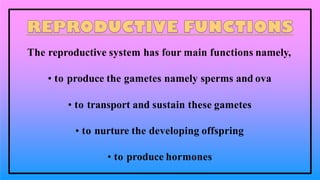
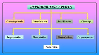
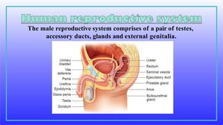
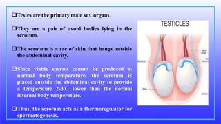
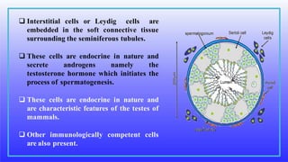
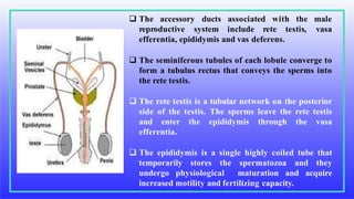
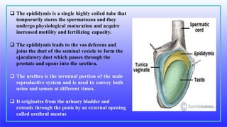
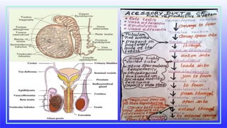
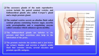
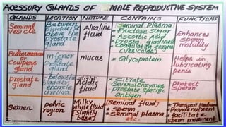
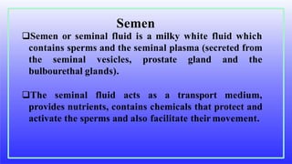
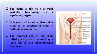
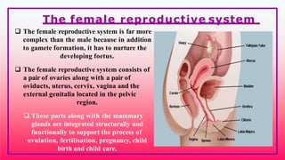
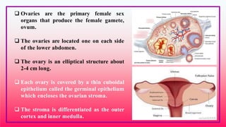
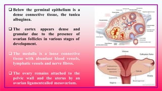
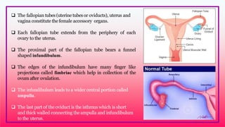
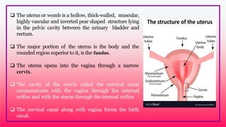
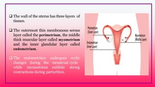
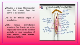
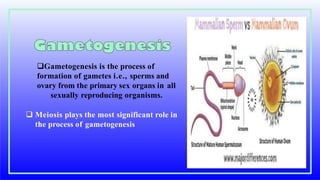
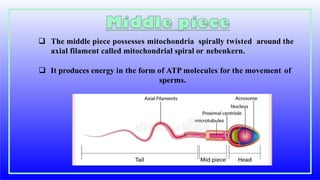
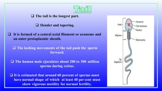
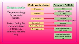
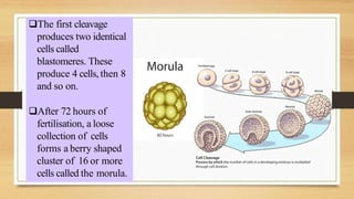
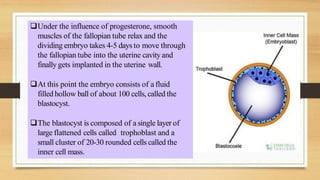
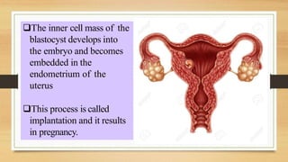
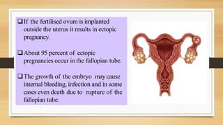
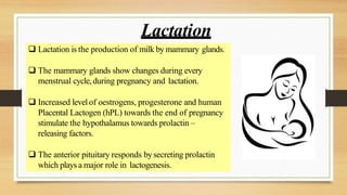
Chapter 2.
HUMAN
REPRODUCTION
 Human reproductive system
 Gametogenesis
 Menstrual cycle
 Fertilisation and implantation
 Maintenance of pregnancy and embryonic
development
 Parturition and lactation
Every organ system in the human body works
continuously to maintain homeostasis for the survival
of the individual. The human reproductive system is
essential for the survival of the species. An individual
may live a long healthy life without producing an
offspring, but reproduction is inevitable for the
existence of a species.
The reproductive system has four main functions namely,
• to produce the gametes namely sperms and ova
• to transport and sustain these gametes
• to nurture the developing offspring
• to produce hormones
Gametogenesis Insemination
REPRODUCTIVE EVENTS
Fertilisation Cleavage
Implantation Placentation Gastrulation Organogenesis
Parturition
• Gametogenesis: Formation of gametes by spermatogenesis and oogenesis.
• Insemination: Transfer of sperms by the male into the female genital tract.
• Fertilisation: Fusion of male and female gametes to form zygote, called
fertilisation.
• Cleavage: Rapid mitotic divisions of the zygote which convert the single celled
zygote into a multicellular structure called blastocyst.
• Implantation: Attachment of blastocyst to the uterine wall.
• Placentation: Formation of placenta which is the intimate
connection between foetus and uterine wall of the mother for
exchange of nutrients.
• Gastrulation: Process by which blastocyst is changed into a
gastrula with three primary germ layers
• Organogenesis: Formation of specific tissues, organs and organ
systems from three germ layers.
• Parturition: Expulsion of the foetus from the mother’s womb
• Reproductive functions are carried out by primary and accessory
reproductive organs
• The primary reproductive organs namely the ovary and testis are
responsible for producing the ova and sperms respectively.
• Hormones secreted by the pituitary gland and the gonads help ,
• development of the secondary sexual characteristics, maturation of
the reproductive system, regulation and normal functioning of the
reproductive system.
• Accessory organs,
transport and to sustain the gametes
nurture the developing offspring.
The male reproductive system comprises of a pair of testes,
accessory ducts, glands and external genitalia.
Testes are the primary male sex organs.
They are a pair of ovoid bodies lying in the
scrotum.
The scrotum is a sac of skin that hangs outside
the abdominal cavity.
Since viable sperms cannot be produced at
normal body temperature, the scrotum is
placed outside the abdominal cavity to provide
a temperature 2-3oC lower than the normal
internal body temperature.
Thus, the scrotum acts as a thermoregulator for
spermatogenesis.
Each testis is covered by an
outermost fibrous tunica albuginea
and is divided by septa into about
200 - 250 lobules.
Each containing 2-4 highly coiled
testicular tubules or seminiferous
tubules.
These highly convoluted tubules
which form 80 percent of the
testicular substance are the sites for
sperm production.
 The stratified epithelium of the
seminiferous tubule is made of two types of
cells namely sertoli cells or nurse cells and
spermatogonic cells or male germ cells.
 Sertoli cells are elongated and pyramidal
and provide nourishment to the sperms till
maturation.
 They also secrete inhibin, a hormone which
is involved in the negative feedback control
of sperm production.
 Spermatogonic cells divide meiotically and
differentiate to produce spermatozoa.
 Interstitial cells or Leydig cells are
embedded in the soft connective tissue
surrounding the seminiferous tubules.
 These cells are endocrine in nature and
secrete androgens namely the
testosterone hormone which initiates the
process of spermatogenesis.
 These cells are endocrine in nature and
are characteristic features of the testes of
mammals.
 Other immunologically competent cells
are also present.
 The accessory
reproductive system include rete testis,
ducts associated with the male
vasa
efferentia, epididymis and vas deferens.
 The seminiferous tubules of each lobule converge to
form a tubulus rectus that conveys the sperms into
the rete testis.
 The rete testis is a tubular network on the posterior
side of the testis. The sperms leave the rete testis
and enter the epididymis through the vasa
efferentia.
 The epididymis is a single highly coiled tube that
temporarily stores the spermatozoa and they
undergo physiological maturation and acquire
increased motility and fertilizing capacity.
 The epididymis is a single highly coiled tube that
temporarily stores the spermatozoa and they
undergo physiological maturation and acquire
increased motility and fertilizing capacity.
 The epididymis leads to the vas deferens and
joins the duct of the seminal vesicle to form the
ejaculatory duct which passes through the
prostate and opens into the urethra.
 The urethra is the terminal portion of the male
reproductive system and is used to convey both
urine and semen at different times.
 It originates from the urinary bladder and
extends through the penis by an external opening
called urethral meatus
 The accessory glands of the male reproductive
system include the paired seminal vesicles and
bulbourethral glands also called Cowper’s gland
and a single prostate gland.
 The seminal vesicles secrete an alkaline fluid called
seminal plasma containing fructose sugar, ascorbic
acid, prostaglandins and a coagulating enzyme
called vesiculase which enhances sperm motility.
prostate and their secretions also help in
 The bulbourethral glands are inferior to the
the
lubrication of the penis.
 The prostate encircles the urethra and is just below
the urinary bladder and secretes a slightly acidic
fluid that contains citrate, several enzymes and
prostate specific antigens.
Semen or seminal fluid is a milky white fluid which
contains sperms and the seminal plasma (secreted from
the seminal vesicles, prostate gland and the
bulbourethal glands).
The seminal fluid acts as a transport medium,
provides nutrients, contains chemicals that protect and
activate the sperms and also facilitate their movement.
Semen
 The penis is the male
genitalia functioning
external
as a
copulatory organ.
 It is made of a special tissue that
helps in the erection of penis to
facilitate insemination.
 The enlarged end of the penis
called glans penis is covered by a
loose fold of skin called foreskin
or prepuce.
 The female reproductive system is far more
complex than the male because in addition
to gamete formation, it has to nurture the
developing foetus.
 The female reproductive system consists of
a pair of ovaries along with a pair of
oviducts, uterus, cervix, vagina and the
external genitalia located in the pelvic
region.
 These parts along with the mammary
glands are integrated structurally and
functionally to support the process of
ovulation, fertilisation, pregnancy, child
birth and child care.
The female reproductive system
 Ovaries are the primary female sex
organs that produce the female gamete,
ovum.
 The ovaries are located one on each side
of the lower abdomen.
 The ovary is an elliptical structure about
2-4 cm long.
 Each ovary is covered by a thin cuboidal
epithelium called the germinal epithelium
which encloses the ovarian stroma.
 The stroma is differentiated as the outer
cortex and inner medulla.
 Below the germinal epithelium is a
dense connective tissue, the tunica
albuginea.
 The cortex appears dense and
granular due to the presence of
ovarian follicles in various stages of
development.
 The medulla is a loose connective
tissue with abundant blood vessels,
lymphatic vessels and nerve fibres.
 The ovary remains attached to the
pelvic wall and the uterus by an
ovarian ligamentcalled mesovarium.
 The fallopian tubes (uterine tubesor oviducts), uterus and
vagina constitute the female accessory organs.
 Each fallopian tube extends from the periphery of each
ovary to the uterus.
 The proximal part of the fallopian tube bears a funnel
shaped infundibulum.
 The edges of the infundibulum have many finger like
projections called fimbriae which help in collection of the
ovum after ovulation.
 The infundibulum leads to a wider central portion called
ampulla.
 The last part of the oviduct is the isthmus which is short
and thick walled connecting the ampulla and infundibulum
to the uterus.
 The uterus or womb is a hollow, thick-walled, muscular,
highly vascular and inverted pear shaped structure lying
in the pelvic cavity between the urinary bladder and
rectum.
 The major portion of the uterus is the body and the
rounded region superior to it, is the fundus.
 The uterus opens into the vagina through a narrow
cervix.
 The cavity of the cervix called the cervical canal
communicates with the vagina through the external
orifice and with the uterus through the internal orifice.
 The cervical canal along with vagina forms the birth
canal.
 The wall of the uterus has three layers of
tissues.
 The outermost thin membranous serous
layer called the perimetrium, the middle
thick muscular layer called myometrium
and the inner glandular layer called
endometrium.
 The endometrium
changes during the
while myometrium
undergoes
menstrual
exhibits
cyclic
cycle
strong
contractions during parturition.
 Vagina is a large fibromuscular
tube that extends from the
cervix to the exterior.
 It is the female organ of
.copulation.
 The female reproductive
structures that lie external to the
vagina are called as the external
genitalia or vulva comprising of
labia majora, labia minora,
hymen and clitoris
 The Bartholin’s glands (also called greater vestibular glands) are
located posterior to the left and right of the opening of the
vagina.
 They secrete mucus to lubricate the vagina and are homologous
to the bulbourethral glands of the male.
 The Skene’s glands are located on the anterior wall of the vagina
and around the lower end of the urethra.
 They secrete a lubricating fluid and are homologous to the
prostate gland of the males.
Aaccessory Glands
 The external opening of the vagina is
partially closed by a thin ring of tissue
called the hymen.
 The hymen is often torn during the first
coitus (physical union). However in some
women it remains intact.
 It can be stretched or torn due to a sudden
fall or jolt and also during
physical
horseback
activities
riding,
such
etc.,
strenuous
as cycling,
and therefore
cannot be considered as an indicator of a
woman’s virginity.
• The mammary glands are modified sweat
glands present both sexes in pairs.
• It is rudimentary in the males and
functional in the females.
• located in the thoracic region.
• It contains glandular tissue and variable
quantities of fat with a median nipple
surrounded by a pigmented area called the
areola.
• Several sebaceous glands called the areolar
glands are found on the surface and they
reduce cracking of the skin of the nipple.
 Internally each mammary gland consists of 2-25
lobes, separated by fat and connective tissues. Each
lobe is made up of lobules which contain acini or
alveoli lined by epithelial cells.
 Cells of the alveoli secrete milk.
 The alveoli open into mammary tubules.
 The tubules of each lobe join to form a mammary
duct.
 Several mammary ducts join to form a wider
mammary ampulla which is connected to the
lactiferous duct in the nipple. Under the nipple, each
lactiferous duct expands to form the lactiferous
sinus which serves as a reservoir of milk. Each
lactiferous duct opens separately by a minute pore
on the surface of the nipple.
 Normal development of the breast begins
at puberty and progresses with changes
during each menstrual cycle.
 In non-pregnant women, the glandular
structure is largely underdeveloped and the
breast size is largely due to amount of fat
deposits.
 The size of the breast does not have an
influence on the efficiency of lactation.
Gametogenesis is the process of
formation of gametes i.e., sperms and
ovary from the primary sex organs in all
sexually reproducing organisms.
 Meiosis plays the most significant role in
the process of gametogenesis
• It is the sequence of events in the
seminiferous tubules of the testes that
produce the male gametes, the sperms.
• During development, the primordial germ
cells migrate into the testes and become
immature germ cells called sperm mother
cells or spermatogonia in the inner surfaces
of the seminiferous tubules.
• The spermatogonia begin to undergo
mitotic division at puberty and continue
throughout life.
• In the first stage of spermatogenesis, the spermatogonia migrate
among sertoli cells towards the central lumen of the seminiferous
tubule and become modified and enlarged to form primary
spermatocytes which are diploid with 23 pairs i.e., 46 chromosomes.
• Some of the primary spermatocytes undergo first meiotic division to
form two secondary spermatocytes which are haploid with 23
chromosomes each.
• The secondary spermatocytes undergo second meiotic division to
produce four haploid spermatids.
• The spermatids are transformed into mature spermatozoa (sperms) by
the process called spermiogenesis.
• Sperms are finally released into the cavity of seminiferous tubules by
a process called spermiation.
• The whole process of spermatogenesis takes about 64 days. At any
given time, different regions of the seminiferous tubules contain
spermatocytes in different stages of development.
• The sperm production remains nearly constant at a rate of about 200
million sperms per day.
 Spermatogenesis starts at the age of puberty and is initiated due to
the increase in the release of Gonadotropin Releasing Hormone
(GnRH) by the hypothalamus.
 GnRH acts on the anterior pituitary gland and stimulates the
secretion of two gonadotropins namely Follicle Stimulating
Hormone (FSH) and Lutenizing Hormone (LH).
 FSH stimulates testicular growth and enhances the production of
Androgen Binding Protein (ABP) by the sertoli cells and helps in the
process of spermiogenesis.
 LH acts on the Leydig stimulates the
testosterone which in
cells and
turn stimulates the
synthesis of
process of
spermatogenesis
 The human sperm is a microscopic, flagellated and motile gamete.
 The whole body of the sperm is enveloped by plasma membrane.
 Body is divisible in to a head, neck and a tail.
 The head comprises of two parts namely acrosome and nucleus.
 Acrosome is a small cap like pointed structure present at the tip of
the nucleus and is formed mainly from the Golgi body of the
spermatid.
 It contains hyaluronidase, a proteolytic enzyme, popularly known
as sperm lysin which helps to penetrate the ovum during
fertilisation.
 The nucleus is flat and oval.
 neck is very short and is present between the head and the middle piece.
 It contains the proximal centriole towards the nucleus which plays a role in the first
division of the zygote.
 The distal centriole gives rise to the axial filament of the sperm.
 The middle piece possesses mitochondria spirally twisted around the
axial filament called mitochondrial spiral or nebenkern.
 It produces energy in the form of ATP molecules for the movement of
sperms.
 The tail is the longest part.
 Slender and tapering.
 It is formed of a central axial filament or axoneme and
an outer protoplasmic sheath.
 The lashing movements of the tail push the sperm
forward.
 The human male ejaculates about 200 to 300 million
sperms during coitus.
 It is estimated that around 60 percent of sperms must
have normal shape of which at least 40 per cent must
show vigorous motility for normal fertility.
CRYPTORCHISM
 The failure of one orboth testes to descend down into the
scrotal sacs is known as cryptorchism (crypto – hidden +
orchis – testicle). It occurs in 1 – 3 percent of new born males.
Asurgical correction at a young age can rectify the defect, else
these individuals may become sterile and are unable to
produce viable sperms.
 The sperm is the smallest human cell
 Males produce more than 500 billion sperms in their life time.
The process of egg
formation in
female.
It starts during the
embryonic stages
of femalei.e.,
inside the mother’s
ovary.
Embryonic stage Primary Follicle
During puberty
1st month 1700
2nd month 6 Lakhs
During birth 2 million
6th month end 7 million
Atricia/ Shrinkage of follicle
Proliferate/ Multiple
Birth to till puberty
60- 80 thousand follicle
ready to start
From 500, one primary
follicle will develop into
egg
Resting phase
Proliferate/ Multiple
Oogenesis
the female
 Oogenesis is the process of development of
gamete or ovum or egg in the ovaries.
 During foetal development, certain cells in the germinal
epithelium of the foetal ovary divide by mitosis and produce
millions of egg mother cells or oogonia.
 No more oogonia are formed or added afterbirth.
 The oogonial cells start dividing and enter into Prophase I of
meiotic division I to form the primary oocytes which are
temporarily arrested at this stage.
 The primary oocytes then get surrounded by a single layer of
granulosa cells to form the primordial or primary follicle.
 A large number of follicles degenerate during the period
from birth to puberty, so at puberty only 60,000 to 80,000
follicles are left in each ovary.
 The primary follicle gets surrounded by many layers of
granulosa cells and a new theca layer to form the
secondary follicle.
 At this time, the primary oocyte within the tertiary follicle
(a fluid filled antrum ) grows in size and completes its first
meiotic division and forms the secondary oocyte.
 When secondary oocyte mature the wall of the tertiary
follicle rupture and release secondary oocyte
 The tertiary follicle eventually becomes a mature follicle or
Graafian follicle.
 During fertilisation, the secondary oocyte undergoes
second meiotic division and produces a large cell, the
ovum and a second polar body.
 At the end of gametogenesis in females, each primary
oocyte gives rise to only one haploid ovum.
• Human ovum is non-cleidoic, alecithal and
microscopic in nature.
• Its cytoplasm called ooplasm contains a
large nucleus called the germinal vesicle.
• The ovum is surrounded by three
coverings namely an inner thin transparent
vitelline membrane, middle thick zona
pellucida and outer thick coat of follicular
cells called corona radiata.
• Between the vitelline membrane and zona
pellucida is a narrow perivitelline space.
Structure of ovum
 The menstrual or ovarian cycle occurs approximately once in every
28/29 days during the reproductive life of the female from menarche
(puberty) to menopause except during pregnancy.
 The cycle of events starting from one menstrual period till the next one
is called the menstrual cycle during which cyclic changes occurs in the
endometrium every month.
 Cyclic menstruation is an indicator of normal reproductive phase.
Menstural cycle
Menstrual cycle comprises
of the following phases
Menstrual phase
Follicular or proliferative phase
Ovulatory phase
Luteal or secretory phase
 The cyclestarts ,menstrual flow occurs and lasts for 3-5
days.
 Menstrual flow is due to the breakdown of
endometrial lining of the uterus, and its blood
vessel due to
decline in the level of progesterone and oestrogen.
 Menstruation occurs only if the released ovum is not
fertilized.
 Absence of menstruation may be an indicator of
pregnancy.
 However it could also be due to stress, hormonal
disorder and anaemia.
 The follicular phase extends from the 5th day of
the cycle until the time of ovulation.
 During this phase, the primary follicle in the
ovary grows to become a fully mature Graafian
follicle and simultaneously, the endometrium
regenerates through proliferation.
 These changes in the ovary and the uterus are
induced by the secretion of gonadotropins like
FSH and LH, which increase gradually during
the follicular phase.
 It stimulates follicular development and
secretion of oestrogen by the folliclecells.
 Both LH and FSH attain peak level in
the middle of the cycle (about the 14th
day).
 Maximum secretion of LH during the mid
cycle called LH surge induces the
rupture of the Graafian follicle and the
release of the ovum (secondary oocyte)
from the ovary wallinto the peritoneal
cavity.
 This process is called as ovulation.
 During luteal phase, the remaining part of the Graafian
follicle is transformed into a transitory endocrine
gland called corpus luteum.
 The corpus luteum secretes large amount of
progesterone which is essential for the maintenance
of theendometrium.
 If fertilisation takes place, it paves way for the
implantation of the fertilizedovum.
 The uterine wall secretes nutritious fluid in the
uterus for the foetus So, this phase is also called as
secretory phase.
 During pregnancy all events of
menstrual cycle stop and there is no
menstruation.
 In the absence of fertilisation, the
corpus luteum degenerates
completely and leaves a scar tissue
called corpus albicans.
 It also initiates the disintegration of
the endometrium leading to
menstruation, marking the next cycle.
o PCOS is a complex endocrine system disorder that affects women in
their reproductive years. Polycystic means ‘many cysts’.
o It refers to many partially formed follicles on the ovaries, which contain
an egg each.
POLY CYSTIC OVARY SYNDROME (PCOS)
o But they do not grow to maturity or produce eggs that can be fertilized.
o Women with PCOS may experience irregular menstrual cycles, increased androgen
levels, excessive facial orbody hairgrowth (hirsutism), acne, obesity, reduced fertility
and increased risk of diabetes.
o Treatment for PCOS includes a healthy lifestyle, weight loss and targeted hormone
therapy.
 Menstrual hygiene is vital for good health, well-being, dignity,
empowerment and productivity of women.
 The impact of poor menstrual hygiene on girls is increased stress
levels, fear and embarrassment during menstruation.
 This can keep girls inactive during such periods leading to
absenteeism from school.
 Clean and safe absorbable clothing materials, sanitary napkins, pads,
tampons and menstrual cups have been identified as materials used
to manage menstruation.
 Changing sanitary material 4-5 hours as per
the requirement, provides comfort,
cleanliness and protection from infections. It
also helps in enhancing the quality of life of
women during this period.
 Used sanitary napkins should be wrapped in
paper and disposed.
 It should not be thrown in open areas or
drain pipe of toilets.
 Flushing of sanitary napkins in the drain
pipes causes choking of the drainage line
leading to water pollution.
• Menopause is the phase in a women’s life when
ovulation and menstruation stops.
• The average age of menopause is 45-50 years.
• It indicates the permanent cessation of the primary
functions of the ovaries.
Fertilisation occurs when a haploid sperm fuses with a haploid ovum to
form a fertilized egg or diploid zygote.
The sperms deposited in the female reproductive tract undergo
capacitation, which is a biochemical event that enables the sperm to
penetrate and fertilise the egg.
Fertilisation occurs only if the ovum and sperms are transported
simultaneously to the ampullary isthmic junction of the fallopian tube.
 Before a sperm can enter the egg, it must
penetrate the multiple layers of granulosa
(follicular) cells which are around the ovum
forming the corona radiata.
 The follicular cells are held together by an
adhesive cementing substance called
hyaluronic acid.
 The acrosomal membrane disintegrates
releasing the proteolytic enzyme, hyaluronidase
during sperm entry through the corona radiata
and zona pellucida.
 This is called acrosomal reaction.
Once fertilisation is accomplished, cortical
granules from the cytoplasm of the ovum form a
barrier called the fertilisation membrane around
the ovum preventing further penetration of other
sperms.
Thus polyspermy is prevented
The first cleavage
produces two identical
cells called
blastomeres. These
produce 4 cells, then 8
and so on.
After 72 hours of
fertilisation, a loose
collection of cells
forms a berry shaped
cluster of 16 or more
cells called the morula.
Under the influence of progesterone, smooth
muscles of the fallopian tube relax and the
dividing embryo takes 4-5 days to move through
the fallopian tube into the uterine cavity and
finally gets implanted in the uterine wall.
At this point the embryo consists of a fluid
filled hollow ball of about 100 cells,called the
blastocyst.
The blastocyst is composed of a single layer of
large flattened cells called trophoblast and a
small cluster of 20-30 rounded cells called the
inner cell mass.
The inner cell mass of the
blastocyst develops into
the embryo and becomes
embedded in the
endometrium of the
uterus
This process is called
implantation and it results
in pregnancy.
If the fertilised ovum is implanted
outside the uterus it results in ectopic
pregnancy.
About 95 percent of ectopic
pregnancies occur in the fallopian tube.
The growth of the embryo may cause
internal bleeding, infection and in some
cases even death due to rupture of the
fallopian tube.
 The inner cell mass in the blastula is differentiated
into epiblast and hypoblast immediately after
implantation.
 The hypoblast is the embryonic endoderm and the
epiblast is the ectoderm.
 The cells remaining in between the epiblast and the
endoderm form the mesoderm.
 Thus the transformation of the blastocyst into a
gastrula with the primary germ layers by the
movement of the blastomeres is called
gastrulation.
 Each germ layer gives rise to specific tissues,
organs and organ systems during organogenesis.
Maintenance of pregnancy and embryonic development
The extra embryonic membranes namely the
amnion, yolk sac, allantois and chorion protect the
embryo from dessication, mechanical shock and
help in the absorption of nutrients and exchange of
gases.
The amnion is a double layered translucent
membrane filled with the amniotic fluid.
It provides a buoyant environment to protect the
developing embryo from injury, regulates the
temperature of the foetus and provides a medium in
which the foetus can move.
The yolksac forms a part of the gut and is the
source of the earliest blood cells and blood vessels.
The allantois forms a small out pocketing of
embryonic tissue at the caudal end of the
yolk sac.
It is the structural base for the umbilical cord
that links the embryo to the placenta and
ultimately it becomes part of the urinary
bladder.
The chorion is the outermost membrane
which encloses the embryo and all other
membranes and also helps in the formation
of theplacenta
The trophoblast cells in the blastocyst send out
several finger like projections called chorionic villi
carrying foetal blood and are surrounded by sinuses
that contain maternal blood.
The chorionic villi and the uterine tissues form the
disc-shaped placenta.
Placenta is a temporary endocrine organ formed
during pregnancy and it connects the foetus to the
uterine wallthrough the umbilical cord.
It is the organ by which the nutritive, respiratoryand
excretory functions are fulfilled.
The embryo’s heart develops during the fourth week
of pregnancy and circulates blood through the
umbilical cord and placenta as well as through its own
tissue
The primary germ layers serve as the primitive tissues from which all body organs
develop.
The ectoderm gives rise to the central nervous system (brain and spinal cord),
peripheral nervous system, epidermis and its derivatives and mammary
glands.
The connective tissue, cartilage and bone, muscles, organs of urinogenital
system (kidney, ureter and gonads) arise from the mesoderm.
The endodermal derivatives are epithelium of gastrointestinal and respiratory
tract, liver, pancreas, thyroid and parathyroids
Human pregnancy lasts for about 280 days or 40 weeks and is called the gestation
period.
It can be divided for convenience into three trimesters of three months each.
The first trimester is the main period of organogenesis, the body organs namely
the heart, limbs, lungs, liver and external genital organs are well developed.
By the end of the second trimester, the face is well formed with features, eyelids
and eyelashes, eyes blink, body is covered with fine hair, muscle tissue
develops and bones become harder.
The foetus is fully developed and is ready for delivery by the end of nine
months (third trimester)
 During pregnancy, the placenta acts as a temporary endocrine gland and produces
large quantities of human Chorionic Gonadotropin (hCG), human Chorionic
Somatomammotropin (hCS) or human Placental Lactogen (hPL), oestrogens
and progesterone which are essential for a normal pregnancy.
 A hormone called relaxin is also secreted during the later phase of pregnancy which
helps in relaxation of the pelvic ligaments at the time of parturition.
 It should be noted that hCG, hPL and relaxin are produced only during pregnancy.
 In addition, during pregnancy the level of other hormones like oestrogen and
progesterone, cortisol, prolactin, thyroxine, etc., is increased several folds in the
maternal blood.
 These hormones are essential for supporting foetal growth.
Parturition is the completion of pregnancy and giving birth to the baby.
The series of events that expels the infant from the uterus is collectively called
“labour”.
Throughout pregnancy the uterus undergoes periodic episodes of weak
and strong contractions.
These contractions called Braxter-Hick’s contractions lead to false labour.
Parturition and lactation
As the pregnancy progresses, increase in the oestrogen concentration promotes
uterine contractions.
These uterine contractions facilitate moulding of the foetus and downward
movement of thefoetus.
The descent of the foetus causes dilation of cervix of the uterus and vaginal canal
resulting in a neurohumoral reflex called Foetal ejection reflex or Ferguson reflex.
This initiates the secretion of oxytocin from the neurohypophysis which in turn
brings about the powerful contraction of the uterine muscles and leads to the
expulsion of the baby through the birth canal.
This sequence of events is called as parturition or childbirth.
 Relaxin is a hormone secreted by the placenta and also found
in the corpus luteum.
 It promotes parturition by relaxing the pelvic joints and by
dilatation of the cervix with continued powerful contractions.
 The amnion ruptures and the amniotic fluid flows out through
the vagina, followed by the foetus.
 The placenta along with the remains of the umbilical cord called
“after birth” is expelled out after delivery.
 Lactation is the production of milk by mammary glands.
 The mammary glands show changes during every
menstrual cycle,during pregnancy and lactation.
 Increased level of oestrogens, progesterone and human
Placental Lactogen (hPL) towards the end of pregnancy
stimulate the hypothalamus towards prolactin –
releasing factors.
 The anterior pituitary responds by secreting prolactin
which plays a major role in lactogenesis.
Lactation
 Oxytocin causes the “Let-Down” reflex- the actual
ejection of milk from the alveoli of the mammary
glands.
 During lactation, oxytocin also stimulates the recently
emptied uterus to contract, helping it to return to pre
- pregnancy size.
Colostrum is a nutrient rich fluid produced by the human
female immediately after giving birth, is loaded with immune,
growth and tissue repair factors.
It acts as a natural antimicrobial agent to actively stimulate the
maturation of the infant’s immune system.
No artificial feed can substitute the first milk, with all its
natural benefits and therefore should be definitely fed to the
baby after birth.
Colostrum
The mammary glands secrete a yellowish fluid called
colostrum during the initial few days after parturition.
It has less lactose than milk and almost no fat, but it
contains more proteins, vitaminAand minerals.
Colostrum is also rich in IgA antibodies.
This helps to protect the infant’s digestive tract against
bacterial infection.
Breast milk is the ideal food for infants as it contains all the
constituents in suitable concentration and is easily
digestible.
It is fully sufficient till about 6 months of age and all infants
must be breast fed by the mother to ensure the growth of a
healthy baby.
Colostrum
 Colostrum, a nutrient rich fluid produced by the human female
immediately after giving birth, is loaded with immune, growth and
tissue repair factors.
 It acts as a natural antimicrobial agent to actively stimulate the
maturation of the infant’s immune system.
 No artificial feed can substitute the first milk, with all its natural
benefits and therefore should be definitely fed to the baby after birth.
Do you know
Twins are two offsprings
produced in the same
pregnancy.
 Monozygotic (Identical)
twins are produced when
a single fertilised egg
splits into two during the
first cleavage. They are of
the same sex, look alike
and share the same genes.
 Dizygotic (Fraternal) twins are produced when two
separate eggs are fertilised by two separate sperms.
The twins may be of the same sex or different sex and
are non-identical.
Siamese (United) twins are the
conjoined twins who are joined during
birth.
Afemale uterus is normally about 3 inches long and 2 inches
wide but can expand 20 times during pregnancy.
Ovum oregg is the largest human cell.
Out of the million eggs women possess during birth, only
about 300 to 400 will ovulate before menopause.
Female uterus contains one of the strongest muscles of the
human body.
More to know
2. Human reproduction.pptx

2. Human reproduction.pptx

  • 1.
  • 2.
     Human reproductivesystem  Gametogenesis  Menstrual cycle  Fertilisation and implantation  Maintenance of pregnancy and embryonic development  Parturition and lactation
  • 3.
    Every organ systemin the human body works continuously to maintain homeostasis for the survival of the individual. The human reproductive system is essential for the survival of the species. An individual may live a long healthy life without producing an offspring, but reproduction is inevitable for the existence of a species.
  • 4.
    The reproductive systemhas four main functions namely, • to produce the gametes namely sperms and ova • to transport and sustain these gametes • to nurture the developing offspring • to produce hormones
  • 5.
    Gametogenesis Insemination REPRODUCTIVE EVENTS FertilisationCleavage Implantation Placentation Gastrulation Organogenesis Parturition
  • 6.
    • Gametogenesis: Formationof gametes by spermatogenesis and oogenesis. • Insemination: Transfer of sperms by the male into the female genital tract. • Fertilisation: Fusion of male and female gametes to form zygote, called fertilisation. • Cleavage: Rapid mitotic divisions of the zygote which convert the single celled zygote into a multicellular structure called blastocyst. • Implantation: Attachment of blastocyst to the uterine wall.
  • 7.
    • Placentation: Formationof placenta which is the intimate connection between foetus and uterine wall of the mother for exchange of nutrients. • Gastrulation: Process by which blastocyst is changed into a gastrula with three primary germ layers • Organogenesis: Formation of specific tissues, organs and organ systems from three germ layers. • Parturition: Expulsion of the foetus from the mother’s womb
  • 8.
    • Reproductive functionsare carried out by primary and accessory reproductive organs • The primary reproductive organs namely the ovary and testis are responsible for producing the ova and sperms respectively. • Hormones secreted by the pituitary gland and the gonads help , • development of the secondary sexual characteristics, maturation of the reproductive system, regulation and normal functioning of the reproductive system. • Accessory organs, transport and to sustain the gametes nurture the developing offspring.
  • 9.
    The male reproductivesystem comprises of a pair of testes, accessory ducts, glands and external genitalia.
  • 10.
    Testes are theprimary male sex organs. They are a pair of ovoid bodies lying in the scrotum. The scrotum is a sac of skin that hangs outside the abdominal cavity. Since viable sperms cannot be produced at normal body temperature, the scrotum is placed outside the abdominal cavity to provide a temperature 2-3oC lower than the normal internal body temperature. Thus, the scrotum acts as a thermoregulator for spermatogenesis.
  • 11.
    Each testis iscovered by an outermost fibrous tunica albuginea and is divided by septa into about 200 - 250 lobules. Each containing 2-4 highly coiled testicular tubules or seminiferous tubules. These highly convoluted tubules which form 80 percent of the testicular substance are the sites for sperm production.
  • 12.
     The stratifiedepithelium of the seminiferous tubule is made of two types of cells namely sertoli cells or nurse cells and spermatogonic cells or male germ cells.  Sertoli cells are elongated and pyramidal and provide nourishment to the sperms till maturation.  They also secrete inhibin, a hormone which is involved in the negative feedback control of sperm production.  Spermatogonic cells divide meiotically and differentiate to produce spermatozoa.
  • 13.
     Interstitial cellsor Leydig cells are embedded in the soft connective tissue surrounding the seminiferous tubules.  These cells are endocrine in nature and secrete androgens namely the testosterone hormone which initiates the process of spermatogenesis.  These cells are endocrine in nature and are characteristic features of the testes of mammals.  Other immunologically competent cells are also present.
  • 14.
     The accessory reproductivesystem include rete testis, ducts associated with the male vasa efferentia, epididymis and vas deferens.  The seminiferous tubules of each lobule converge to form a tubulus rectus that conveys the sperms into the rete testis.  The rete testis is a tubular network on the posterior side of the testis. The sperms leave the rete testis and enter the epididymis through the vasa efferentia.  The epididymis is a single highly coiled tube that temporarily stores the spermatozoa and they undergo physiological maturation and acquire increased motility and fertilizing capacity.
  • 15.
     The epididymisis a single highly coiled tube that temporarily stores the spermatozoa and they undergo physiological maturation and acquire increased motility and fertilizing capacity.  The epididymis leads to the vas deferens and joins the duct of the seminal vesicle to form the ejaculatory duct which passes through the prostate and opens into the urethra.  The urethra is the terminal portion of the male reproductive system and is used to convey both urine and semen at different times.  It originates from the urinary bladder and extends through the penis by an external opening called urethral meatus
  • 17.
     The accessoryglands of the male reproductive system include the paired seminal vesicles and bulbourethral glands also called Cowper’s gland and a single prostate gland.  The seminal vesicles secrete an alkaline fluid called seminal plasma containing fructose sugar, ascorbic acid, prostaglandins and a coagulating enzyme called vesiculase which enhances sperm motility. prostate and their secretions also help in  The bulbourethral glands are inferior to the the lubrication of the penis.  The prostate encircles the urethra and is just below the urinary bladder and secretes a slightly acidic fluid that contains citrate, several enzymes and prostate specific antigens.
  • 19.
    Semen or seminalfluid is a milky white fluid which contains sperms and the seminal plasma (secreted from the seminal vesicles, prostate gland and the bulbourethal glands). The seminal fluid acts as a transport medium, provides nutrients, contains chemicals that protect and activate the sperms and also facilitate their movement. Semen
  • 20.
     The penisis the male genitalia functioning external as a copulatory organ.  It is made of a special tissue that helps in the erection of penis to facilitate insemination.  The enlarged end of the penis called glans penis is covered by a loose fold of skin called foreskin or prepuce.
  • 21.
     The femalereproductive system is far more complex than the male because in addition to gamete formation, it has to nurture the developing foetus.  The female reproductive system consists of a pair of ovaries along with a pair of oviducts, uterus, cervix, vagina and the external genitalia located in the pelvic region.  These parts along with the mammary glands are integrated structurally and functionally to support the process of ovulation, fertilisation, pregnancy, child birth and child care. The female reproductive system
  • 22.
     Ovaries arethe primary female sex organs that produce the female gamete, ovum.  The ovaries are located one on each side of the lower abdomen.  The ovary is an elliptical structure about 2-4 cm long.  Each ovary is covered by a thin cuboidal epithelium called the germinal epithelium which encloses the ovarian stroma.  The stroma is differentiated as the outer cortex and inner medulla.
  • 23.
     Below thegerminal epithelium is a dense connective tissue, the tunica albuginea.  The cortex appears dense and granular due to the presence of ovarian follicles in various stages of development.  The medulla is a loose connective tissue with abundant blood vessels, lymphatic vessels and nerve fibres.  The ovary remains attached to the pelvic wall and the uterus by an ovarian ligamentcalled mesovarium.
  • 24.
     The fallopiantubes (uterine tubesor oviducts), uterus and vagina constitute the female accessory organs.  Each fallopian tube extends from the periphery of each ovary to the uterus.  The proximal part of the fallopian tube bears a funnel shaped infundibulum.  The edges of the infundibulum have many finger like projections called fimbriae which help in collection of the ovum after ovulation.  The infundibulum leads to a wider central portion called ampulla.  The last part of the oviduct is the isthmus which is short and thick walled connecting the ampulla and infundibulum to the uterus.
  • 25.
     The uterusor womb is a hollow, thick-walled, muscular, highly vascular and inverted pear shaped structure lying in the pelvic cavity between the urinary bladder and rectum.  The major portion of the uterus is the body and the rounded region superior to it, is the fundus.  The uterus opens into the vagina through a narrow cervix.  The cavity of the cervix called the cervical canal communicates with the vagina through the external orifice and with the uterus through the internal orifice.  The cervical canal along with vagina forms the birth canal.
  • 26.
     The wallof the uterus has three layers of tissues.  The outermost thin membranous serous layer called the perimetrium, the middle thick muscular layer called myometrium and the inner glandular layer called endometrium.  The endometrium changes during the while myometrium undergoes menstrual exhibits cyclic cycle strong contractions during parturition.
  • 27.
     Vagina isa large fibromuscular tube that extends from the cervix to the exterior.  It is the female organ of .copulation.  The female reproductive structures that lie external to the vagina are called as the external genitalia or vulva comprising of labia majora, labia minora, hymen and clitoris
  • 28.
     The Bartholin’sglands (also called greater vestibular glands) are located posterior to the left and right of the opening of the vagina.  They secrete mucus to lubricate the vagina and are homologous to the bulbourethral glands of the male.  The Skene’s glands are located on the anterior wall of the vagina and around the lower end of the urethra.  They secrete a lubricating fluid and are homologous to the prostate gland of the males. Aaccessory Glands
  • 29.
     The externalopening of the vagina is partially closed by a thin ring of tissue called the hymen.  The hymen is often torn during the first coitus (physical union). However in some women it remains intact.  It can be stretched or torn due to a sudden fall or jolt and also during physical horseback activities riding, such etc., strenuous as cycling, and therefore cannot be considered as an indicator of a woman’s virginity.
  • 30.
    • The mammaryglands are modified sweat glands present both sexes in pairs. • It is rudimentary in the males and functional in the females. • located in the thoracic region. • It contains glandular tissue and variable quantities of fat with a median nipple surrounded by a pigmented area called the areola. • Several sebaceous glands called the areolar glands are found on the surface and they reduce cracking of the skin of the nipple.
  • 31.
     Internally eachmammary gland consists of 2-25 lobes, separated by fat and connective tissues. Each lobe is made up of lobules which contain acini or alveoli lined by epithelial cells.  Cells of the alveoli secrete milk.  The alveoli open into mammary tubules.  The tubules of each lobe join to form a mammary duct.  Several mammary ducts join to form a wider mammary ampulla which is connected to the lactiferous duct in the nipple. Under the nipple, each lactiferous duct expands to form the lactiferous sinus which serves as a reservoir of milk. Each lactiferous duct opens separately by a minute pore on the surface of the nipple.
  • 32.
     Normal developmentof the breast begins at puberty and progresses with changes during each menstrual cycle.  In non-pregnant women, the glandular structure is largely underdeveloped and the breast size is largely due to amount of fat deposits.  The size of the breast does not have an influence on the efficiency of lactation.
  • 33.
    Gametogenesis is theprocess of formation of gametes i.e., sperms and ovary from the primary sex organs in all sexually reproducing organisms.  Meiosis plays the most significant role in the process of gametogenesis
  • 34.
    • It isthe sequence of events in the seminiferous tubules of the testes that produce the male gametes, the sperms. • During development, the primordial germ cells migrate into the testes and become immature germ cells called sperm mother cells or spermatogonia in the inner surfaces of the seminiferous tubules. • The spermatogonia begin to undergo mitotic division at puberty and continue throughout life.
  • 35.
    • In thefirst stage of spermatogenesis, the spermatogonia migrate among sertoli cells towards the central lumen of the seminiferous tubule and become modified and enlarged to form primary spermatocytes which are diploid with 23 pairs i.e., 46 chromosomes. • Some of the primary spermatocytes undergo first meiotic division to form two secondary spermatocytes which are haploid with 23 chromosomes each. • The secondary spermatocytes undergo second meiotic division to produce four haploid spermatids. • The spermatids are transformed into mature spermatozoa (sperms) by the process called spermiogenesis. • Sperms are finally released into the cavity of seminiferous tubules by a process called spermiation. • The whole process of spermatogenesis takes about 64 days. At any given time, different regions of the seminiferous tubules contain spermatocytes in different stages of development. • The sperm production remains nearly constant at a rate of about 200 million sperms per day.
  • 36.
     Spermatogenesis startsat the age of puberty and is initiated due to the increase in the release of Gonadotropin Releasing Hormone (GnRH) by the hypothalamus.  GnRH acts on the anterior pituitary gland and stimulates the secretion of two gonadotropins namely Follicle Stimulating Hormone (FSH) and Lutenizing Hormone (LH).  FSH stimulates testicular growth and enhances the production of Androgen Binding Protein (ABP) by the sertoli cells and helps in the process of spermiogenesis.  LH acts on the Leydig stimulates the testosterone which in cells and turn stimulates the synthesis of process of spermatogenesis
  • 37.
     The humansperm is a microscopic, flagellated and motile gamete.  The whole body of the sperm is enveloped by plasma membrane.  Body is divisible in to a head, neck and a tail.  The head comprises of two parts namely acrosome and nucleus.  Acrosome is a small cap like pointed structure present at the tip of the nucleus and is formed mainly from the Golgi body of the spermatid.  It contains hyaluronidase, a proteolytic enzyme, popularly known as sperm lysin which helps to penetrate the ovum during fertilisation.  The nucleus is flat and oval.
  • 38.
     neck isvery short and is present between the head and the middle piece.  It contains the proximal centriole towards the nucleus which plays a role in the first division of the zygote.  The distal centriole gives rise to the axial filament of the sperm.
  • 39.
     The middlepiece possesses mitochondria spirally twisted around the axial filament called mitochondrial spiral or nebenkern.  It produces energy in the form of ATP molecules for the movement of sperms.
  • 40.
     The tailis the longest part.  Slender and tapering.  It is formed of a central axial filament or axoneme and an outer protoplasmic sheath.  The lashing movements of the tail push the sperm forward.  The human male ejaculates about 200 to 300 million sperms during coitus.  It is estimated that around 60 percent of sperms must have normal shape of which at least 40 per cent must show vigorous motility for normal fertility.
  • 41.
    CRYPTORCHISM  The failureof one orboth testes to descend down into the scrotal sacs is known as cryptorchism (crypto – hidden + orchis – testicle). It occurs in 1 – 3 percent of new born males. Asurgical correction at a young age can rectify the defect, else these individuals may become sterile and are unable to produce viable sperms.  The sperm is the smallest human cell  Males produce more than 500 billion sperms in their life time.
  • 42.
    The process ofegg formation in female. It starts during the embryonic stages of femalei.e., inside the mother’s ovary. Embryonic stage Primary Follicle During puberty 1st month 1700 2nd month 6 Lakhs During birth 2 million 6th month end 7 million Atricia/ Shrinkage of follicle Proliferate/ Multiple Birth to till puberty 60- 80 thousand follicle ready to start From 500, one primary follicle will develop into egg Resting phase Proliferate/ Multiple
  • 43.
    Oogenesis the female  Oogenesisis the process of development of gamete or ovum or egg in the ovaries.  During foetal development, certain cells in the germinal epithelium of the foetal ovary divide by mitosis and produce millions of egg mother cells or oogonia.  No more oogonia are formed or added afterbirth.  The oogonial cells start dividing and enter into Prophase I of meiotic division I to form the primary oocytes which are temporarily arrested at this stage.  The primary oocytes then get surrounded by a single layer of granulosa cells to form the primordial or primary follicle.  A large number of follicles degenerate during the period from birth to puberty, so at puberty only 60,000 to 80,000 follicles are left in each ovary.
  • 44.
     The primaryfollicle gets surrounded by many layers of granulosa cells and a new theca layer to form the secondary follicle.  At this time, the primary oocyte within the tertiary follicle (a fluid filled antrum ) grows in size and completes its first meiotic division and forms the secondary oocyte.  When secondary oocyte mature the wall of the tertiary follicle rupture and release secondary oocyte  The tertiary follicle eventually becomes a mature follicle or Graafian follicle.  During fertilisation, the secondary oocyte undergoes second meiotic division and produces a large cell, the ovum and a second polar body.  At the end of gametogenesis in females, each primary oocyte gives rise to only one haploid ovum.
  • 45.
    • Human ovumis non-cleidoic, alecithal and microscopic in nature. • Its cytoplasm called ooplasm contains a large nucleus called the germinal vesicle. • The ovum is surrounded by three coverings namely an inner thin transparent vitelline membrane, middle thick zona pellucida and outer thick coat of follicular cells called corona radiata. • Between the vitelline membrane and zona pellucida is a narrow perivitelline space. Structure of ovum
  • 46.
     The menstrualor ovarian cycle occurs approximately once in every 28/29 days during the reproductive life of the female from menarche (puberty) to menopause except during pregnancy.  The cycle of events starting from one menstrual period till the next one is called the menstrual cycle during which cyclic changes occurs in the endometrium every month.  Cyclic menstruation is an indicator of normal reproductive phase. Menstural cycle
  • 47.
    Menstrual cycle comprises ofthe following phases Menstrual phase Follicular or proliferative phase Ovulatory phase Luteal or secretory phase
  • 48.
     The cyclestarts,menstrual flow occurs and lasts for 3-5 days.  Menstrual flow is due to the breakdown of endometrial lining of the uterus, and its blood vessel due to decline in the level of progesterone and oestrogen.  Menstruation occurs only if the released ovum is not fertilized.  Absence of menstruation may be an indicator of pregnancy.  However it could also be due to stress, hormonal disorder and anaemia.
  • 49.
     The follicularphase extends from the 5th day of the cycle until the time of ovulation.  During this phase, the primary follicle in the ovary grows to become a fully mature Graafian follicle and simultaneously, the endometrium regenerates through proliferation.  These changes in the ovary and the uterus are induced by the secretion of gonadotropins like FSH and LH, which increase gradually during the follicular phase.  It stimulates follicular development and secretion of oestrogen by the folliclecells.
  • 50.
     Both LHand FSH attain peak level in the middle of the cycle (about the 14th day).  Maximum secretion of LH during the mid cycle called LH surge induces the rupture of the Graafian follicle and the release of the ovum (secondary oocyte) from the ovary wallinto the peritoneal cavity.  This process is called as ovulation.
  • 51.
     During lutealphase, the remaining part of the Graafian follicle is transformed into a transitory endocrine gland called corpus luteum.  The corpus luteum secretes large amount of progesterone which is essential for the maintenance of theendometrium.  If fertilisation takes place, it paves way for the implantation of the fertilizedovum.  The uterine wall secretes nutritious fluid in the uterus for the foetus So, this phase is also called as secretory phase.
  • 53.
     During pregnancyall events of menstrual cycle stop and there is no menstruation.  In the absence of fertilisation, the corpus luteum degenerates completely and leaves a scar tissue called corpus albicans.  It also initiates the disintegration of the endometrium leading to menstruation, marking the next cycle.
  • 54.
    o PCOS isa complex endocrine system disorder that affects women in their reproductive years. Polycystic means ‘many cysts’. o It refers to many partially formed follicles on the ovaries, which contain an egg each. POLY CYSTIC OVARY SYNDROME (PCOS)
  • 55.
    o But theydo not grow to maturity or produce eggs that can be fertilized. o Women with PCOS may experience irregular menstrual cycles, increased androgen levels, excessive facial orbody hairgrowth (hirsutism), acne, obesity, reduced fertility and increased risk of diabetes. o Treatment for PCOS includes a healthy lifestyle, weight loss and targeted hormone therapy.
  • 56.
     Menstrual hygieneis vital for good health, well-being, dignity, empowerment and productivity of women.  The impact of poor menstrual hygiene on girls is increased stress levels, fear and embarrassment during menstruation.  This can keep girls inactive during such periods leading to absenteeism from school.  Clean and safe absorbable clothing materials, sanitary napkins, pads, tampons and menstrual cups have been identified as materials used to manage menstruation.
  • 57.
     Changing sanitarymaterial 4-5 hours as per the requirement, provides comfort, cleanliness and protection from infections. It also helps in enhancing the quality of life of women during this period.  Used sanitary napkins should be wrapped in paper and disposed.  It should not be thrown in open areas or drain pipe of toilets.  Flushing of sanitary napkins in the drain pipes causes choking of the drainage line leading to water pollution.
  • 58.
    • Menopause isthe phase in a women’s life when ovulation and menstruation stops. • The average age of menopause is 45-50 years. • It indicates the permanent cessation of the primary functions of the ovaries.
  • 59.
    Fertilisation occurs whena haploid sperm fuses with a haploid ovum to form a fertilized egg or diploid zygote. The sperms deposited in the female reproductive tract undergo capacitation, which is a biochemical event that enables the sperm to penetrate and fertilise the egg. Fertilisation occurs only if the ovum and sperms are transported simultaneously to the ampullary isthmic junction of the fallopian tube.
  • 60.
     Before asperm can enter the egg, it must penetrate the multiple layers of granulosa (follicular) cells which are around the ovum forming the corona radiata.  The follicular cells are held together by an adhesive cementing substance called hyaluronic acid.  The acrosomal membrane disintegrates releasing the proteolytic enzyme, hyaluronidase during sperm entry through the corona radiata and zona pellucida.  This is called acrosomal reaction.
  • 61.
    Once fertilisation isaccomplished, cortical granules from the cytoplasm of the ovum form a barrier called the fertilisation membrane around the ovum preventing further penetration of other sperms. Thus polyspermy is prevented
  • 63.
    The first cleavage producestwo identical cells called blastomeres. These produce 4 cells, then 8 and so on. After 72 hours of fertilisation, a loose collection of cells forms a berry shaped cluster of 16 or more cells called the morula.
  • 64.
    Under the influenceof progesterone, smooth muscles of the fallopian tube relax and the dividing embryo takes 4-5 days to move through the fallopian tube into the uterine cavity and finally gets implanted in the uterine wall. At this point the embryo consists of a fluid filled hollow ball of about 100 cells,called the blastocyst. The blastocyst is composed of a single layer of large flattened cells called trophoblast and a small cluster of 20-30 rounded cells called the inner cell mass.
  • 65.
    The inner cellmass of the blastocyst develops into the embryo and becomes embedded in the endometrium of the uterus This process is called implantation and it results in pregnancy.
  • 66.
    If the fertilisedovum is implanted outside the uterus it results in ectopic pregnancy. About 95 percent of ectopic pregnancies occur in the fallopian tube. The growth of the embryo may cause internal bleeding, infection and in some cases even death due to rupture of the fallopian tube.
  • 67.
     The innercell mass in the blastula is differentiated into epiblast and hypoblast immediately after implantation.  The hypoblast is the embryonic endoderm and the epiblast is the ectoderm.  The cells remaining in between the epiblast and the endoderm form the mesoderm.  Thus the transformation of the blastocyst into a gastrula with the primary germ layers by the movement of the blastomeres is called gastrulation.  Each germ layer gives rise to specific tissues, organs and organ systems during organogenesis. Maintenance of pregnancy and embryonic development
  • 68.
    The extra embryonicmembranes namely the amnion, yolk sac, allantois and chorion protect the embryo from dessication, mechanical shock and help in the absorption of nutrients and exchange of gases. The amnion is a double layered translucent membrane filled with the amniotic fluid. It provides a buoyant environment to protect the developing embryo from injury, regulates the temperature of the foetus and provides a medium in which the foetus can move. The yolksac forms a part of the gut and is the source of the earliest blood cells and blood vessels.
  • 69.
    The allantois formsa small out pocketing of embryonic tissue at the caudal end of the yolk sac. It is the structural base for the umbilical cord that links the embryo to the placenta and ultimately it becomes part of the urinary bladder. The chorion is the outermost membrane which encloses the embryo and all other membranes and also helps in the formation of theplacenta
  • 70.
    The trophoblast cellsin the blastocyst send out several finger like projections called chorionic villi carrying foetal blood and are surrounded by sinuses that contain maternal blood. The chorionic villi and the uterine tissues form the disc-shaped placenta. Placenta is a temporary endocrine organ formed during pregnancy and it connects the foetus to the uterine wallthrough the umbilical cord. It is the organ by which the nutritive, respiratoryand excretory functions are fulfilled. The embryo’s heart develops during the fourth week of pregnancy and circulates blood through the umbilical cord and placenta as well as through its own tissue
  • 72.
    The primary germlayers serve as the primitive tissues from which all body organs develop. The ectoderm gives rise to the central nervous system (brain and spinal cord), peripheral nervous system, epidermis and its derivatives and mammary glands. The connective tissue, cartilage and bone, muscles, organs of urinogenital system (kidney, ureter and gonads) arise from the mesoderm. The endodermal derivatives are epithelium of gastrointestinal and respiratory tract, liver, pancreas, thyroid and parathyroids
  • 73.
    Human pregnancy lastsfor about 280 days or 40 weeks and is called the gestation period. It can be divided for convenience into three trimesters of three months each. The first trimester is the main period of organogenesis, the body organs namely the heart, limbs, lungs, liver and external genital organs are well developed. By the end of the second trimester, the face is well formed with features, eyelids and eyelashes, eyes blink, body is covered with fine hair, muscle tissue develops and bones become harder. The foetus is fully developed and is ready for delivery by the end of nine months (third trimester)
  • 74.
     During pregnancy,the placenta acts as a temporary endocrine gland and produces large quantities of human Chorionic Gonadotropin (hCG), human Chorionic Somatomammotropin (hCS) or human Placental Lactogen (hPL), oestrogens and progesterone which are essential for a normal pregnancy.  A hormone called relaxin is also secreted during the later phase of pregnancy which helps in relaxation of the pelvic ligaments at the time of parturition.  It should be noted that hCG, hPL and relaxin are produced only during pregnancy.  In addition, during pregnancy the level of other hormones like oestrogen and progesterone, cortisol, prolactin, thyroxine, etc., is increased several folds in the maternal blood.  These hormones are essential for supporting foetal growth.
  • 75.
    Parturition is thecompletion of pregnancy and giving birth to the baby. The series of events that expels the infant from the uterus is collectively called “labour”. Throughout pregnancy the uterus undergoes periodic episodes of weak and strong contractions. These contractions called Braxter-Hick’s contractions lead to false labour. Parturition and lactation
  • 76.
    As the pregnancyprogresses, increase in the oestrogen concentration promotes uterine contractions. These uterine contractions facilitate moulding of the foetus and downward movement of thefoetus. The descent of the foetus causes dilation of cervix of the uterus and vaginal canal resulting in a neurohumoral reflex called Foetal ejection reflex or Ferguson reflex. This initiates the secretion of oxytocin from the neurohypophysis which in turn brings about the powerful contraction of the uterine muscles and leads to the expulsion of the baby through the birth canal. This sequence of events is called as parturition or childbirth.
  • 77.
     Relaxin isa hormone secreted by the placenta and also found in the corpus luteum.  It promotes parturition by relaxing the pelvic joints and by dilatation of the cervix with continued powerful contractions.  The amnion ruptures and the amniotic fluid flows out through the vagina, followed by the foetus.  The placenta along with the remains of the umbilical cord called “after birth” is expelled out after delivery.
  • 78.
     Lactation isthe production of milk by mammary glands.  The mammary glands show changes during every menstrual cycle,during pregnancy and lactation.  Increased level of oestrogens, progesterone and human Placental Lactogen (hPL) towards the end of pregnancy stimulate the hypothalamus towards prolactin – releasing factors.  The anterior pituitary responds by secreting prolactin which plays a major role in lactogenesis. Lactation
  • 79.
     Oxytocin causesthe “Let-Down” reflex- the actual ejection of milk from the alveoli of the mammary glands.  During lactation, oxytocin also stimulates the recently emptied uterus to contract, helping it to return to pre - pregnancy size.
  • 80.
    Colostrum is anutrient rich fluid produced by the human female immediately after giving birth, is loaded with immune, growth and tissue repair factors. It acts as a natural antimicrobial agent to actively stimulate the maturation of the infant’s immune system. No artificial feed can substitute the first milk, with all its natural benefits and therefore should be definitely fed to the baby after birth. Colostrum
  • 81.
    The mammary glandssecrete a yellowish fluid called colostrum during the initial few days after parturition. It has less lactose than milk and almost no fat, but it contains more proteins, vitaminAand minerals. Colostrum is also rich in IgA antibodies.
  • 82.
    This helps toprotect the infant’s digestive tract against bacterial infection. Breast milk is the ideal food for infants as it contains all the constituents in suitable concentration and is easily digestible. It is fully sufficient till about 6 months of age and all infants must be breast fed by the mother to ensure the growth of a healthy baby.
  • 83.
    Colostrum  Colostrum, anutrient rich fluid produced by the human female immediately after giving birth, is loaded with immune, growth and tissue repair factors.  It acts as a natural antimicrobial agent to actively stimulate the maturation of the infant’s immune system.  No artificial feed can substitute the first milk, with all its natural benefits and therefore should be definitely fed to the baby after birth. Do you know
  • 84.
    Twins are twooffsprings produced in the same pregnancy.  Monozygotic (Identical) twins are produced when a single fertilised egg splits into two during the first cleavage. They are of the same sex, look alike and share the same genes.
  • 85.
     Dizygotic (Fraternal)twins are produced when two separate eggs are fertilised by two separate sperms. The twins may be of the same sex or different sex and are non-identical.
  • 86.
    Siamese (United) twinsare the conjoined twins who are joined during birth.
  • 87.
    Afemale uterus isnormally about 3 inches long and 2 inches wide but can expand 20 times during pregnancy. Ovum oregg is the largest human cell. Out of the million eggs women possess during birth, only about 300 to 400 will ovulate before menopause. Female uterus contains one of the strongest muscles of the human body. More to know