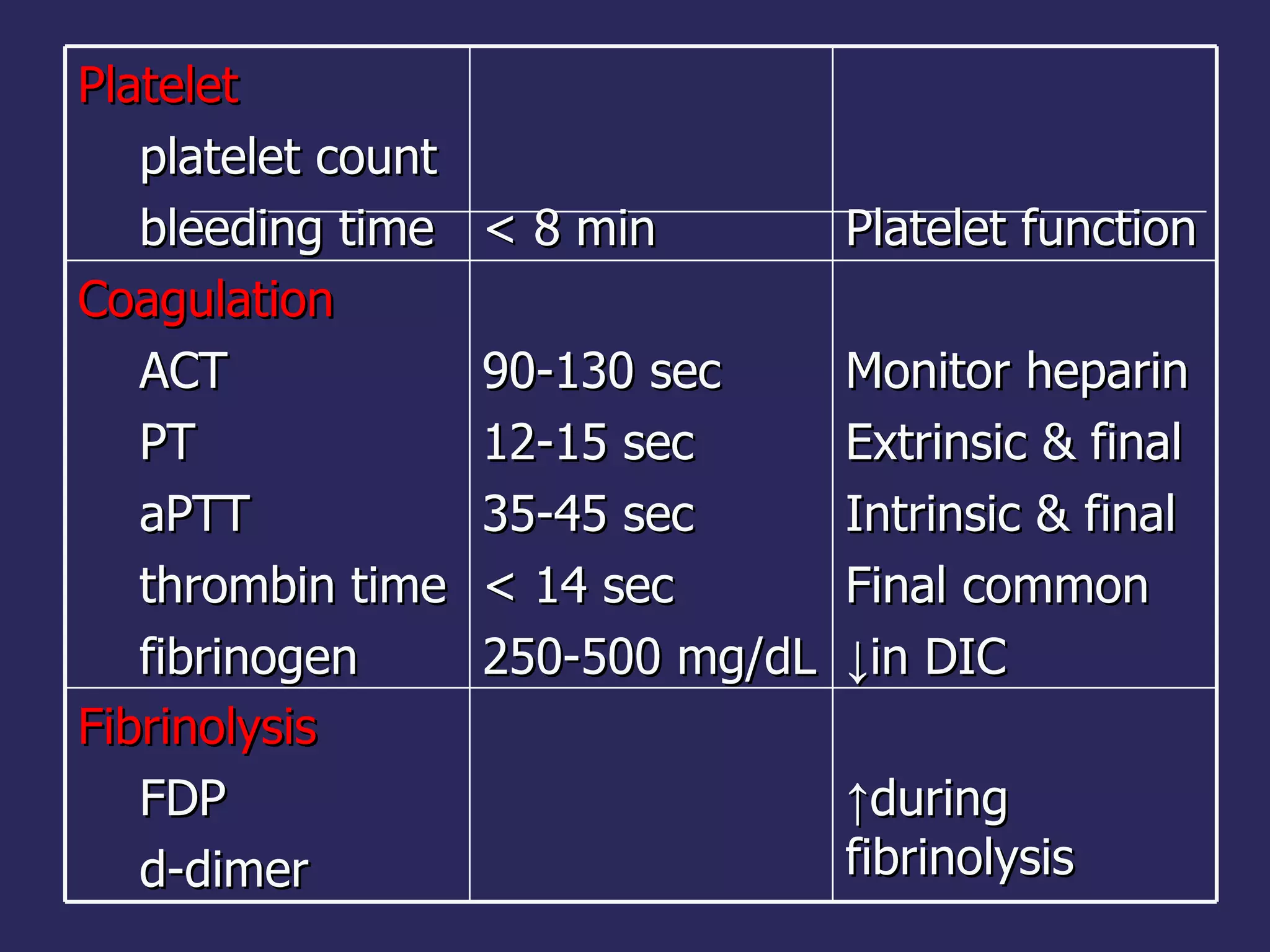
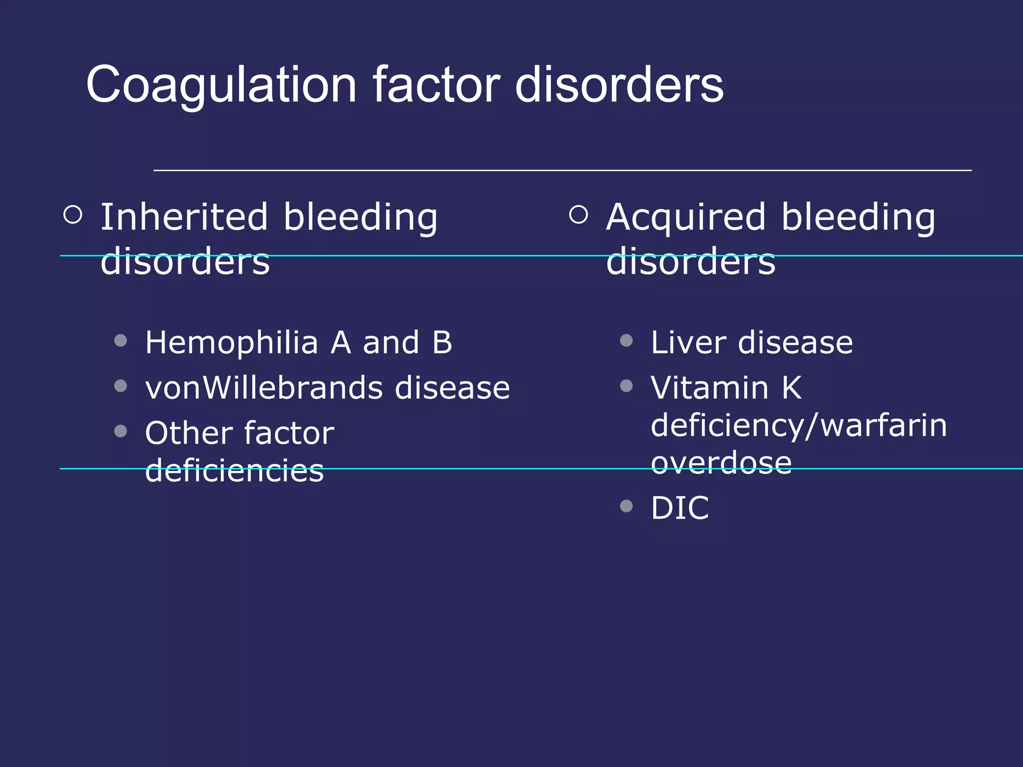
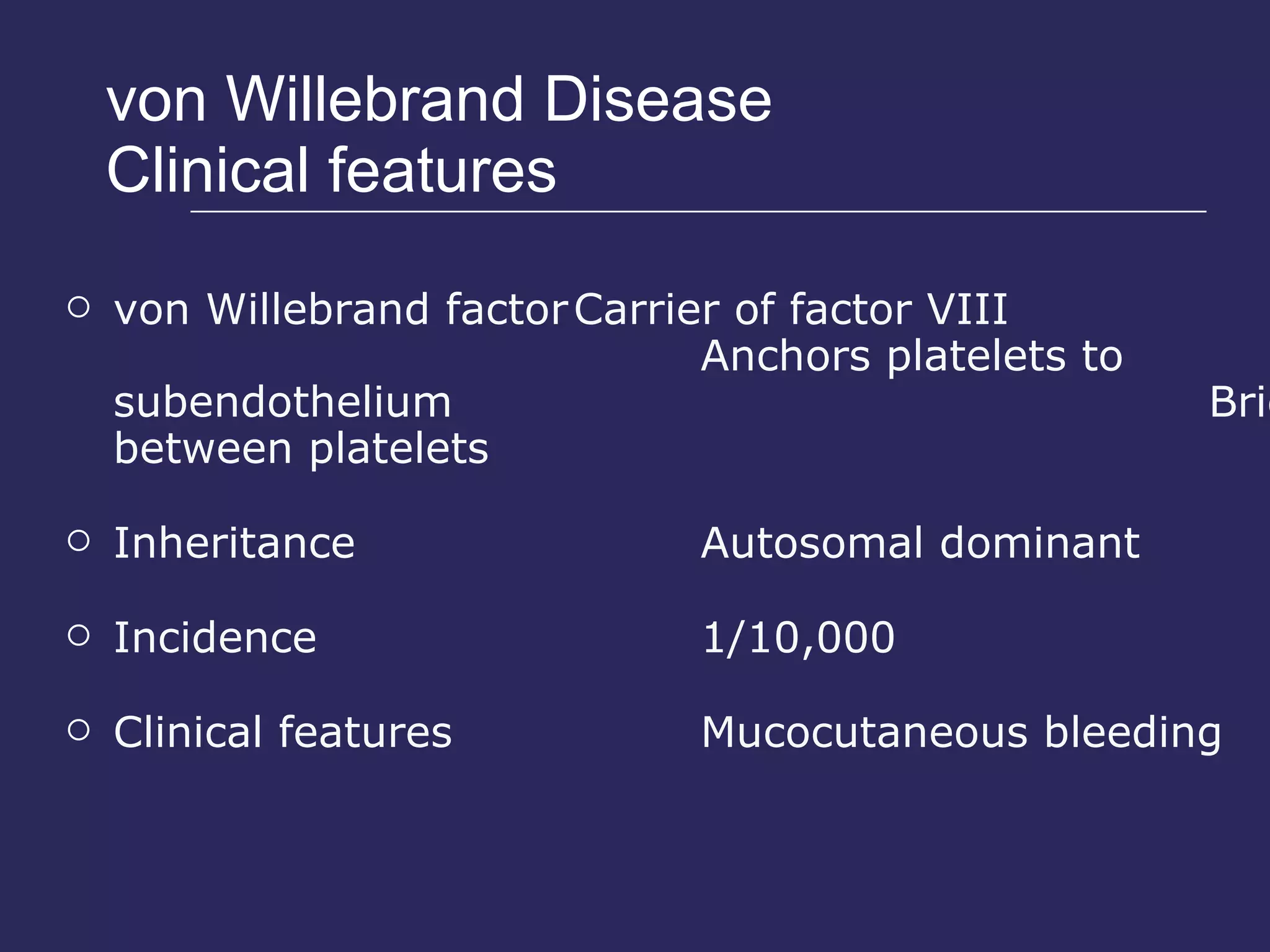
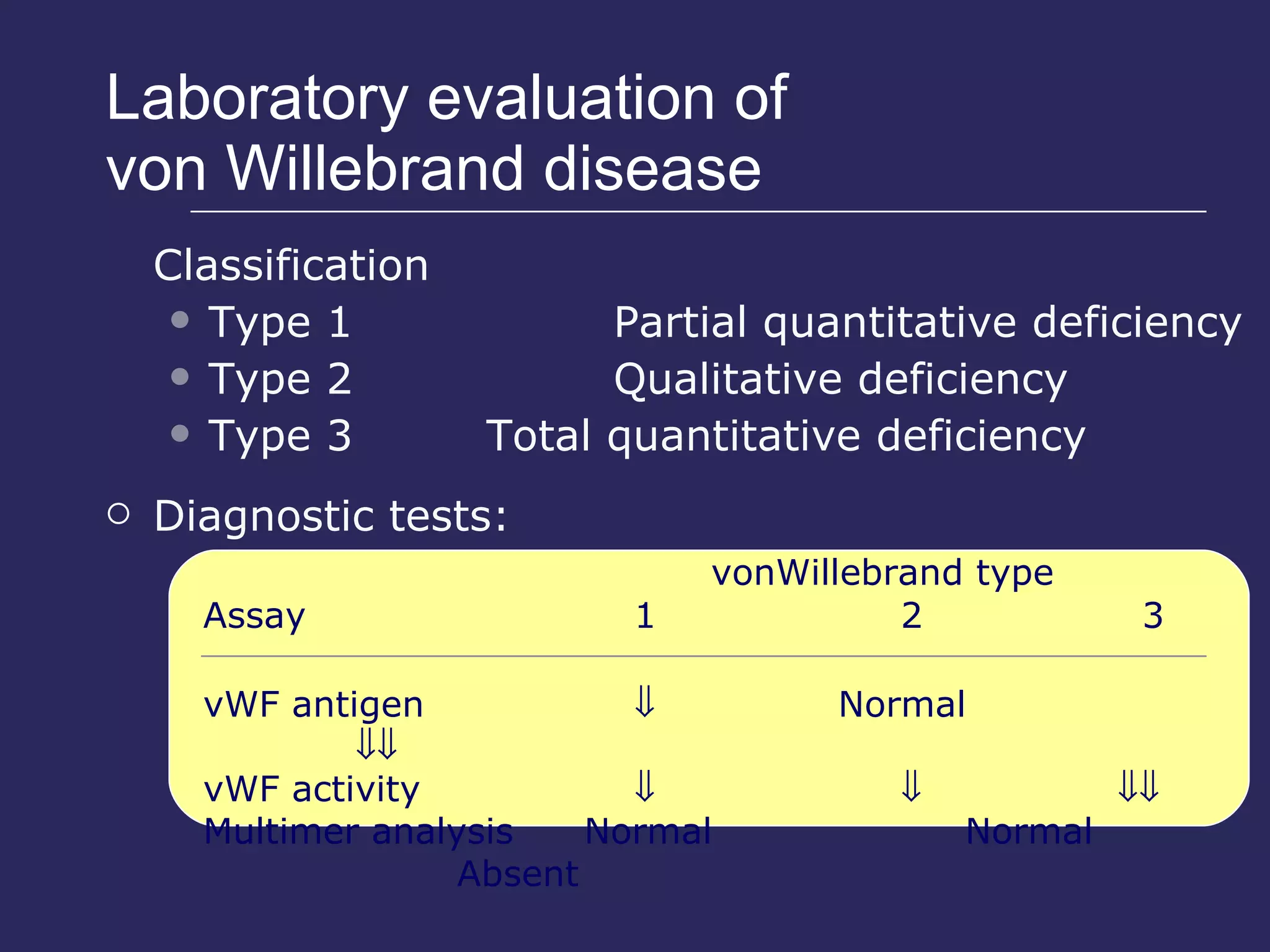
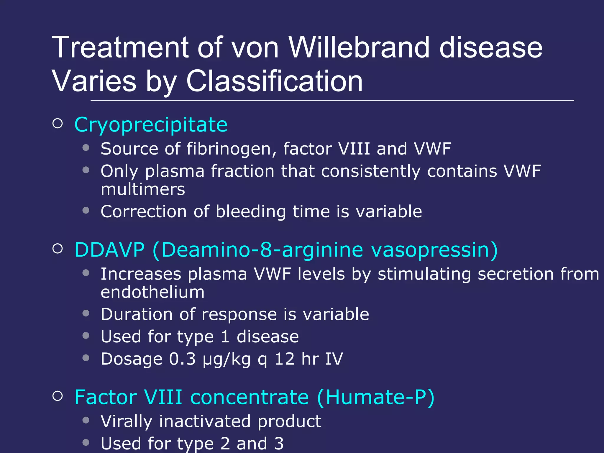
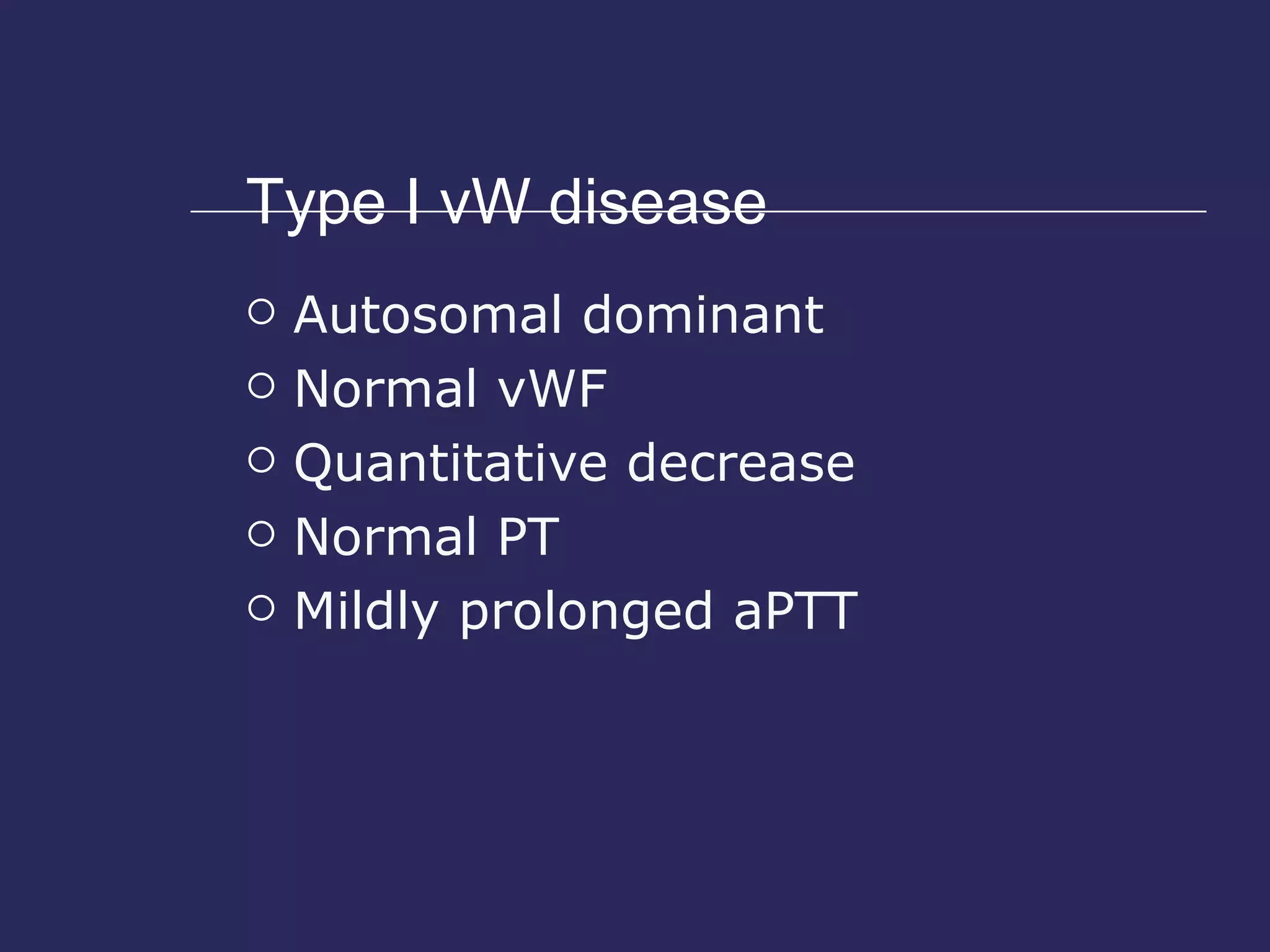
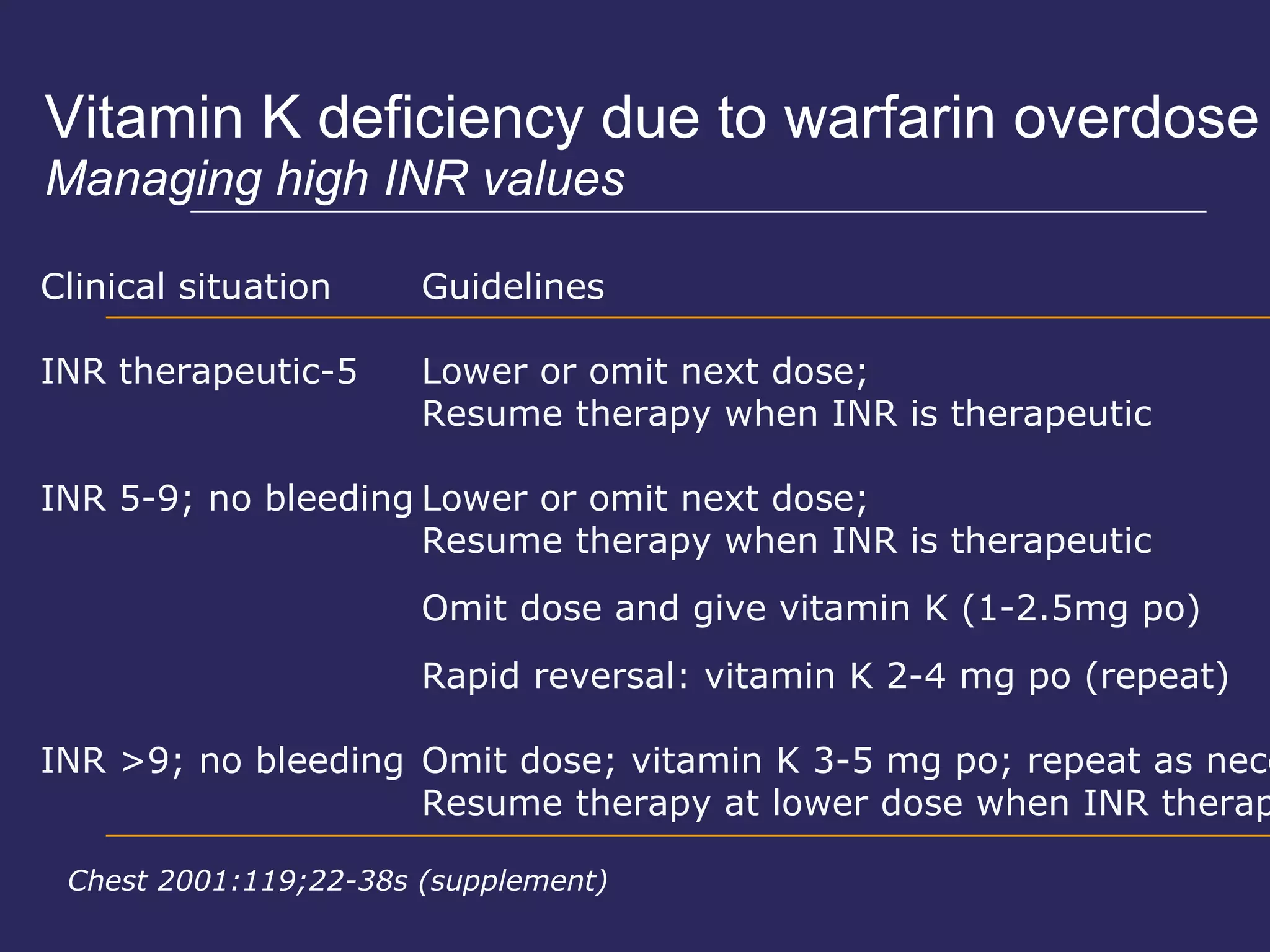
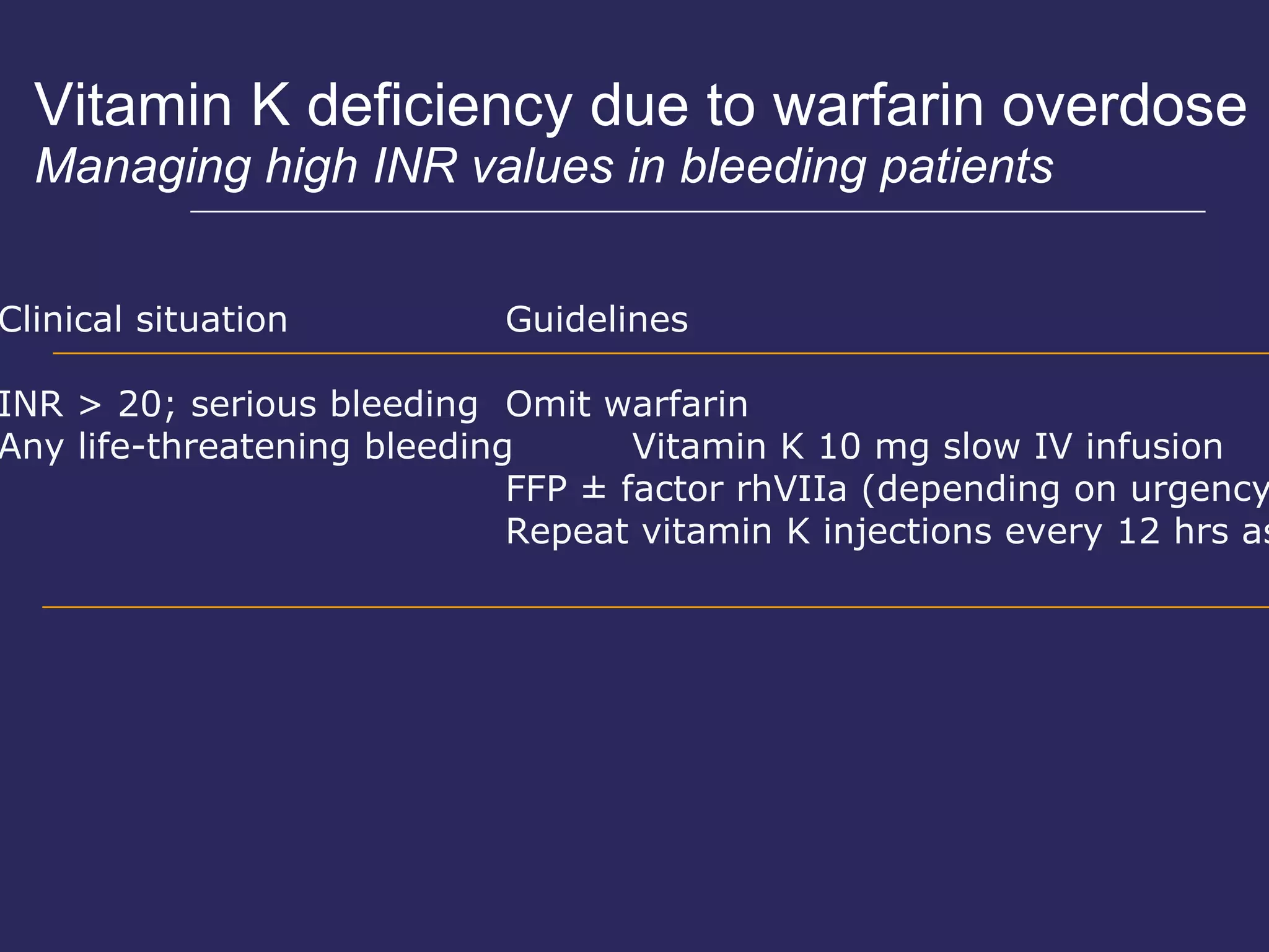
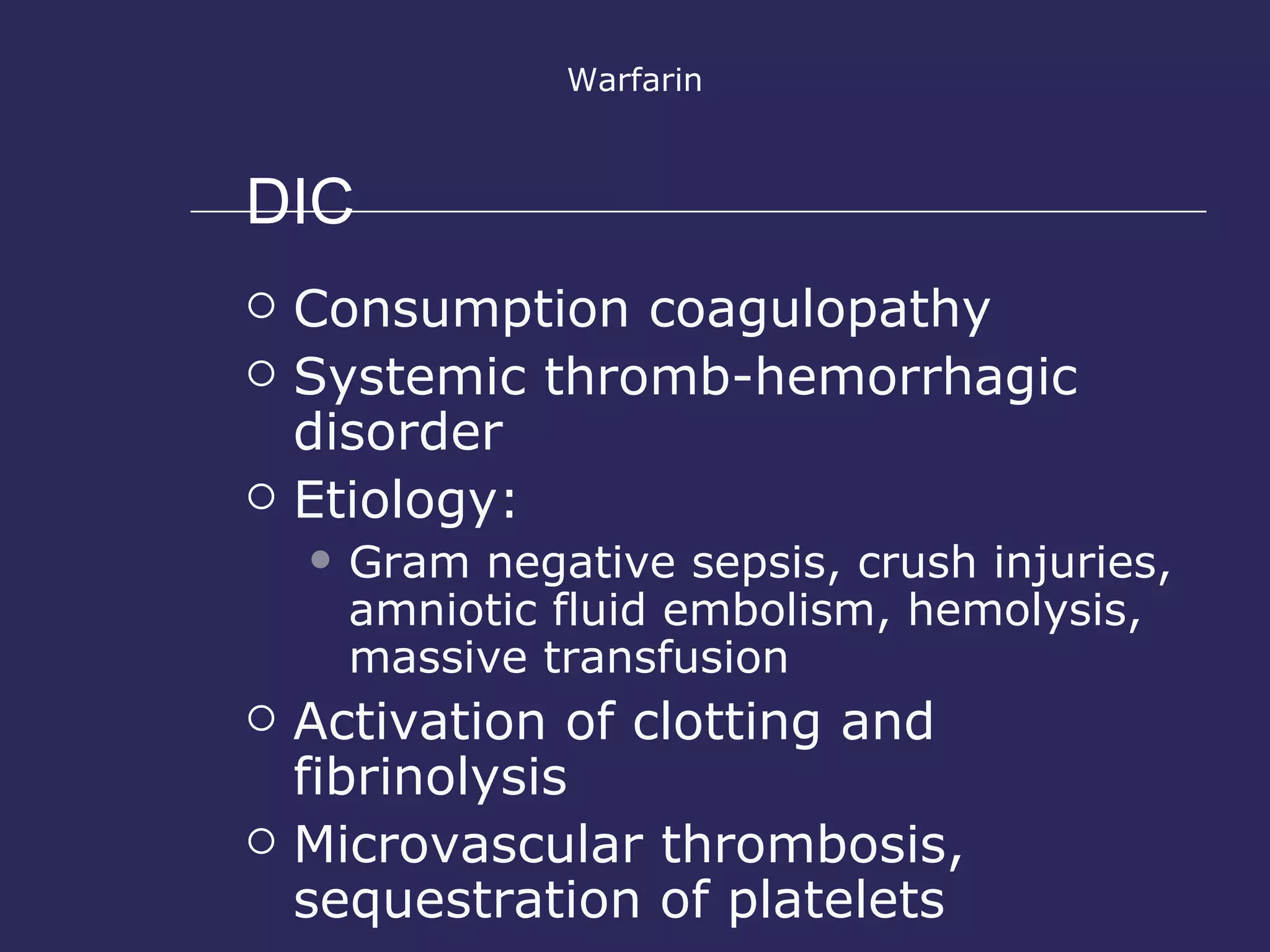
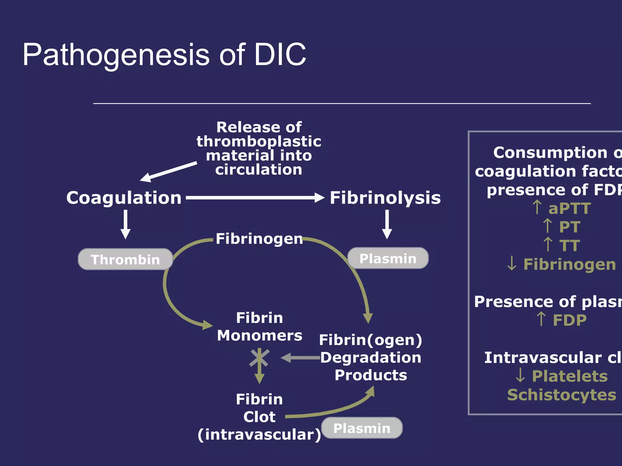
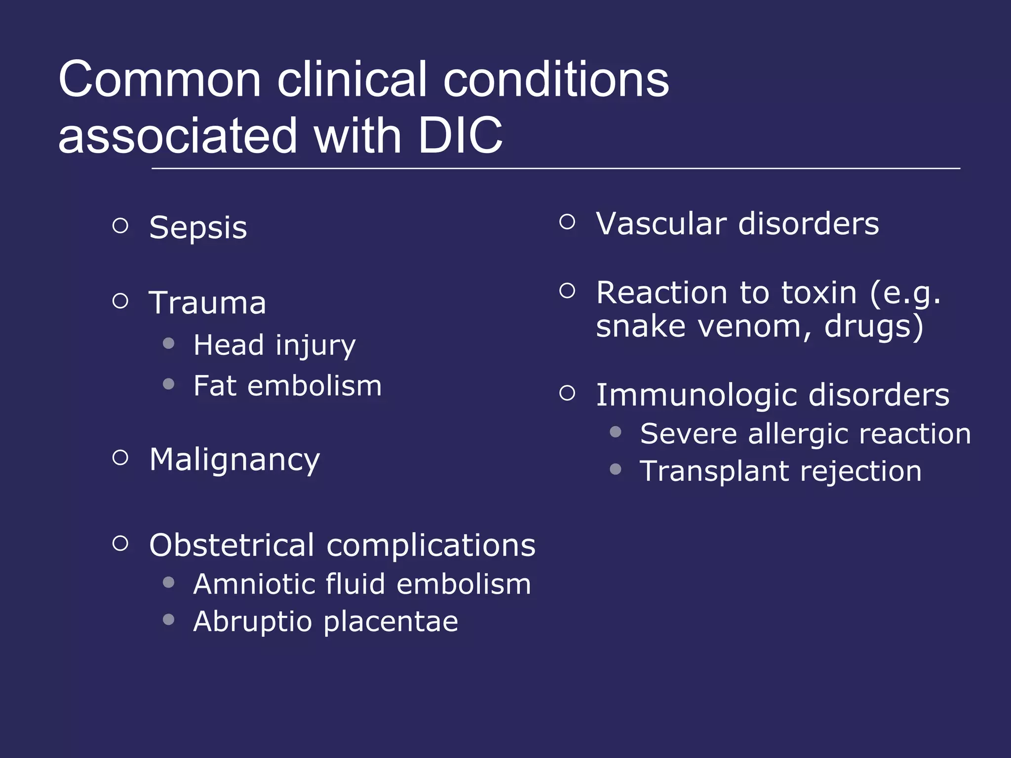
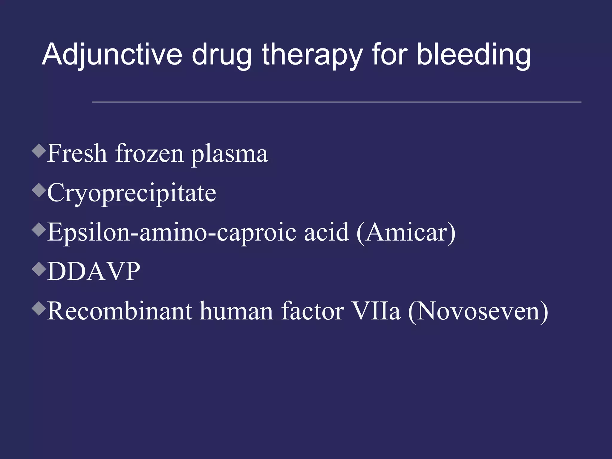
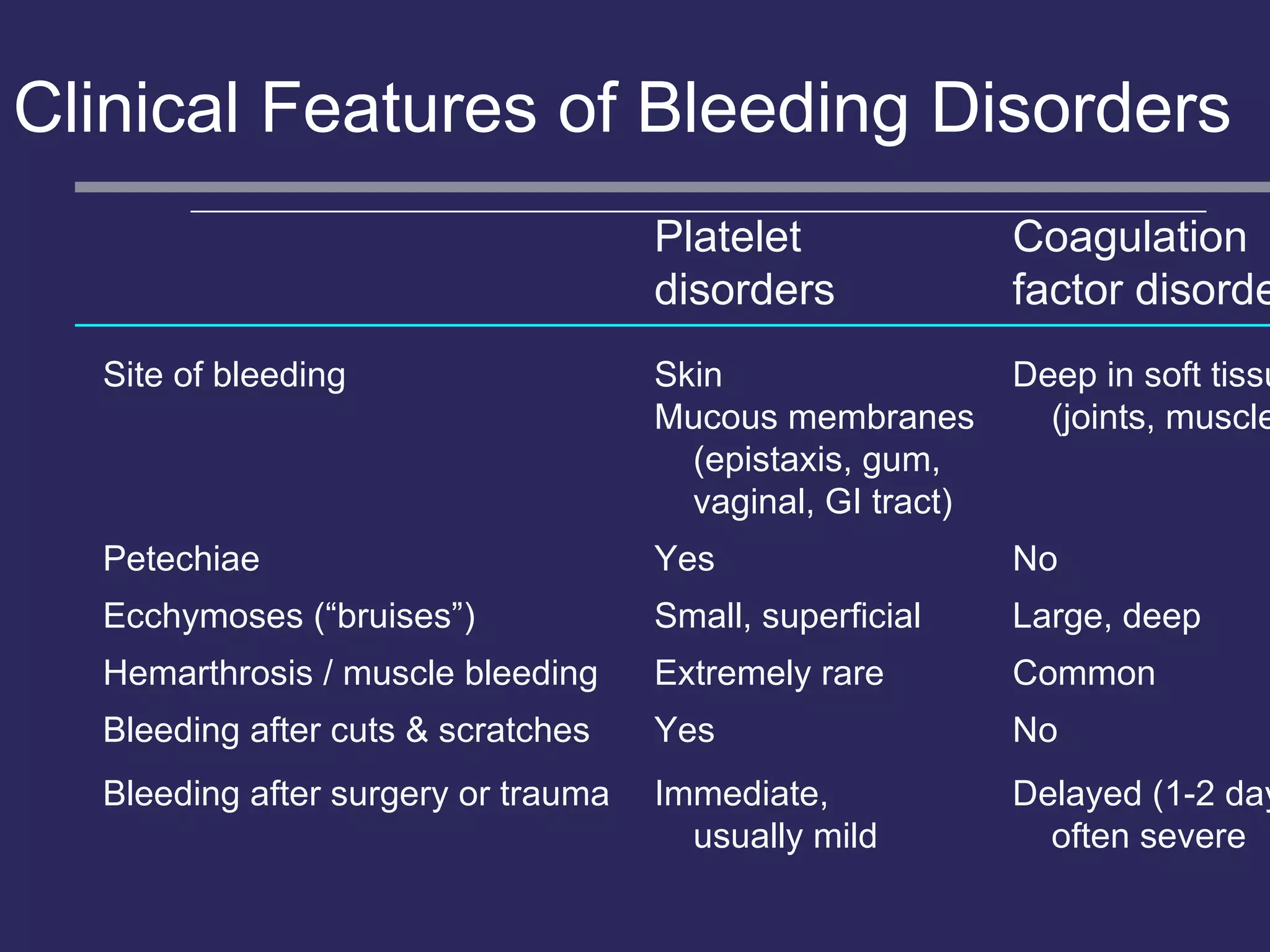
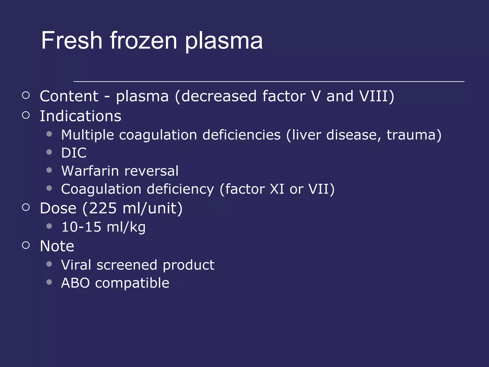
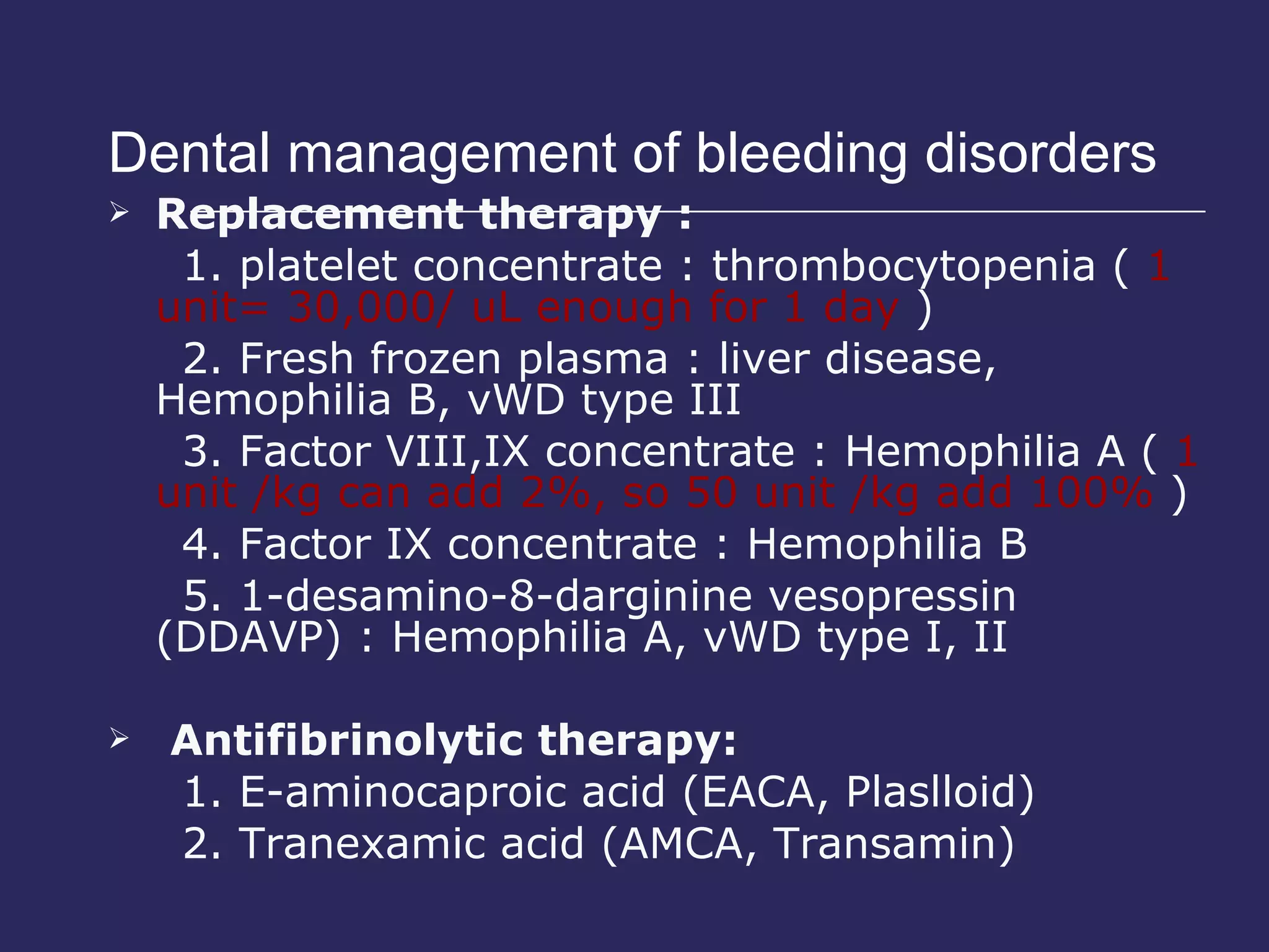
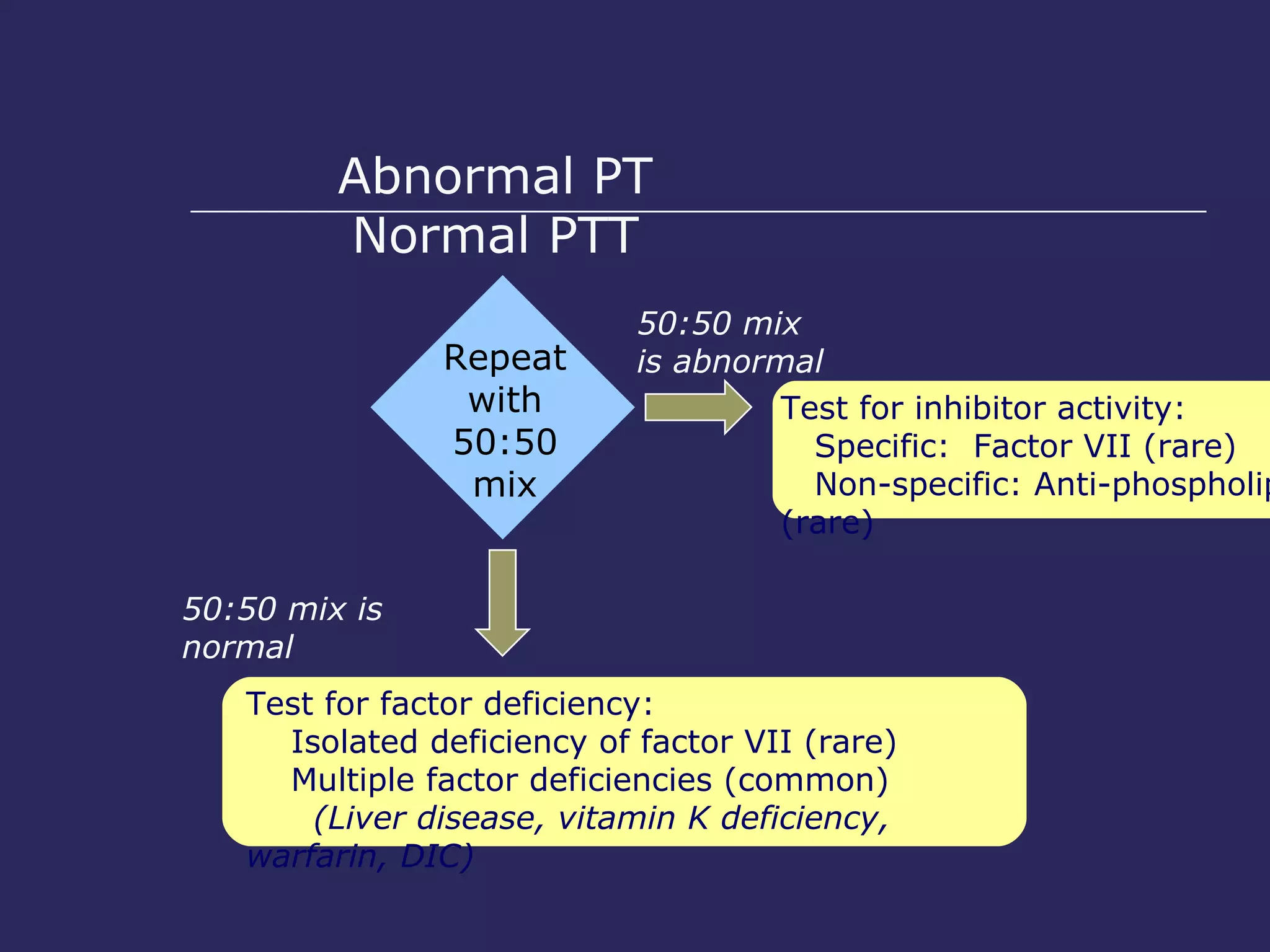
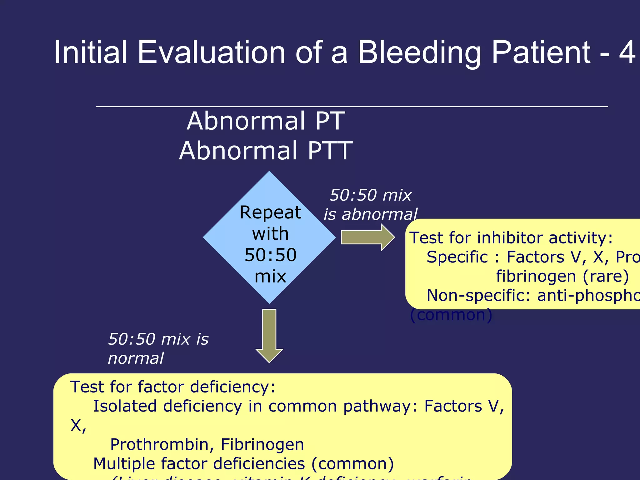
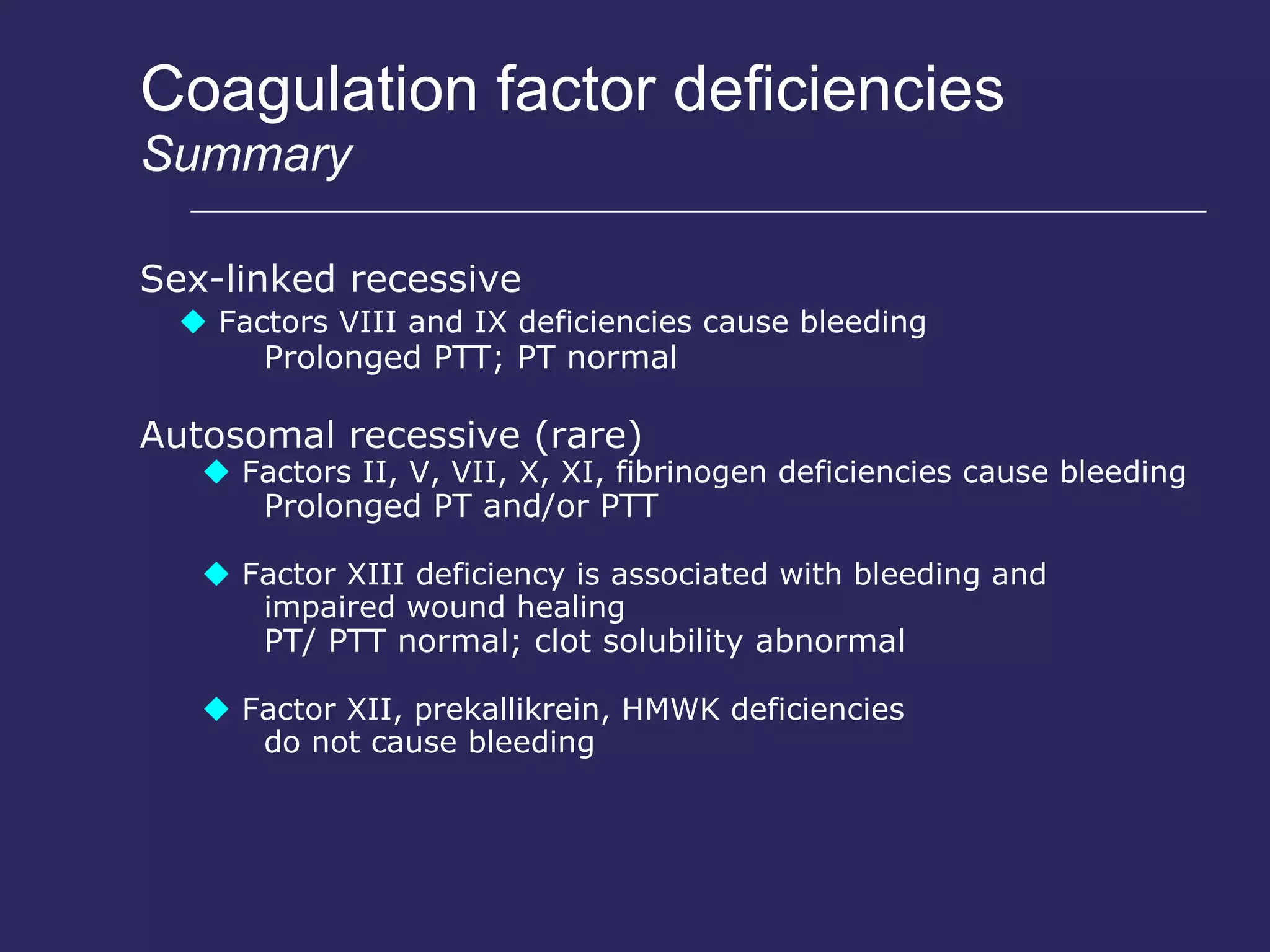
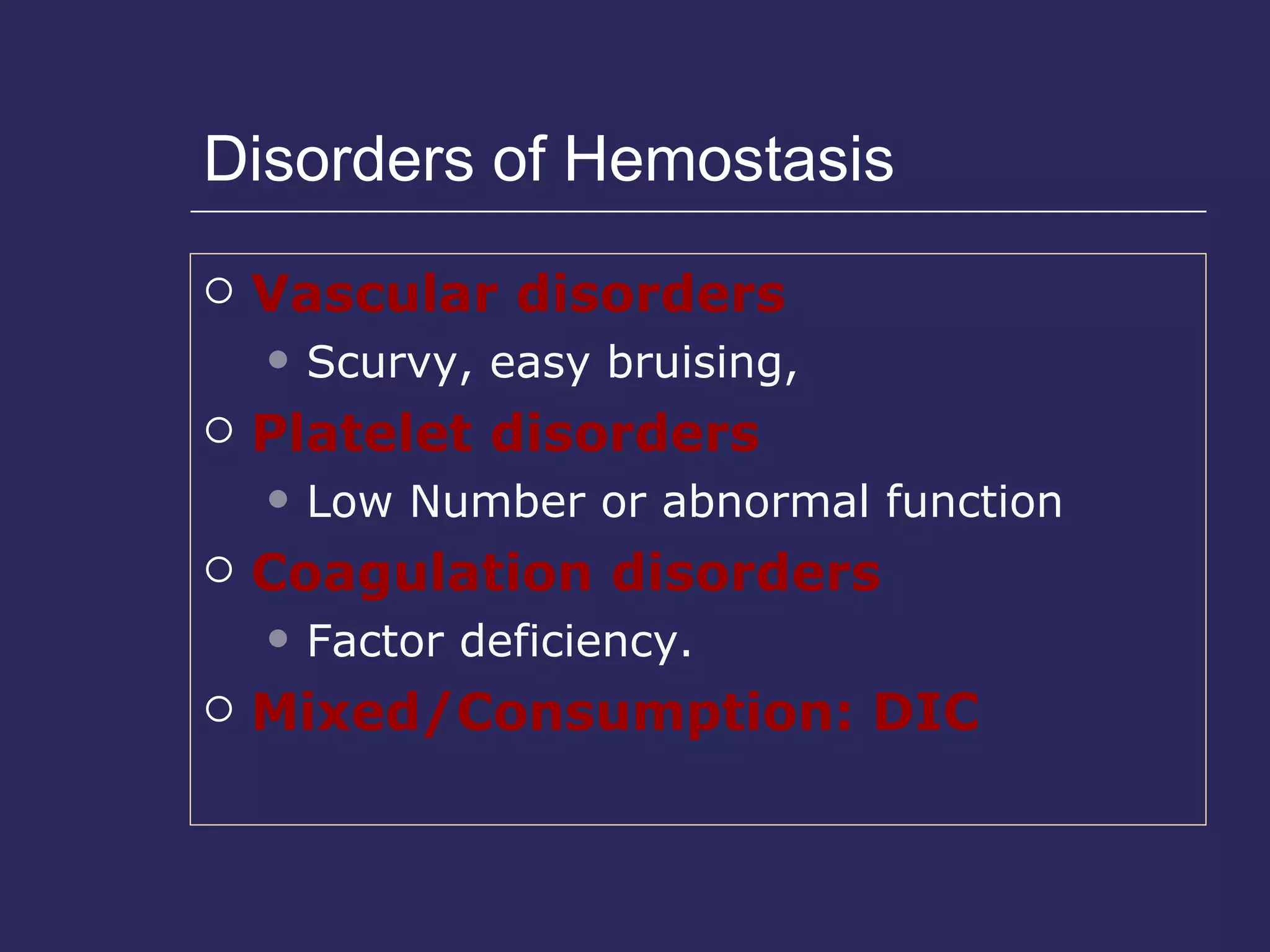
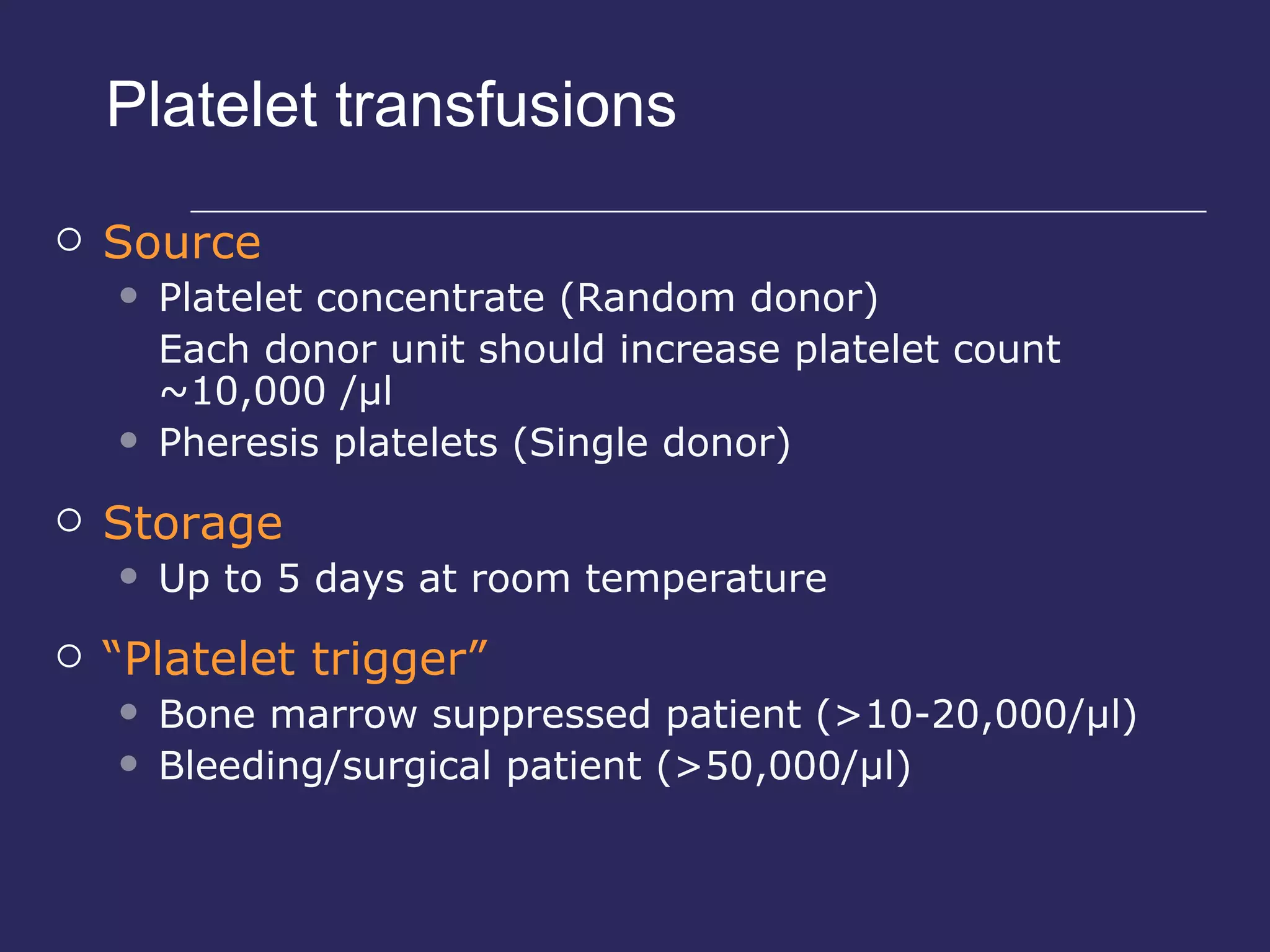
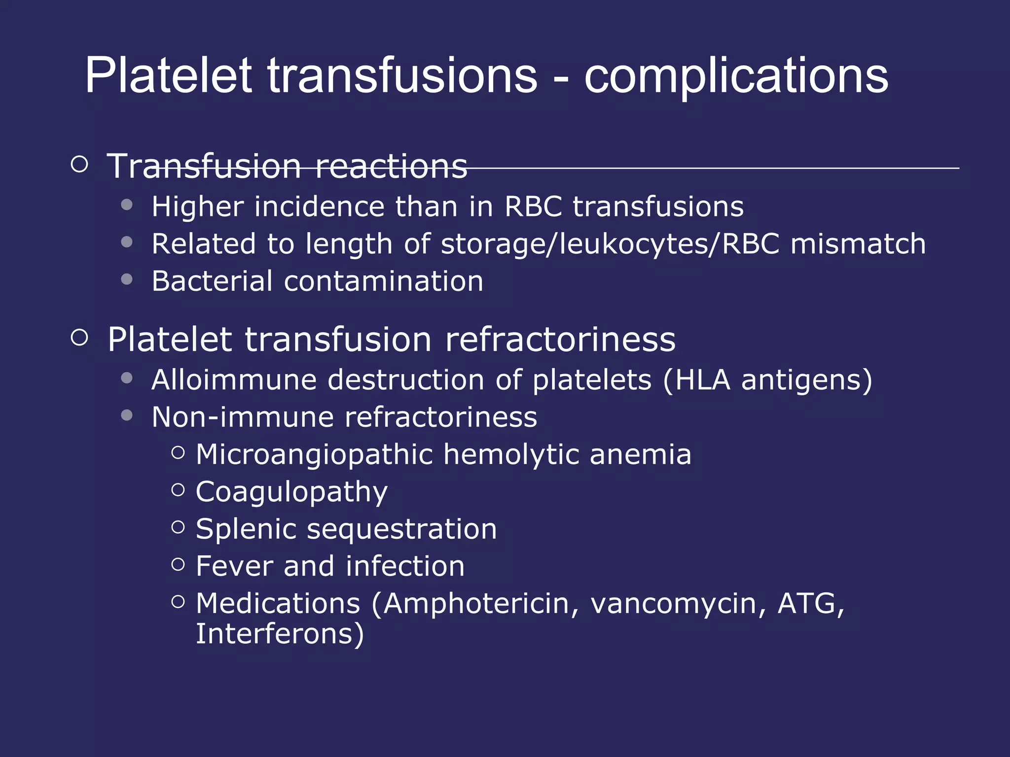
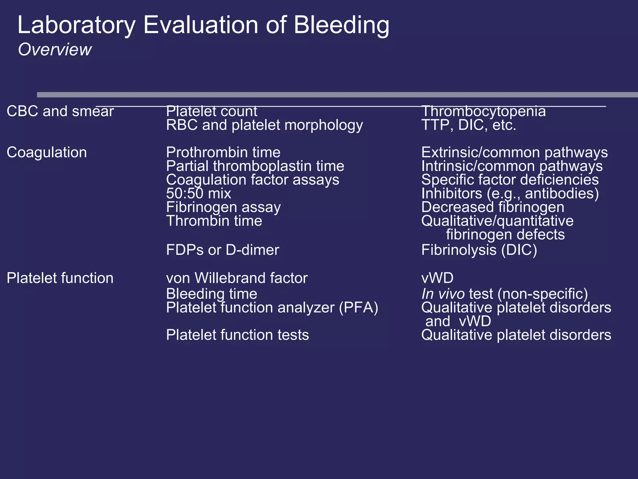
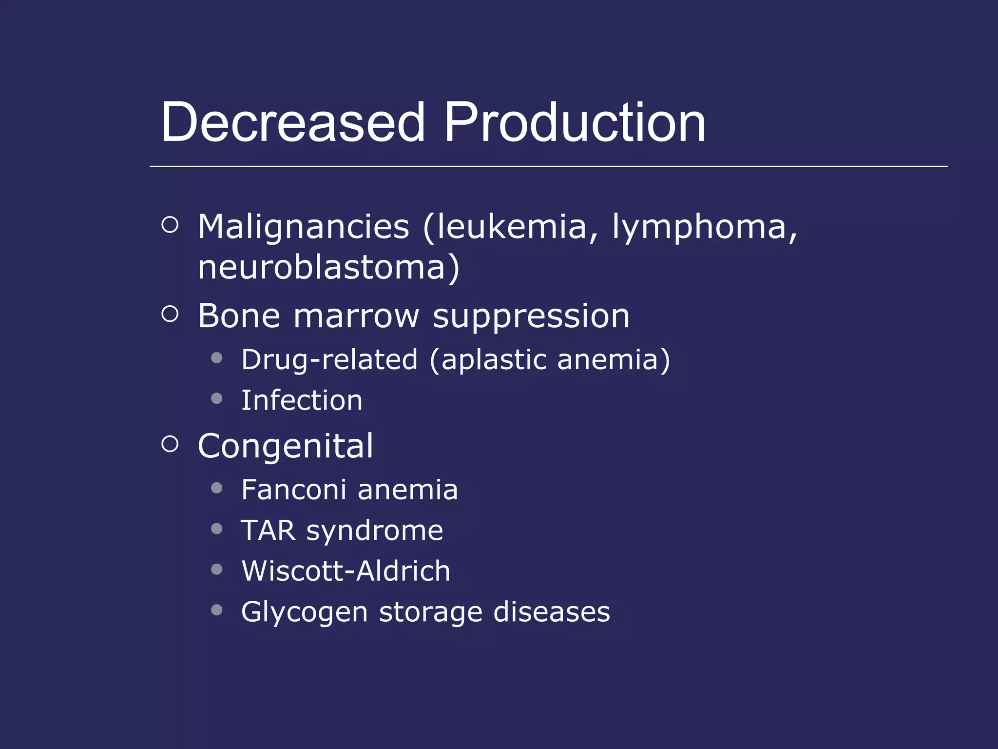
Notable Quote: Smile ………..Is the key for every one S-----------Smile M------------Make shack hand I-------------Introduce your self L---------------- Learn your partner Name E------------Eye contact
Haemostasis &Bleeding disorders BY Prof.Dr .  Mervat Atfy Mohamed
Objectives/Outline Definitions Review pathophysiology of bleeding  Provide a framework for differential diagnosis of purpura Brief discussion of some of the more common presentations and their management
Purpura Hemorrhage into skin or mucous membrane Due to: Loss of vascular integrity Platelet problems Number Function Clotting factor deficiency
Purpuric lesions Petechiae  capillary hemorrhages, purple macular lesions     2mm develop in areas of increased venous pressure Purpura – 2mm-1cm Ecchymoses – > 1cm, often tender Non-blanching, may be raised
Platelet platelet count bleeding time < 8 min Platelet function Coagulation ACT PT aPTT thrombin time fibrinogen 90-130 sec 12-15 sec 35-45 sec < 14 sec 250-500 mg/dL Monitor heparin Extrinsic & final Intrinsic & final Final common ↓ in DIC Fibrinolysis FDP d-dimer ↑ during fibrinolysis
Hemostasis requires normal: vessels platelets  coagulation pathway components
Coagulation factor disorders Inherited bleeding disorders Hemophilia A and B vonWillebrands disease Other factor deficiencies Acquired bleeding disorders Liver disease Vitamin K deficiency/warfarin overdose DIC
Hemostasis Arteriolar vasoconstriction Formation of platelet plug Activation of coagulation cascade Formation of permanent plug Secondary Hemostasis Primary Hemostasis
 
 
Four reasons for Cell based model 1. Congenital deficiency of XII, HMW kininogen and pre-kallikrein have prolonged PTT but NO BLEEDING PHENOTYPE.  2. These components are NOT required for hemostasis  in vivo 3. TF/VIIa  activates not only X but also VIII and IX 4. XI deficiency is not associated with bleeding phenotype
Cell Based coagulation model TF bearing cells Adventital cells Myo-epithelial cells Endothelial cells 3 phases Initiation Priming Propagation
Initiation TF bearing cells exposed to plasma VII binds to TF and is activated VIIa    Xa TF/VIIa    IXa TF/VIIa/Xa binds Va    converts Prothrombin to Thrombin Small amount of Thrombin Activate platelets accelerant
Priming Release of granules containing V Cleaved to Va Formation of VIIIa Propagation Activated platelets rapidly bind V/VIII/IX Formation of VIII/IXa complex MAJOR activator of X Major prothrombin activator
Screening tests for bleeding disorders Prothrombin time Measures function of VII, X, Prothrombin, fibrinogen Prolonged when V, VII, X fall below 50% And when prothrombin level falls to <30% Vit K dependent proteins: Prothrombin, V, VII, X,  Proteins C and S Warfarin, Liver Failure
Screening tests for bleeding disorders aPTT Detects decrease in XII, XI, IX, VIII AS WELL AS Fibrinogen, prothrombin, V and X Heparin
Hemophilia X linked recessive Deficiency of Factor VIII Degrees of severity: Mild : 5% - 30% Moderate : 2% - 5% Severe : <2% Prolonged PTT. Normal PT, vWf, BT
Hemophilia A and B Hemophilia A Hemophilia B Coagulation factor deficiency Factor VIII Factor IX Inheritance X-linked X-linked recessive  recessive Incidence 1/10,000 males 1/50,000 males Severity Related to factor level <1% - Severe - spontaneous bleeding 1-5% - Moderate - bleeding with mild injury 5-25% - Mild - bleeding with surgery or trauma Complications Soft tissue bleeding
Hemophilia Clinical manifestations  (hemophilia A & B indistinguishable) Hemarthrosis  (most common) Fixed joints Soft tissue hematomas (e.g., muscle) Muscle atrophy Shortened tendons Other sites of bleeding Urinary tract CNS, neck (may be life-threatening) Prolonged bleeding after surgery or dental extractions
Treatment of hemophilia A Intermediate purity plasma products Virucidally treated May contain von Willebrand factor High purity (monoclonal) plasma products Virucidally treated No functional von Willebrand factor Recombinant factor VIII Virus free/No apparent risk No functional von Willebrand factor
Dosing guidelines for hemophilia A Mild bleeding Target: 30% dosing q8-12h; 1-2 days (15U/kg) Hemarthrosis, oropharyngeal or dental, epistaxis, hematuria Major bleeding Target: 80-100% q8-12h; 7-14 days (50U/kg) CNS trauma, hemorrhage, lumbar puncture Surgery Retroperitoneal hemorrhage GI bleeding Adjunctive therapy    amino caproic acid (Amicar) or DDAVP (for mild disease only)
Complications of  therapy Formation of inhibitors (antibodies) 10-15% of severe hemophilia A patients 1-2% of severe hemophilia B patients Viral infections Hepatitis B Human parvovirus Hepatitis C Hepatitis A HIV Other
Treatment of hemophilia B Agent  High purity factor IX Recombinant human factor IX Dose Initial dose: 100U/kg Subsequent: 50 U/kg every 24 hours
Von Willebrand disease Most common congenital bleeding disorder Bleeding is usually mild Etiology of bleeding Platelet aggregation Major carrier for VIII Types
von Willebrand Disease Clinical features von Willebrand factor Carrier of factor VIII Anchors platelets to subendothelium Bridge between platelets Inheritance Autosomal dominant Incidence  1/10,000 Clinical features Mucocutaneous bleeding
Laboratory evaluation of  von Willebrand disease Classification Type 1  Partial quantitative deficiency Type 2  Qualitative deficiency Type 3 Total quantitative deficiency Diagnostic tests: vonWillebrand type Assay      1   2   3 vWF antigen      Normal      vWF activity              Multimer analysis Normal Normal Absent
Treatment of von Willebrand disease Varies by Classification Cryoprecipitate Source of fibrinogen, factor VIII and VWF Only plasma fraction that consistently contains VWF multimers Correction of bleeding time is variable DDAVP (Deamino-8-arginine vasopressin) Increases plasma VWF levels by stimulating secretion from endothelium Duration of response is variable Used for type 1 disease Dosage 0.3 µg/kg q 12 hr IV Factor VIII concentrate (Humate-P) Virally inactivated product Used for type 2 and 3
Type I vW disease Autosomal dominant Normal vWF Quantitative decrease Normal PT Mildly prolonged aPTT
Rx of vW disease DDAVP Cryoprecipitate Monitor VIII levels
DDAVP 1-Deamino, 8 D-arginine vasopressin Rx for mild Hemophilia A , vW disease Increases plasma fVIII, vWf from endogenous sorces Amount of increase = factor transfusion Useful for platelet dysfunction secondary to aspirin, dextran, ticlopidine 0.3- 0.4 mcg/kg Onset 30 min, peak 90-120 min
Cryoprecipitate useful in treating factor deficiency (hemophilia A), von Willebrand's disease, and hypofibrinogenemia  uremic bleeding Each 5- to 15-mL unit contains more than 80 units of factor VIII and about 200 mg of fibrinogen. Because the proteins mentioned previously are in relatively high concentration, a smaller volume may be given than would be required if plasma were used. Cryoprecipitate is usually administered as a transfusion of 10 single units.
NovoSeven Recombinant f VIIa Activates X and IX (conversion of prothrombin to thrombin) $$$$ Not FDA approved Anecdotal miraculous rescues
 
Heparin Blocks Xa by binding to AT-III and thrombin. Load 80 mg/kg  Infusion 18 mg/kg Cleared from blood in 6hrs Neutralized by protamine  100u of Heparin = 1mg of protamine
HIT Heparin induced thrombocytopenia Formation of IgG against Heparin- PF4 complexes Upto 5% of patients Onset 4-5 days, but earlier if prior exposure Can occur with flushes, heparin bonded catheters also
LMW Heparins More selective anti-Xa activity Less bleeding complications Do not affect PT Measure anti Xa activity <1% incidence of HIT
Enoxaparin LMW heparin Porcine intestinal submucosa Binds to and accelerates AT-III activity
Fondaparinux (Erixtra) Synthetic pentasaccharide 5 sugar AT-III binding site similar to Heparin 1000x more potent Very low- no incidence of HIT Cannot be given in CRI
Vitamin K Necessary for addition of carboxyglutamate residues to the clotting factors synthesized in the liver. Sites of Calcium binding Prothrombin, VII, IX, X drugs- cephalosporins, quinolones 5mg iv slowly corrects deficit in 6 hrs 10-25 mcg /day sq or im x 3days
Vitamin K deficiency Source of vitamin K  Green vegetables Synthesized by intestinal flora Required for synthesis Factors II, VII, IX ,X Protein C and S Causes of deficiency Malnutrition Biliary obstruction Malabsorption Antibiotic therapy Treatment Vitamin K Fresh frozen plasma
Vitamin K deficiency due to warfarin overdose Managing high INR values Clinical situation Guidelines INR therapeutic-5 Lower or omit next dose; Resume therapy when INR is therapeutic INR 5-9; no bleeding Lower or omit next dose; Resume therapy when INR is therapeutic Omit dose and give vitamin K (1-2.5mg po) Rapid reversal: vitamin K 2-4 mg po (repeat) INR >9; no bleeding Omit dose; vitamin K 3-5 mg po; repeat as necessary Resume therapy at lower dose when INR therapeutic Chest 2001:119;22-38s (supplement)
Vitamin K deficiency due to warfarin overdose Managing high INR values in bleeding patients Clinical situation Guidelines INR > 20; serious bleeding Omit warfarin Any life-threatening bleeding Vitamin K 10 mg slow IV infusion FFP ± factor rhVIIa (depending on urgency) Repeat vitamin K injections every 12 hrs as needed
DIC Warfarin Consumption coagulopathy Systemic thromb-hemorrhagic disorder Etiology: Gram negative sepsis, crush injuries, amniotic fluid embolism, hemolysis, massive transfusion Activation of clotting and fibrinolysis Microvascular thrombosis, sequestration of platelets
Pathogenesis of DIC Coagulation Fibrinolysis Fibrinogen Fibrin Monomers Fibrin Clot (intravascular) Fibrin(ogen) Degradation Products Plasmin Thrombin Plasmin Release of thromboplastic material into circulation Consumption of coagulation factors; presence of FDPs    aPTT    PT    TT    Fibrinogen Presence of plasmin    FDP Intravascular clot    Platelets Schistocytes
Common clinical conditions associated with DIC Sepsis Trauma Head injury Fat embolism Malignancy Obstetrical complications Amniotic fluid embolism Abruptio placentae Vascular disorders Reaction to toxin (e.g. snake venom, drugs) Immunologic disorders Severe allergic reaction Transplant rejection
DIC Low fibrinogen High FDP Low platelets Elevated D-dimer
Disseminated Intravascular Coagulation (DIC) Mechanism Systemic activation of coagulation Intravascular deposition of fibrin Depletion of platelets and coagulation factors Bleeding Thrombosis of small and midsize vessels with organ failure
DIC Treatment approaches Treatment of underlying disorder Anticoagulation with heparin Platelet transfusion Fresh frozen plasma
Liver Disease Decreased synthesis of  II, VII, IX, X, XI, and fibrinogen Prolongation of PT, aPTT and Thrombin Time Often complicated by Gastritis, esophageal varices, DIC Treatment Fresh-frozen plasma infusion  (immediate but temporary effect) Vitamin K (usually ineffective)
Stopping the bleeding Direct pressure. More direct pressure. Pack. Pack. Pack. Electrocautery. Ligate vessel
Methylcellulose Gelfoam Absorbable Liquefies in 2-5 days Serves as a scaffold for coagulation
Microfibrillar collagen Decellularized bovine source Stimulates latelet adhesion Stops venous ooze Absorbed in 90 days
Thrombin + Gelfoam + CaCl Thrombin for cleavage/activation Gelfoam as matrix Very useful in vascular surgery
Fibrin glue Tiseel FDA approved in 1998 Concentrated fibrinogen and f VIII Thrombin and calcium Aprotinin to prevent clot dissolution Takes time to prepare Good for diffuse oozing, needle punctures, parenchymal injuries
Hemostasis: Lab Tests CBC- Plt BT ,(CT) PT PTT Plt Study Morphology Function Antibody BV  Injury Platelet Aggregation Platelet Activation Blood Vessel   Constriction Coagulation   Cascade Stable  Hemostatic Plug Fibrin formation Reduced Blood flow Tissue Factor Primary hemostatic plug Neural
Hemostasis 1. Vascular phase : vasoconstriction,  immediately 2. Platelet phase : adhesion & aggregation,  several seconds after 3. Coagulation phase : later, contains  extrinsic & intrinsic pathways  4. Metabolic (fibrinolytic) phase:  release antithrombotic agent primary secondary
Hemostasis The intrinsic pathway : Initiated through surface contact  activation of factor XII  by exposed subendothelial tissues--collagen The extrinsic pathway : Initiated through tissue thromboplastin released by injured tissue, which activates factor VII Antihemophilic factor; von Willebrand factor, VWF Plasma thromboplastin component , PTC; Christmas factor
 
Antihemophilic factor; von Willebrand factor, VWF
Gla domain: activated by vit. K and NADH
Antithrombotic agent Postaglandin: secretion by  endothelial cell Antithrombin III (AT III): main Antithrombotic agent Protein C: inactivate  factor V and VIII  with  protein S Plasmin: activated by urokinase and streptokinase from plasminogen primary secondary
Etiology of bleeding disorder  1. Nonthrombocytopenia 2. Thrombocytopenia purpuras 3. Disorders of coagulation
Etiology of bleeding disorder  Nonthrombocytopenia 1. vascular wall alteration : infection, chemical, allergy 2. Disorder of platelet function : Genetic defects (bernard-soulier disease:  glycoprotein, GP-Ib dysfunction with VWF ) Aspirin, NSAIDs, broad-spectrum antibiotics(Ampicillin, Penicillin, Gentamycin, Vancomycin) Autoimmue disease
Adjunctive drug therapy for bleeding Fresh frozen plasma Cryoprecipitate Epsilon-amino-caproic acid (Amicar) DDAVP Recombinant human factor VIIa (Novoseven)
Clinical Features of Bleeding Disorders Platelet Coagulation  disorders factor disorders Site of bleeding Skin Deep in soft tissues Mucous membranes   (joints, muscles)   (epistaxis, gum,   vaginal, GI tract) Petechiae Yes No Ecchymoses (“bruises”) Small, superficial Large, deep Hemarthrosis / muscle bleeding Extremely rare Common Bleeding after cuts & scratches Yes No Bleeding after surgery or trauma Immediate, Delayed (1-2 days),   usually mild   often severe
 
Correct surgical bleeding ACT elevated normal Protamine 25-50mg PLT 0.1 u/kg ACT elevated normal Heparin level yes no protamine Coagulation profile FDP >32mcg/kg  TEG abnormal clot lysis fibrinolysis FFP 5-10ml/kg  CP 0.2-0.4u/kg EACA PLT<70000 thrombocytopenia PLT 0.1u/kg bleeding time >20min PLT dysfunction Fibrinogen  < 100mg/dL  TEG abnormal hypofibrinogenemia FFP 5-10ml/kg  CP 0.2-0.4u/kg aPTT >1.3x  PT >1.5x Anesthesiologist ’ s manual of surgical procedures
Fresh frozen plasma Content - plasma (decreased factor V and VIII) Indications Multiple coagulation deficiencies (liver disease, trauma) DIC Warfarin reversal Coagulation deficiency (factor XI or VII) Dose (225 ml/unit) 10-15 ml/kg Note Viral screened product ABO compatible
Cryoprecipitate Prepared from FFP Content Factor VIII, von Willebrand factor, fibrinogen Indications Fibrinogen deficiency Uremia von Willebrand disease Dose (1 unit = 1 bag) 1-2 units/10 kg body weight
Etiology of bleeding disorders  Thrombotic Thrombocytopenia purpuras (TTP) : 血栓性血小板低下紫斑 1. primary 2. secondary : Chemicals, ex: mitomycin C Physical agent (radiation) Systemic disease (leukemia ) Character: 1.Thrombocytopenia 2. Micro-angiopathic hemolytic anemia(MAHA) 3. Fever 4. Hyporenal function 5. Neural systemic desturbance due to ischemic Considered to be an emergency  Tx: plasma exchange ( 血漿置換術 ) and glucocortisone application
Etiology of bleeding disorders  Disorders of coagulation 1. Inherited : Hemophilia A Christmas disease von Willebrandis Disease 2. Acquired : Liver disease Vitamin K deficiency Anticoagulation drugs (heparin, coumarin) Anemia
Etiology of bleeding disorders  Thrombotic Thrombocytopenia purpuras (TTP) : 血栓性血小板低下紫斑 1. primary 2. secondary : Chemicals, ex: mitomycin C Physical agent (radiation) Systemic disease (leukemia ) Character: 1.Thrombocytopenia 2. Micro-angiopathic hemolytic anemia(MAHA) 3. Fever 4. Hyporenal function 5. Neural systemic desturbance due to ischemic Considered to be an emergency  Tx: plasma exchange ( 血漿置換術 ) and glucocortisone application
Evaluation of bleeding disorders  1. Take history 2. Physical examination 3. Screening clinical laboratory tests 4. Observation of excessive bleeding following a surgical  procedure
Physical examination Jaundice ( 黃疸 ) Petechiae ( 淤點 ) : < 0.2 cm Purpura ( 紫斑 ) : 0.2 cm-1 cm Eccymoses ( 淤斑 ) : > 1 cm Spider angioma ( 蜘蛛斑 ) Oral ulcer Hyperplasia of gingiva Hemarthrosis ( 關節血腫 )
Platelet count  Test platelet phase: evaluation of platelet function Normal (140,000 to 400,000/mm3)  Thrombocytopenia : < 140,000/mm3 Clinical bleeding problem : <50,000/mm3 Spontaneous bleeding with life theartening : <20,000/mm3
PT (Prothrombin Time)  Activated by  tissue thromboplastin Tests  extrinsic  ( factor VII ) and common ( I,II,V,X ) pathways Normal (  11-15sec  ) Coumarin  therapy- PT at  1.5 to 2.5 time International normalized ratio= INR,  (1) surgery can be done under INR< 3.0 (2) when INR=3.0-3.5, consultation is needed (3) delay surgery when INR>3.5
Activated PTT (aPTT) Activated by contact activator (kaolin) Tests  intrinsic  and common pathway Normal (  25-35 sec  ) Heparin  therapy- PTT in  50-65  sec range by promote  AT III
TT (Thrombin Time)  Activated by thrombin Tests ability to form initial clot from fibrinogen Normal (  9 to 13 seconds  )
1. No historical bleeding problem 3.  Aspirin therapy 4. Coumarin therapy 6. Possible liver disease 7. Chronic leukemia 8.  Long term antibiotic therapy 5.  Renal dialysis (heparin) 2. History bleeding problem 9. Vascular wall alteration 10.  Cancer (fibrinogenolysis)   Following surgical procedure PT, aPTT, TT, BT BT, aPTT PT aPTT BT, PT BT PT BT TT Dental management of the medically compromised patient
condition 8. thrombocytopenia 2. Coumarin therapy 4.  Liver disease 7. Vascular wall defect 9. hemophilia 1. Aspirin therapy Platelet count 5. leukemia 6. Long term  antibiotic 3. Heparin therapy 10. fibrinogenolysis + - + + + - - ++ - - BT + - + + + - + ++ - - PTT + ++ ++ ++ - ++ - - ++ + PT + ++ - ++ - ++ - - - + TT - - - ++ - ++ - - - ++ -: normal, +: may be abnormal, ++: abnormal
Patient at low risk 1. patient with no history of bleeding disorders, normal  examinations, no medications associated with bleeding  disorders and normal bleeding parameters 2. patients with nonspecific history of excessive bleeding  with normal bleeding parameters (PT, PTT, BT,  platelet  count, are within normal time)
Patient at moderate risk  1. patients in chronic oral anticoagulant therapy  (coumadin) 2. patients on chronic aspirin therapy
Patient at high risk  1. patients with known bleeding disorders  Thrombocytopenia Thrombocytopathy Clotting factor defects 2. Patient without known bleeding disorders found to  have abnormal , platelet count, BT, PT, PTT
Dental management of bleeding disorders  Replacement therapy : 1. platelet concentrate : thrombocytopenia (  1 unit= 30,000/ uL enough for 1 day  ) 2. Fresh frozen plasma : liver disease, Hemophilia B, vWD type III 3. Factor VIII,IX concentrate : Hemophilia A (  1 unit /kg can add 2%, so 50 unit /kg add 100%  ) 4. Factor IX concentrate : Hemophilia B 5. 1-desamino-8-darginine vesopressin (DDAVP) : Hemophilia A, vWD type I, II Antifibrinolytic therapy: 1. E-aminocaproic acid (EACA, Plaslloid) 2. Tranexamic acid (AMCA, Transamin)
Heparin (anticoagulant)  Complex inhibited (  IXa, Xa, XIa, XIIa  ) Used in deep vein thrombosis , renal dialysis Rapid onset, Duration  4-6hrs  ( given IV ) Monitoring by aPTT:  50-65 sec Discontinue  6 hrs  before surgery then reinstituting therapy  6-12hrs post –op Protamine sulfate can reverse the effect
Coumarin (Vit k anatagonist)  Inhibit Vit K action (Factor II,VII,IX,X) Used venous thrombosis, cerebrovascular disease Duration  haft-life 40hrs Monitored by PT : INR 1.5-2.5 PT>2.5, reduction coumarin dosage (  2-3 days  ) Vit. K can reverse the effect
Local hemostatic methods  splints, pressure packs, sutures; gelfoam with thrombin, surgicel, oxycel, microfibrillar collagen(avitene), topical AHF
Aspirin (antiplatelet)  Inhibit cycloxygenase, TxA2 formation Analgesic drug impairs platelet function Aterial thrombosis, MI Tests-BT, aPTT If tests are abnormal , MD should be consulted before dental surgery is done Stop aspirin for  5 days , substitute alternative drug in consultation with MD
Thrombocytopenia  Disease in number of circulation platelets Idiopathic thrombocytopenia, secondary thrombocytopenia TX : is none indicated unless platelets<20000/mm3, or excessive bleeding TX : Steroid, platelet transfusion
Von Willebrandis Disease Gene mutation on Von Willebrandis factor; most common Inherited disease in America ( 1% ) Type I : 70%-80%, partial loss on quantity Type II : poor on quality  Type III : severe loss on quantity, inactive to DDAVP
Laboratory evaluation of  von Willebrand disease Classification Type 1  Partial quantitative deficiency Type 2  Qualitative deficiency Type 3 Total quantitative deficiency Diagnostic tests: vonWillebrand type Assay      1   2   3 vWF antigen      Normal      vWF activity              Multimer analysis Normal Normal Absent
Hemophilia  Sex-linked recessive trait, X chromosome, male > female Prolong aPTT, normal BT,PT Hemophilia A (factor VIII deficiency) Hemophilia B or Christmas disease (factor IX deficiency) Severity of disorder : severe<1%, moderate 1-5%,  mild 6-30% TX : Replacement factors, antifibrinilytic agents, steroids
Hemophilia-dental management  Preventive dentistry 1. tooth brushing, flossing, rubber cup prophylaxis &  topical fluoride, supragingival scaling 2. without prior replacement therapy Pain control  1. block anesthesia: factor level>50% 2. Avoid aspirin, NSAIDs
Hemophilia-dental management  Orthodontic treatment : 1.  no contraindication  in well-motivated patients 2. care with placement of bands and wires Operative dentistry 1.  rubber dam  to protect tissue against accidental  laceration 2. wedges should be place to protect and retract  papilla
Hemophilia-dental management  Pulp therapy 1. Preferable to extraction 2. Avoid overinstrumentation and overfilling Periodontal therapy 1.  no contraindication  of probing and supragingival  scaling 2. deep scaling, curettage, surgery need replacement  therapy
Hemophilia-dental management  Oral surgery : 1. Dental extraction:  40%-50% level 2. Maxillofacial surgery (including surgery  extraction of impaction teeth):  80-100% 3. Antifribrinilytic therapy & local hemastatic  measure 4. do not open  lingual tissue  in lower molar regions to  avoid hemorrhage track down a  endanger airway
Summary  History, PE, Lab data Consultation with physician Antibiotics to prevent post-op infection Avoid aspirin and NSAIDs Local hemostatic measure is very important
Haemostasis
Hemostasis 1° hemostasis Vasoconstriction &  retraction of cut ends of blood vessels Release of FVIII-vWF  Platelets adhere to endothelium granules release ADP, Ca & ThromboplastinA 2   aggregation Stabilization of platelet plug


Coagulation Cascade: Intrinsic Path (12,11,9,8) Extrinsic Path (7) Fibrinogen    Fibrin Common Path   (5,2) (PT) (aPTT) (TT) (F & FDP) (Factor 10) (Thrombin)
Coagulation cascade Vitamin K dependant factors XIIa IIa Intrinsic system (surface contact) XII XI XIa Tissue factor IX IXa VIIa VII VIII VIIIa Extrinsic system (tissue damage) X V Va II Fibrinogen Fibrin (Thrombin) IIa Xa
Laboratory Evaluation of the Coagulation Pathways Partial thromboplastin time (PTT) Prothrombin time (PT) Intrinsic pathway Extrinsic pathway Common pathway Thrombin time Thrombin Surface activating agent (Ellagic acid, kaolin) Phospholipid Calcium Thromboplastin Tissue factor Phospholipid Calcium Fibrin clot
Pre-analytic errors Problems with blue-top tube Partial fill tubes Vacuum leak and citrate evaporation Problems with phlebotomy Heparin contamination Wrong label Slow fill Underfill Vigorous shaking Biological effects Hct ≥55 or ≤15 Lipemia, hyperbilirubinemia, hemolysis Laboratory errors Delay in testing Prolonged incubation at 37°C Freeze/thaw deterioration
Initial Evaluation of a Bleeding Patient - 1 Normal PT Normal PTT Consider evaluating for: Mild factor deficiency  Monoclonal gammopathy Abnormal fibrinolysis Platelet disorder (  2 anti-plasmin def) Vascular disorder Elevated FDPs Urea solubility Normal Abnormal Factor XIII deficiency
Initial Evaluation of a Bleeding Patient - 2 Normal PT Abnormal PTT Test for factor deficiency: Isolated deficiency in intrinsic pathway (factors VIII, IX, XI) Multiple factor deficiencies (rare) Repeat with 50:50 mix 50:50 mix is normal 50:50 mix is abnormal Test for inhibitor activity: Specific factors: VIII,IX, XI Non-specific (anti-phospholipid Ab)
Abnormal PT Normal PTT Test for factor deficiency: Isolated deficiency of factor VII (rare) Multiple factor deficiencies (common) (Liver disease, vitamin K deficiency, warfarin, DIC) Repeat with 50:50 mix 50:50 mix is normal 50:50 mix is abnormal Test for inhibitor activity: Specific:  Factor VII (rare) Non-specific: Anti-phospholipid (rare)
Initial Evaluation of a Bleeding Patient - 4 Abnormal PT Abnormal PTT Test for factor deficiency: Isolated deficiency in common pathway: Factors V, X,  Prothrombin, Fibrinogen Multiple factor deficiencies (common) (Liver disease, vitamin K deficiency, warfarin, DIC) Repeat with 50:50 mix 50:50 mix is normal 50:50 mix is abnormal Test for inhibitor activity: Specific : Factors V, X, Prothrombin,    fibrinogen (rare) Non-specific: anti-phospholipid (common)
Coagulation factor deficiencies Summary Sex-linked recessive    Factors VIII and IX deficiencies cause bleeding Prolonged  PTT;  PT normal Autosomal recessive  (rare)    Factors II, V, VII, X, XI, fibrinogen deficiencies cause bleeding Prolonged  PT  and/or  PTT    Factor XIII deficiency is associated with bleeding and impaired wound healing PT/ PTT normal;  clot solubility  abnormal    Factor XII, prekallikrein, HMWK deficiencies do not cause bleeding
Disorders of Hemostasis Vascular disorders Scurvy, easy bruising,  Platelet disorders Low Number or abnormal function Coagulation disorders Factor deficiency. Mixed/Consumption: DIC
Purpura - Manifestations Platelet  petechiae, mucosal bleeding  CNS bleeding in severe cases Coagulation disorders  ecchymoses or hemarthroses Vascular contusion, ecchymosis, hemorrhage palpable purpura
Clinical manifestations of disordered hemostasis Clinical Characteristic Platelet defect Clotting factor deficiency Site of bleeding Skin, mucous membranes Deep in soft tissues Bleeding after minor cuts Yes Unusual Petechiae Yes No Ecchymoses Small, superficial Large, palpable Hemarthroses, muscle hematomas Rare  Common Bleeding after surgery Immediate, mild Delayed, severe
Platelet Coagulation Petechiae, Purpura   Hematoma, Hemarthrosis
Thrombocytopenias Decreased production  Platelet pooling and splenic sequestration  Increased destruction Plt <100 000 Spontaneous bleeding uncommmon with plt > 20 000 Bleeding time prolonged, PT/PTT normal
Sites of bleeding in thrombocytopenia Skin and mucous membranes Petechiae Ecchymosis Hemorrhagic vesicles Gingival bleeding and epistaxis Menorrhagia Gastrointestinal bleeding Intracranial bleeding
Classification of platelet disorders Quantitative disorders Abnormal distribution Dilution effect Decreased production Increased destruction Qualitative disorders Inherited disorders (rare) Acquired disorders Medications Chronic renal failure Cardiopulmonary bypass
Associated with bleeding Immune-mediated thrombocytopenia (ITP) Most drug-induced thrombocytopenias Most others Associated with  thrombosis Thrombotic thrombocytopenic purpura DIC Trousseau’s syndrome Heparin-associated thrombocytopenia Acquired thrombocytopenia with  shortened platelet survival
Approach to the thrombocytopenic patient History Is the patient bleeding? Are there symptoms of a secondary illness? (neoplasm, infection, autoimmune disease) Is there a history of medications, alcohol use, or recent transfusion? Are there risk factors for HIV infection? Is there a family history of thrombocytopenia? Do the sites of bleeding suggest a platelet defect? Assess the number and function of platelets CBC with peripheral smear Platelet function study
Platelet function  screen Results Epi ADP Interpretation Normal Normal Normal platelet function Abnormal Normal “Aspirin effect” Abnormal Abnormal Abnormal platelet function Valvular heart disease Renal failure Von Willebrand disease
Platelet transfusions Source Platelet concentrate (Random donor) Each donor unit should increase platelet count ~10,000 /µl Pheresis platelets (Single donor) Storage Up to 5 days at room temperature “ Platelet trigger” Bone marrow suppressed patient (>10-20,000/µl) Bleeding/surgical patient (>50,000/µl)
Platelet transfusions - complications Transfusion reactions Higher incidence than in RBC transfusions Related to length of storage/leukocytes/RBC mismatch Bacterial contamination Platelet transfusion refractoriness Alloimmune destruction of platelets (HLA antigens) Non-immune refractoriness  Microangiopathic hemolytic anemia Coagulopathy Splenic sequestration Fever and infection Medications (Amphotericin, vancomycin, ATG, Interferons)
Laboratory Evaluation of Bleeding Overview CBC and smear Platelet count Thrombocytopenia RBC and platelet morphology TTP, DIC, etc. Coagulation Prothrombin time Extrinsic/common pathways Partial thromboplastin time Intrinsic/common pathways Coagulation factor assays Specific factor deficiencies 50:50 mix Inhibitors (e.g., antibodies) Fibrinogen assay Decreased fibrinogen Thrombin time Qualitative/quantitative   fibrinogen defects FDPs or D-dimer Fibrinolysis (DIC) Platelet function von Willebrand factor vWD Bleeding time In vivo  test (non-specific) Platelet function analyzer (PFA) Qualitative platelet disorders   and  vWD Platelet function tests Qualitative platelet disorders
Decreased Production Malignancies (leukemia, lymphoma, neuroblastoma) Bone marrow suppression Drug-related (aplastic anemia) Infection  Congenital  Fanconi anemia TAR syndrome Wiscott-Aldrich Glycogen storage diseases
Platelet Dysfunction Congenital Glanzmanns thrombasthenia (GP 11b/IIIa  deficiency) Bernard Soulier (vWF receptor deficiency) Acquired Drugs  (ASA/NSAIDs, lasix, nitrofurantoin) Renal disease Liver disease
Increased Platelet Destruction Immunologic  ITP SLE Isoimmunization  (post-transfusion, neonatal) Drug related  (heparin, quinine, dig, sulphonamides, phenytoin) Infection  Sepsis Mechanical  DIC  HUS-TTP Giant hemangioma (Kasabach-Merritt) Burns Trauma
Thrombocytopenia (cont’d) Sequestration Hypersplenism Sickle cell anemia Dilutional Massive transfusion
Loss of Vascular Integrity Congenital  Disorders of connective tissue  Ehlers-Danlos syndrome  Osteogenesis imperfecta Marfan’s Disorders of blood vessels  Hemorrhagic telangiectasia Acquired  Trauma/NAI Scurvy  Steroid induced Increased intravascular pressure coughing vomiting straining
Vasculitis Drug-related Infection Viral Coxsackie A9, B3 Echoviruses 4,9 Atypical measles Bacterial Meningococcemia Streptococcal pharyngitis Septic emboli SBE Gonococcus Rickettsial Rocky Mountain spotted fever Immune-mediated HSP SLE Serum sickness Dysgammaglobulinemia
Coagulation Factor Deficiency Usually cause larger ecchymoses, deep tissue hemorrhage, and mucosal bleeding Clotting factor deficiencies Congenital von Willebrand’s disease Hemophilias Protein C and S deficiencies Acquired Vitamin K deficiency Liver disease Malabsorption
Diagnosis Careful history and focused physical exam are keys to diagnosis Laboratory tests are confirmatory
History Present history Onset Location Associated symptoms Fever Abdominal pain Arthralgias/joint swelling meningismus
History Recent Viral illness Ill contacts Trauma  Medications  Immunization Travel Past history Easy bruising or bleeding Epistaxis, menorrhagia Excessive bleeding following circumcision or dental extraction Family History Bleeding disorders
Physical Exam Sick vs. not sick Fever Lymph nodes Liver, spleen Joints – arthritis, hemarthrosis
Physical exam - Skin Are lesions petechial, purpuric or ecchymotic? Palpable purpura  Vasculitis – HSP, RMSF, SLE Embolic lesions, meningococcemia, bacterial endocarditis Location  Isolated petechiae above nipple line often associated with crying, coughing, vomiting Purpura in dependent areas with HSP Ecchymoses on upper extremities,, or with recognizable pattern –
Investigations - initial CBC, peripheral smear Hgb - Blood loss, hemolysis Plt - thrombocytopenia WBC - Sepsis, leukemia Prothrombin Time  extrinsic and common pathway  Factors II, V,  VII , X PTT intrinsic and common pathway all factors except VII and XIII
Specific tests Factor Level Assays Fibrin, FSP, D-dimer Bleeding time - test of platelet function platelet aggregation - ADP, epinephrine, collagen, and ristocetin
Cases
Case1 - 2 year old presents with purpuric rash Acting otherwise well, eating normally No fever, no weight loss, no systemic Sx Recovered from a URI  Rash is not itchy and is flat No regular medications No hospitalizations or chronic illnesses
Idiopathic Thrombocytopenic Purpura Most common platelet disorder in children (5/100,000 prevalence) Isolated thrombocytopenia (often < 20 000) No associated conditions that may cause thrombocytopenia  Peak age 2-4 Male = female
ITP -pathophysiology Development of IgG Abs to plt membrane glycoproteins as a result of an unbalanced response to an infectious agent or autoimmunity Sequestration by splenic macrophages 70% cases occur 1-4 weeks following viral illness VZV, EBV, influenza common causes
ITP - Clinical presentation Usually acute, sudden onset with history of viral illness in the several weeks preceeding onset Petechiae, purpura, and easy bruising Epistaxis, gingival bleeding and menorrhagia are common ICH very rare
Diagnosis Hx, PE, CBC and peripheral smear Isolated thrombocytopenia Bone marrow ex will show megakaryocytes  hyperplasia (increased plt production)
Clinical course Self limiting in 80-90% cases Normalize within months ↑   plt 5-7 days Serious bleeding in 2-4%  Epistaxis, GI, hematuria, menorrhagia ICH  - 0.1-0.5%  Plt<10,000, NSAIDs, head trauma,  Chronic 10-20%
ED Management – if severe bleeding No controlled studies to guide management Transfusions of PRBC to HGB > 10 Transfusions of platelets (0.2unit/ kg) Corticosteroids Prednisone 1-2mg/kg/d x 1-2wk, then taper Methylprednisolone 30mg/kg (max 1g) IV over 30 min QD x3 IgG  IvIG 1g/kg/d over 3 hrs x 2days Anti-Rh D (only if Rh+) Rhogam  40-80ug/kg single dose over 5 min Splenectomy
Management if not bleeding 80% resolve spontaneously Treatment does shorten duration of profound thrombocytopenia, but doesn’t alter clinical course
HSP Vasculitis
Case 2  6 yo boy c/o periumbilical abdominal pain and rash x 2/7 Also c/o knee and ankle pain Recently recovered from Grp A Strep infection
Henoch Sch ö nlein Purpura Palpable purpura  Arthritis Abdominal pain +/-Glomerulonephritis Most common in spring  75% cases occur between ages 2-11 Often follows URI (grp A strep, mycoplasma, VZV, EBV, parvovirus B19) Insect stings
HSP - pathophysiology Exact cause unknown IgA mediated systemic vasculitis of small vessels of skin, GIT, kidneys, synovium
HSP – Clinical features Gradual or acute onset Blanching macules or papules on buttocks and lower extremities that evolve into palpable purpura Younger children - face, torso and extremities Arthritis of large joints (65-85%) 50% will have GI symptoms Crampy abdominal pain +/- hematochezia Intussusception, bowel infarct, pancreatitis 25-50% develop glomerulonephritis Self limited, CRF in 1%
HSP - Diagnosis Clinical diagnosis CBC, Urea, Cr, urine analysis DDx Meningococcemia Rheumatic fever Rocky Mountain spotted fever Bacterial endocarditis Juvenile rheumatoid arthritis Systemic lupus erythematosis Reactive arthritis
HSP- Treatment No specific Tx Majority resolve over 2-4 wks Symptomatic treatment of arthritis, malaise, fever – acetaminophen or NSAIDs* If severe abdo pain: prednisone 1-2mg/kg/d
Case 3  13 year boy c/o fever,, myalgias Developed rash that started on hands and feet, now all over Recent travel to camp in Colorado
Rocky Mountain Spotted Fever 1-2 weeks after bite by tick infected with Rickettsia rickettsii fever, headache, myalgias, nausea, vomiting petechial rash that begins on day 2-6 of fever Starts on palms and soles and spreads centripedally over the torso within hours Vasculitis 2º to rickettsial invasion of endothelial cells
RMSF - Epidemiology SE and South Central US Between April and October Most common in under 15 yrs Not contagious
RMSF - Treatment Tx based on clinical suspicion Abx, supportive care, +/- steroids <8 chloramphenicol 50mg/kg/day x 7-10 d    8 doxycycline 100mg PO BID x 7-10 d Delayed treatment may result in DIC, shock, encephalopathy, gastrointestinal bleeding and myocarditis
Meningococcemia most often seen in children younger than 5 years old peak attack rate in infants < 6 months gram-negative diplococcus spread by respiratory droplets outbreaks are common after index case exposes others day care centers, schools, and colleges
Meningococcemia begins abruptly with fever, lethargy, and rash Initially the rash may be maculopapular or urticarial Rapidly becomes purpuric as the disease progresses 15-25% of patients  -purpura fulminans syndrome characterized by very large purpuric lesions secondary to cutaneous hemorrhage and necrosis with DIC
Meningococcemia -Treatment ABCs, supportive care Abx: Ceftriaxone 200mg/kg/d IV q6H Vancomycin 60 mg/kg/d IV q6H If severe B-lactam allergy: Chloramphenicol 75-100mg/kg/d IV q6H Vancomycin 60 mg/kg/d IV q6H
Case 4 5yo male with 3/7 hx watery diarrhea after attending a birthday party Today developed fever and  petechial rash Looks unwell
Hemolytic Uremic Syndrome Acute renal failure Microangiopathic hemolytic anemia Thrombocytopenia Fever 90% of cases follow a diarrheal illness Most often  E. coli  0157:H7 Also  Shigella, Salmonella, Yersinia , and  Campylobacter  species  From undercooked meats, unpasteurized dairy
HUS - pathophysiology Verotoxins from bacteria    endothelial cell injury    cascade of events leading to hyaline microthrombi formation and consumptive thrombocytopenia TTP seems to have similar pathophysiology but has predominately CNS effects cf predominately renal involvement with HUS
HUS-Tx Diagnosis is clinical with supportive lab investigations CBC and smear  - MAHA, thrombocytopenia PT/PTT, fibrinogen – normal Elevated BUN, Cr E. coli 0157:H7 on stool culture Treatment  -  consultation with hematology and nephrology early dialysis, plasma exchange, treatment of hypertension Mortality 5-15%
Disorders of Coagulation
von Willebrand’s Disease Most common inherited bleeding disorder 2 functions of vWF: plt aggregation at site of injury carrier protein for FVIII 3 phenotypes: Type I -  ↓  amount vWF (60-80% cases) Type II – vWF abnormal (10-30%) Type III – no vWF (1-5%) patients typically present with bruising, menorrhagia, epistaxis or excessive bleeding after surgical procedures
von Willebrand’s Disease Difficult to diagnose Platelets normal Prolonged bleeding time PTT – may be prolonged vWF activity, levels Factor VIII levels
von Willebrand’s Disease - Tx DDAVP  0.3 ugkg IV over 30 min or SC 150ug nasal inh Releases FVIII and vWF from endothelial cells (3-5x  ↑ level)* Factor VIII/vWF concentrates Cryoprecipitate 1 bag/ 10kg Aminocaproic acid (Amicar®) Tranexamic acid (Cyclokapron®)
Hemophilia A and B X-linked recessive Deficiency factor VIII (A) and IX (B) Multiple cutaneous bruises, muscular bleeding and hemarthroses Prolonged  PTT  but normal PT and bleeding time Tx - FFP or factor replacement – Life long
Summary Bleeding caused by vascular, platelet or clotting factor problems Broad differential diagnosis Relatively benign to potentially fatal Most causes can be diagnosed by good history, physical exam and simple laboratory tests
Infections Viral Atypical measles Congenital rubella Cytomegalovirus Enterovirus HIV Hemorrhagic varicella Bacterial Meningococcemia Gonnococcemia Pneumococcal sepsis Haemophilus influenzae sepsis Pseudomonas aeruginosa sepsis Rickettsial Rocky Mountain spotted fever Protozoal Malaria
BV Injury Platelet Aggregation Platelet Activation Blood Vessel   Constriction Coagulation   Cascade Stable  Hemostatic Plug Fibrin formation Reduced Blood flow Contact/ Tissue Factor Primary hemostatic plug Neural
Questions?
 
1. What is the mechanism for the thrombocytopenia in ITP?  2. What is the classic triad associated with hemolytic uremic syndrome?  3. How is hemophilia inherited?  4. Describe some indications for factor VIII administration in a patient with hemophilia A.  5. What are the functions of von Willebrand factor?
6. What combination of laboratory tests are good screening studies for von Willebrand disease?  7. Why is it important to test for blood type in a person with suspected von Willebrand disease?  8. Name the vitamin K dependent factors.  9. Explain why the addition of normal plasma to a patient's PTT test, will help to identify a circulating anticoagulant such as the lupus anticoagulant.
1. True/False: Newborns with Down syndrome and elevated white counts and immature forms frequently progress to leukemia.  2. True/False: Factor VIII deficiency is on the vitamin K dependent factors leading to Hemorrhagic disease of the newborn.  3. Rh antibodies in mothers can result from:  . . . . . a. previous mismatched transfusions  . . . . . b. prior miscarriages  . . . . . c. fetal maternal transfusion  . . . . . d. all of the above.
4. True/False: Red cell problems are usually seen with abnormalities of white cells and platelets.  5. True/False: Neonatal immune thrombocytopenia can result from maternal auto sensitization or fetal maternal transfusion.  6. True/False: Thalassemia and hemoglobinopathies can present in the neonatal period with severe anemia.


Homestasis Surgery

  • 1.
    Notable Quote: Smile………..Is the key for every one S-----------Smile M------------Make shack hand I-------------Introduce your self L---------------- Learn your partner Name E------------Eye contact
  • 2.
    Haemostasis &Bleeding disordersBY Prof.Dr . Mervat Atfy Mohamed
  • 3.
    Objectives/Outline Definitions Reviewpathophysiology of bleeding Provide a framework for differential diagnosis of purpura Brief discussion of some of the more common presentations and their management
  • 4.
    Purpura Hemorrhage intoskin or mucous membrane Due to: Loss of vascular integrity Platelet problems Number Function Clotting factor deficiency
  • 5.
    Purpuric lesions Petechiae capillary hemorrhages, purple macular lesions  2mm develop in areas of increased venous pressure Purpura – 2mm-1cm Ecchymoses – > 1cm, often tender Non-blanching, may be raised
  • 6.
    Platelet platelet countbleeding time < 8 min Platelet function Coagulation ACT PT aPTT thrombin time fibrinogen 90-130 sec 12-15 sec 35-45 sec < 14 sec 250-500 mg/dL Monitor heparin Extrinsic & final Intrinsic & final Final common ↓ in DIC Fibrinolysis FDP d-dimer ↑ during fibrinolysis
  • 7.
    Hemostasis requires normal:vessels platelets coagulation pathway components
  • 8.
    Coagulation factor disordersInherited bleeding disorders Hemophilia A and B vonWillebrands disease Other factor deficiencies Acquired bleeding disorders Liver disease Vitamin K deficiency/warfarin overdose DIC
  • 9.
    Hemostasis Arteriolar vasoconstrictionFormation of platelet plug Activation of coagulation cascade Formation of permanent plug Secondary Hemostasis Primary Hemostasis
  • 10.
  • 11.
  • 12.
    Four reasons forCell based model 1. Congenital deficiency of XII, HMW kininogen and pre-kallikrein have prolonged PTT but NO BLEEDING PHENOTYPE. 2. These components are NOT required for hemostasis in vivo 3. TF/VIIa activates not only X but also VIII and IX 4. XI deficiency is not associated with bleeding phenotype
  • 13.
    Cell Based coagulationmodel TF bearing cells Adventital cells Myo-epithelial cells Endothelial cells 3 phases Initiation Priming Propagation
  • 14.
    Initiation TF bearingcells exposed to plasma VII binds to TF and is activated VIIa  Xa TF/VIIa  IXa TF/VIIa/Xa binds Va  converts Prothrombin to Thrombin Small amount of Thrombin Activate platelets accelerant
  • 15.
    Priming Release ofgranules containing V Cleaved to Va Formation of VIIIa Propagation Activated platelets rapidly bind V/VIII/IX Formation of VIII/IXa complex MAJOR activator of X Major prothrombin activator
  • 16.
    Screening tests forbleeding disorders Prothrombin time Measures function of VII, X, Prothrombin, fibrinogen Prolonged when V, VII, X fall below 50% And when prothrombin level falls to <30% Vit K dependent proteins: Prothrombin, V, VII, X, Proteins C and S Warfarin, Liver Failure
  • 17.
    Screening tests forbleeding disorders aPTT Detects decrease in XII, XI, IX, VIII AS WELL AS Fibrinogen, prothrombin, V and X Heparin
  • 18.
    Hemophilia X linkedrecessive Deficiency of Factor VIII Degrees of severity: Mild : 5% - 30% Moderate : 2% - 5% Severe : <2% Prolonged PTT. Normal PT, vWf, BT
  • 19.
    Hemophilia A andB Hemophilia A Hemophilia B Coagulation factor deficiency Factor VIII Factor IX Inheritance X-linked X-linked recessive recessive Incidence 1/10,000 males 1/50,000 males Severity Related to factor level <1% - Severe - spontaneous bleeding 1-5% - Moderate - bleeding with mild injury 5-25% - Mild - bleeding with surgery or trauma Complications Soft tissue bleeding
  • 20.
    Hemophilia Clinical manifestations (hemophilia A & B indistinguishable) Hemarthrosis (most common) Fixed joints Soft tissue hematomas (e.g., muscle) Muscle atrophy Shortened tendons Other sites of bleeding Urinary tract CNS, neck (may be life-threatening) Prolonged bleeding after surgery or dental extractions
  • 21.
    Treatment of hemophiliaA Intermediate purity plasma products Virucidally treated May contain von Willebrand factor High purity (monoclonal) plasma products Virucidally treated No functional von Willebrand factor Recombinant factor VIII Virus free/No apparent risk No functional von Willebrand factor
  • 22.
    Dosing guidelines forhemophilia A Mild bleeding Target: 30% dosing q8-12h; 1-2 days (15U/kg) Hemarthrosis, oropharyngeal or dental, epistaxis, hematuria Major bleeding Target: 80-100% q8-12h; 7-14 days (50U/kg) CNS trauma, hemorrhage, lumbar puncture Surgery Retroperitoneal hemorrhage GI bleeding Adjunctive therapy  amino caproic acid (Amicar) or DDAVP (for mild disease only)
  • 23.
    Complications of therapy Formation of inhibitors (antibodies) 10-15% of severe hemophilia A patients 1-2% of severe hemophilia B patients Viral infections Hepatitis B Human parvovirus Hepatitis C Hepatitis A HIV Other
  • 24.
    Treatment of hemophiliaB Agent High purity factor IX Recombinant human factor IX Dose Initial dose: 100U/kg Subsequent: 50 U/kg every 24 hours
  • 25.
    Von Willebrand diseaseMost common congenital bleeding disorder Bleeding is usually mild Etiology of bleeding Platelet aggregation Major carrier for VIII Types
  • 26.
    von Willebrand DiseaseClinical features von Willebrand factor Carrier of factor VIII Anchors platelets to subendothelium Bridge between platelets Inheritance Autosomal dominant Incidence 1/10,000 Clinical features Mucocutaneous bleeding
  • 27.
    Laboratory evaluation of von Willebrand disease Classification Type 1 Partial quantitative deficiency Type 2 Qualitative deficiency Type 3 Total quantitative deficiency Diagnostic tests: vonWillebrand type Assay 1 2 3 vWF antigen  Normal  vWF activity    Multimer analysis Normal Normal Absent
  • 28.
    Treatment of vonWillebrand disease Varies by Classification Cryoprecipitate Source of fibrinogen, factor VIII and VWF Only plasma fraction that consistently contains VWF multimers Correction of bleeding time is variable DDAVP (Deamino-8-arginine vasopressin) Increases plasma VWF levels by stimulating secretion from endothelium Duration of response is variable Used for type 1 disease Dosage 0.3 µg/kg q 12 hr IV Factor VIII concentrate (Humate-P) Virally inactivated product Used for type 2 and 3
  • 29.
    Type I vWdisease Autosomal dominant Normal vWF Quantitative decrease Normal PT Mildly prolonged aPTT
  • 30.
    Rx of vWdisease DDAVP Cryoprecipitate Monitor VIII levels
  • 31.
    DDAVP 1-Deamino, 8D-arginine vasopressin Rx for mild Hemophilia A , vW disease Increases plasma fVIII, vWf from endogenous sorces Amount of increase = factor transfusion Useful for platelet dysfunction secondary to aspirin, dextran, ticlopidine 0.3- 0.4 mcg/kg Onset 30 min, peak 90-120 min
  • 32.
    Cryoprecipitate useful intreating factor deficiency (hemophilia A), von Willebrand's disease, and hypofibrinogenemia uremic bleeding Each 5- to 15-mL unit contains more than 80 units of factor VIII and about 200 mg of fibrinogen. Because the proteins mentioned previously are in relatively high concentration, a smaller volume may be given than would be required if plasma were used. Cryoprecipitate is usually administered as a transfusion of 10 single units.
  • 33.
    NovoSeven Recombinant fVIIa Activates X and IX (conversion of prothrombin to thrombin) $$$$ Not FDA approved Anecdotal miraculous rescues
  • 34.
  • 35.
    Heparin Blocks Xaby binding to AT-III and thrombin. Load 80 mg/kg Infusion 18 mg/kg Cleared from blood in 6hrs Neutralized by protamine 100u of Heparin = 1mg of protamine
  • 36.
    HIT Heparin inducedthrombocytopenia Formation of IgG against Heparin- PF4 complexes Upto 5% of patients Onset 4-5 days, but earlier if prior exposure Can occur with flushes, heparin bonded catheters also
  • 37.
    LMW Heparins Moreselective anti-Xa activity Less bleeding complications Do not affect PT Measure anti Xa activity <1% incidence of HIT
  • 38.
    Enoxaparin LMW heparinPorcine intestinal submucosa Binds to and accelerates AT-III activity
  • 39.
    Fondaparinux (Erixtra) Syntheticpentasaccharide 5 sugar AT-III binding site similar to Heparin 1000x more potent Very low- no incidence of HIT Cannot be given in CRI
  • 40.
    Vitamin K Necessaryfor addition of carboxyglutamate residues to the clotting factors synthesized in the liver. Sites of Calcium binding Prothrombin, VII, IX, X drugs- cephalosporins, quinolones 5mg iv slowly corrects deficit in 6 hrs 10-25 mcg /day sq or im x 3days
  • 41.
    Vitamin K deficiencySource of vitamin K Green vegetables Synthesized by intestinal flora Required for synthesis Factors II, VII, IX ,X Protein C and S Causes of deficiency Malnutrition Biliary obstruction Malabsorption Antibiotic therapy Treatment Vitamin K Fresh frozen plasma
  • 42.
    Vitamin K deficiencydue to warfarin overdose Managing high INR values Clinical situation Guidelines INR therapeutic-5 Lower or omit next dose; Resume therapy when INR is therapeutic INR 5-9; no bleeding Lower or omit next dose; Resume therapy when INR is therapeutic Omit dose and give vitamin K (1-2.5mg po) Rapid reversal: vitamin K 2-4 mg po (repeat) INR >9; no bleeding Omit dose; vitamin K 3-5 mg po; repeat as necessary Resume therapy at lower dose when INR therapeutic Chest 2001:119;22-38s (supplement)
  • 43.
    Vitamin K deficiencydue to warfarin overdose Managing high INR values in bleeding patients Clinical situation Guidelines INR > 20; serious bleeding Omit warfarin Any life-threatening bleeding Vitamin K 10 mg slow IV infusion FFP ± factor rhVIIa (depending on urgency) Repeat vitamin K injections every 12 hrs as needed
  • 44.
    DIC Warfarin Consumptioncoagulopathy Systemic thromb-hemorrhagic disorder Etiology: Gram negative sepsis, crush injuries, amniotic fluid embolism, hemolysis, massive transfusion Activation of clotting and fibrinolysis Microvascular thrombosis, sequestration of platelets
  • 45.
    Pathogenesis of DICCoagulation Fibrinolysis Fibrinogen Fibrin Monomers Fibrin Clot (intravascular) Fibrin(ogen) Degradation Products Plasmin Thrombin Plasmin Release of thromboplastic material into circulation Consumption of coagulation factors; presence of FDPs  aPTT  PT  TT  Fibrinogen Presence of plasmin  FDP Intravascular clot  Platelets Schistocytes
  • 46.
    Common clinical conditionsassociated with DIC Sepsis Trauma Head injury Fat embolism Malignancy Obstetrical complications Amniotic fluid embolism Abruptio placentae Vascular disorders Reaction to toxin (e.g. snake venom, drugs) Immunologic disorders Severe allergic reaction Transplant rejection
  • 47.
    DIC Low fibrinogenHigh FDP Low platelets Elevated D-dimer
  • 48.
    Disseminated Intravascular Coagulation(DIC) Mechanism Systemic activation of coagulation Intravascular deposition of fibrin Depletion of platelets and coagulation factors Bleeding Thrombosis of small and midsize vessels with organ failure
  • 49.
    DIC Treatment approachesTreatment of underlying disorder Anticoagulation with heparin Platelet transfusion Fresh frozen plasma
  • 50.
    Liver Disease Decreasedsynthesis of II, VII, IX, X, XI, and fibrinogen Prolongation of PT, aPTT and Thrombin Time Often complicated by Gastritis, esophageal varices, DIC Treatment Fresh-frozen plasma infusion (immediate but temporary effect) Vitamin K (usually ineffective)
  • 51.
    Stopping the bleedingDirect pressure. More direct pressure. Pack. Pack. Pack. Electrocautery. Ligate vessel
  • 52.
    Methylcellulose Gelfoam AbsorbableLiquefies in 2-5 days Serves as a scaffold for coagulation
  • 53.
    Microfibrillar collagen Decellularizedbovine source Stimulates latelet adhesion Stops venous ooze Absorbed in 90 days
  • 54.
    Thrombin + Gelfoam+ CaCl Thrombin for cleavage/activation Gelfoam as matrix Very useful in vascular surgery
  • 55.
    Fibrin glue TiseelFDA approved in 1998 Concentrated fibrinogen and f VIII Thrombin and calcium Aprotinin to prevent clot dissolution Takes time to prepare Good for diffuse oozing, needle punctures, parenchymal injuries
  • 56.
    Hemostasis: Lab TestsCBC- Plt BT ,(CT) PT PTT Plt Study Morphology Function Antibody BV Injury Platelet Aggregation Platelet Activation Blood Vessel Constriction Coagulation Cascade Stable Hemostatic Plug Fibrin formation Reduced Blood flow Tissue Factor Primary hemostatic plug Neural
  • 57.
    Hemostasis 1. Vascularphase : vasoconstriction, immediately 2. Platelet phase : adhesion & aggregation, several seconds after 3. Coagulation phase : later, contains extrinsic & intrinsic pathways 4. Metabolic (fibrinolytic) phase: release antithrombotic agent primary secondary
  • 58.
    Hemostasis The intrinsicpathway : Initiated through surface contact activation of factor XII by exposed subendothelial tissues--collagen The extrinsic pathway : Initiated through tissue thromboplastin released by injured tissue, which activates factor VII Antihemophilic factor; von Willebrand factor, VWF Plasma thromboplastin component , PTC; Christmas factor
  • 59.
  • 60.
    Antihemophilic factor; vonWillebrand factor, VWF
  • 61.
    Gla domain: activatedby vit. K and NADH
  • 62.
    Antithrombotic agent Postaglandin:secretion by endothelial cell Antithrombin III (AT III): main Antithrombotic agent Protein C: inactivate factor V and VIII with protein S Plasmin: activated by urokinase and streptokinase from plasminogen primary secondary
  • 63.
    Etiology of bleedingdisorder 1. Nonthrombocytopenia 2. Thrombocytopenia purpuras 3. Disorders of coagulation
  • 64.
    Etiology of bleedingdisorder Nonthrombocytopenia 1. vascular wall alteration : infection, chemical, allergy 2. Disorder of platelet function : Genetic defects (bernard-soulier disease: glycoprotein, GP-Ib dysfunction with VWF ) Aspirin, NSAIDs, broad-spectrum antibiotics(Ampicillin, Penicillin, Gentamycin, Vancomycin) Autoimmue disease
  • 65.
    Adjunctive drug therapyfor bleeding Fresh frozen plasma Cryoprecipitate Epsilon-amino-caproic acid (Amicar) DDAVP Recombinant human factor VIIa (Novoseven)
  • 66.
    Clinical Features ofBleeding Disorders Platelet Coagulation disorders factor disorders Site of bleeding Skin Deep in soft tissues Mucous membranes (joints, muscles) (epistaxis, gum, vaginal, GI tract) Petechiae Yes No Ecchymoses (“bruises”) Small, superficial Large, deep Hemarthrosis / muscle bleeding Extremely rare Common Bleeding after cuts & scratches Yes No Bleeding after surgery or trauma Immediate, Delayed (1-2 days), usually mild often severe
  • 67.
  • 68.
    Correct surgical bleedingACT elevated normal Protamine 25-50mg PLT 0.1 u/kg ACT elevated normal Heparin level yes no protamine Coagulation profile FDP >32mcg/kg TEG abnormal clot lysis fibrinolysis FFP 5-10ml/kg CP 0.2-0.4u/kg EACA PLT<70000 thrombocytopenia PLT 0.1u/kg bleeding time >20min PLT dysfunction Fibrinogen < 100mg/dL TEG abnormal hypofibrinogenemia FFP 5-10ml/kg CP 0.2-0.4u/kg aPTT >1.3x PT >1.5x Anesthesiologist ’ s manual of surgical procedures
  • 69.
    Fresh frozen plasmaContent - plasma (decreased factor V and VIII) Indications Multiple coagulation deficiencies (liver disease, trauma) DIC Warfarin reversal Coagulation deficiency (factor XI or VII) Dose (225 ml/unit) 10-15 ml/kg Note Viral screened product ABO compatible
  • 70.
    Cryoprecipitate Prepared fromFFP Content Factor VIII, von Willebrand factor, fibrinogen Indications Fibrinogen deficiency Uremia von Willebrand disease Dose (1 unit = 1 bag) 1-2 units/10 kg body weight
  • 71.
    Etiology of bleedingdisorders Thrombotic Thrombocytopenia purpuras (TTP) : 血栓性血小板低下紫斑 1. primary 2. secondary : Chemicals, ex: mitomycin C Physical agent (radiation) Systemic disease (leukemia ) Character: 1.Thrombocytopenia 2. Micro-angiopathic hemolytic anemia(MAHA) 3. Fever 4. Hyporenal function 5. Neural systemic desturbance due to ischemic Considered to be an emergency Tx: plasma exchange ( 血漿置換術 ) and glucocortisone application
  • 72.
    Etiology of bleedingdisorders Disorders of coagulation 1. Inherited : Hemophilia A Christmas disease von Willebrandis Disease 2. Acquired : Liver disease Vitamin K deficiency Anticoagulation drugs (heparin, coumarin) Anemia
  • 73.
    Etiology of bleedingdisorders Thrombotic Thrombocytopenia purpuras (TTP) : 血栓性血小板低下紫斑 1. primary 2. secondary : Chemicals, ex: mitomycin C Physical agent (radiation) Systemic disease (leukemia ) Character: 1.Thrombocytopenia 2. Micro-angiopathic hemolytic anemia(MAHA) 3. Fever 4. Hyporenal function 5. Neural systemic desturbance due to ischemic Considered to be an emergency Tx: plasma exchange ( 血漿置換術 ) and glucocortisone application
  • 74.
    Evaluation of bleedingdisorders 1. Take history 2. Physical examination 3. Screening clinical laboratory tests 4. Observation of excessive bleeding following a surgical procedure
  • 75.
    Physical examination Jaundice( 黃疸 ) Petechiae ( 淤點 ) : < 0.2 cm Purpura ( 紫斑 ) : 0.2 cm-1 cm Eccymoses ( 淤斑 ) : > 1 cm Spider angioma ( 蜘蛛斑 ) Oral ulcer Hyperplasia of gingiva Hemarthrosis ( 關節血腫 )
  • 76.
    Platelet count Test platelet phase: evaluation of platelet function Normal (140,000 to 400,000/mm3) Thrombocytopenia : < 140,000/mm3 Clinical bleeding problem : <50,000/mm3 Spontaneous bleeding with life theartening : <20,000/mm3
  • 77.
    PT (Prothrombin Time) Activated by tissue thromboplastin Tests extrinsic ( factor VII ) and common ( I,II,V,X ) pathways Normal ( 11-15sec ) Coumarin therapy- PT at 1.5 to 2.5 time International normalized ratio= INR, (1) surgery can be done under INR< 3.0 (2) when INR=3.0-3.5, consultation is needed (3) delay surgery when INR>3.5
  • 78.
    Activated PTT (aPTT)Activated by contact activator (kaolin) Tests intrinsic and common pathway Normal ( 25-35 sec ) Heparin therapy- PTT in 50-65 sec range by promote AT III
  • 79.
    TT (Thrombin Time) Activated by thrombin Tests ability to form initial clot from fibrinogen Normal ( 9 to 13 seconds )
  • 80.
    1. No historicalbleeding problem 3. Aspirin therapy 4. Coumarin therapy 6. Possible liver disease 7. Chronic leukemia 8. Long term antibiotic therapy 5. Renal dialysis (heparin) 2. History bleeding problem 9. Vascular wall alteration 10. Cancer (fibrinogenolysis) Following surgical procedure PT, aPTT, TT, BT BT, aPTT PT aPTT BT, PT BT PT BT TT Dental management of the medically compromised patient
  • 81.
    condition 8. thrombocytopenia2. Coumarin therapy 4. Liver disease 7. Vascular wall defect 9. hemophilia 1. Aspirin therapy Platelet count 5. leukemia 6. Long term antibiotic 3. Heparin therapy 10. fibrinogenolysis + - + + + - - ++ - - BT + - + + + - + ++ - - PTT + ++ ++ ++ - ++ - - ++ + PT + ++ - ++ - ++ - - - + TT - - - ++ - ++ - - - ++ -: normal, +: may be abnormal, ++: abnormal
  • 82.
    Patient at lowrisk 1. patient with no history of bleeding disorders, normal examinations, no medications associated with bleeding disorders and normal bleeding parameters 2. patients with nonspecific history of excessive bleeding with normal bleeding parameters (PT, PTT, BT, platelet count, are within normal time)
  • 83.
    Patient at moderaterisk 1. patients in chronic oral anticoagulant therapy (coumadin) 2. patients on chronic aspirin therapy
  • 84.
    Patient at highrisk 1. patients with known bleeding disorders Thrombocytopenia Thrombocytopathy Clotting factor defects 2. Patient without known bleeding disorders found to have abnormal , platelet count, BT, PT, PTT
  • 85.
    Dental management ofbleeding disorders Replacement therapy : 1. platelet concentrate : thrombocytopenia ( 1 unit= 30,000/ uL enough for 1 day ) 2. Fresh frozen plasma : liver disease, Hemophilia B, vWD type III 3. Factor VIII,IX concentrate : Hemophilia A ( 1 unit /kg can add 2%, so 50 unit /kg add 100% ) 4. Factor IX concentrate : Hemophilia B 5. 1-desamino-8-darginine vesopressin (DDAVP) : Hemophilia A, vWD type I, II Antifibrinolytic therapy: 1. E-aminocaproic acid (EACA, Plaslloid) 2. Tranexamic acid (AMCA, Transamin)
  • 86.
    Heparin (anticoagulant) Complex inhibited ( IXa, Xa, XIa, XIIa ) Used in deep vein thrombosis , renal dialysis Rapid onset, Duration 4-6hrs ( given IV ) Monitoring by aPTT: 50-65 sec Discontinue 6 hrs before surgery then reinstituting therapy 6-12hrs post –op Protamine sulfate can reverse the effect
  • 87.
    Coumarin (Vit kanatagonist) Inhibit Vit K action (Factor II,VII,IX,X) Used venous thrombosis, cerebrovascular disease Duration haft-life 40hrs Monitored by PT : INR 1.5-2.5 PT>2.5, reduction coumarin dosage ( 2-3 days ) Vit. K can reverse the effect
  • 88.
    Local hemostatic methods splints, pressure packs, sutures; gelfoam with thrombin, surgicel, oxycel, microfibrillar collagen(avitene), topical AHF
  • 89.
    Aspirin (antiplatelet) Inhibit cycloxygenase, TxA2 formation Analgesic drug impairs platelet function Aterial thrombosis, MI Tests-BT, aPTT If tests are abnormal , MD should be consulted before dental surgery is done Stop aspirin for 5 days , substitute alternative drug in consultation with MD
  • 90.
    Thrombocytopenia Diseasein number of circulation platelets Idiopathic thrombocytopenia, secondary thrombocytopenia TX : is none indicated unless platelets<20000/mm3, or excessive bleeding TX : Steroid, platelet transfusion
  • 91.
    Von Willebrandis DiseaseGene mutation on Von Willebrandis factor; most common Inherited disease in America ( 1% ) Type I : 70%-80%, partial loss on quantity Type II : poor on quality Type III : severe loss on quantity, inactive to DDAVP
  • 92.
    Laboratory evaluation of von Willebrand disease Classification Type 1 Partial quantitative deficiency Type 2 Qualitative deficiency Type 3 Total quantitative deficiency Diagnostic tests: vonWillebrand type Assay 1 2 3 vWF antigen  Normal  vWF activity    Multimer analysis Normal Normal Absent
  • 93.
    Hemophilia Sex-linkedrecessive trait, X chromosome, male > female Prolong aPTT, normal BT,PT Hemophilia A (factor VIII deficiency) Hemophilia B or Christmas disease (factor IX deficiency) Severity of disorder : severe<1%, moderate 1-5%, mild 6-30% TX : Replacement factors, antifibrinilytic agents, steroids
  • 94.
    Hemophilia-dental management Preventive dentistry 1. tooth brushing, flossing, rubber cup prophylaxis & topical fluoride, supragingival scaling 2. without prior replacement therapy Pain control 1. block anesthesia: factor level>50% 2. Avoid aspirin, NSAIDs
  • 95.
    Hemophilia-dental management Orthodontic treatment : 1. no contraindication in well-motivated patients 2. care with placement of bands and wires Operative dentistry 1. rubber dam to protect tissue against accidental laceration 2. wedges should be place to protect and retract papilla
  • 96.
    Hemophilia-dental management Pulp therapy 1. Preferable to extraction 2. Avoid overinstrumentation and overfilling Periodontal therapy 1. no contraindication of probing and supragingival scaling 2. deep scaling, curettage, surgery need replacement therapy
  • 97.
    Hemophilia-dental management Oral surgery : 1. Dental extraction: 40%-50% level 2. Maxillofacial surgery (including surgery extraction of impaction teeth): 80-100% 3. Antifribrinilytic therapy & local hemastatic measure 4. do not open lingual tissue in lower molar regions to avoid hemorrhage track down a endanger airway
  • 98.
    Summary History,PE, Lab data Consultation with physician Antibiotics to prevent post-op infection Avoid aspirin and NSAIDs Local hemostatic measure is very important
  • 99.
  • 100.
    Hemostasis 1° hemostasisVasoconstriction & retraction of cut ends of blood vessels Release of FVIII-vWF Platelets adhere to endothelium granules release ADP, Ca & ThromboplastinA 2  aggregation Stabilization of platelet plug
  • 101.
  • 102.
  • 103.
  • 104.
  • 105.
  • 106.
  • 107.
  • 108.
    Coagulation Cascade: IntrinsicPath (12,11,9,8) Extrinsic Path (7) Fibrinogen  Fibrin Common Path (5,2) (PT) (aPTT) (TT) (F & FDP) (Factor 10) (Thrombin)
  • 109.
    Coagulation cascade VitaminK dependant factors XIIa IIa Intrinsic system (surface contact) XII XI XIa Tissue factor IX IXa VIIa VII VIII VIIIa Extrinsic system (tissue damage) X V Va II Fibrinogen Fibrin (Thrombin) IIa Xa
  • 110.
    Laboratory Evaluation ofthe Coagulation Pathways Partial thromboplastin time (PTT) Prothrombin time (PT) Intrinsic pathway Extrinsic pathway Common pathway Thrombin time Thrombin Surface activating agent (Ellagic acid, kaolin) Phospholipid Calcium Thromboplastin Tissue factor Phospholipid Calcium Fibrin clot
  • 111.
    Pre-analytic errors Problemswith blue-top tube Partial fill tubes Vacuum leak and citrate evaporation Problems with phlebotomy Heparin contamination Wrong label Slow fill Underfill Vigorous shaking Biological effects Hct ≥55 or ≤15 Lipemia, hyperbilirubinemia, hemolysis Laboratory errors Delay in testing Prolonged incubation at 37°C Freeze/thaw deterioration
  • 112.
    Initial Evaluation ofa Bleeding Patient - 1 Normal PT Normal PTT Consider evaluating for: Mild factor deficiency Monoclonal gammopathy Abnormal fibrinolysis Platelet disorder (  2 anti-plasmin def) Vascular disorder Elevated FDPs Urea solubility Normal Abnormal Factor XIII deficiency
  • 113.
    Initial Evaluation ofa Bleeding Patient - 2 Normal PT Abnormal PTT Test for factor deficiency: Isolated deficiency in intrinsic pathway (factors VIII, IX, XI) Multiple factor deficiencies (rare) Repeat with 50:50 mix 50:50 mix is normal 50:50 mix is abnormal Test for inhibitor activity: Specific factors: VIII,IX, XI Non-specific (anti-phospholipid Ab)
  • 114.
    Abnormal PT NormalPTT Test for factor deficiency: Isolated deficiency of factor VII (rare) Multiple factor deficiencies (common) (Liver disease, vitamin K deficiency, warfarin, DIC) Repeat with 50:50 mix 50:50 mix is normal 50:50 mix is abnormal Test for inhibitor activity: Specific: Factor VII (rare) Non-specific: Anti-phospholipid (rare)
  • 115.
    Initial Evaluation ofa Bleeding Patient - 4 Abnormal PT Abnormal PTT Test for factor deficiency: Isolated deficiency in common pathway: Factors V, X, Prothrombin, Fibrinogen Multiple factor deficiencies (common) (Liver disease, vitamin K deficiency, warfarin, DIC) Repeat with 50:50 mix 50:50 mix is normal 50:50 mix is abnormal Test for inhibitor activity: Specific : Factors V, X, Prothrombin, fibrinogen (rare) Non-specific: anti-phospholipid (common)
  • 116.
    Coagulation factor deficienciesSummary Sex-linked recessive  Factors VIII and IX deficiencies cause bleeding Prolonged PTT; PT normal Autosomal recessive (rare)  Factors II, V, VII, X, XI, fibrinogen deficiencies cause bleeding Prolonged PT and/or PTT  Factor XIII deficiency is associated with bleeding and impaired wound healing PT/ PTT normal; clot solubility abnormal  Factor XII, prekallikrein, HMWK deficiencies do not cause bleeding
  • 117.
    Disorders of HemostasisVascular disorders Scurvy, easy bruising, Platelet disorders Low Number or abnormal function Coagulation disorders Factor deficiency. Mixed/Consumption: DIC
  • 118.
    Purpura - ManifestationsPlatelet petechiae, mucosal bleeding CNS bleeding in severe cases Coagulation disorders ecchymoses or hemarthroses Vascular contusion, ecchymosis, hemorrhage palpable purpura
  • 119.
    Clinical manifestations ofdisordered hemostasis Clinical Characteristic Platelet defect Clotting factor deficiency Site of bleeding Skin, mucous membranes Deep in soft tissues Bleeding after minor cuts Yes Unusual Petechiae Yes No Ecchymoses Small, superficial Large, palpable Hemarthroses, muscle hematomas Rare Common Bleeding after surgery Immediate, mild Delayed, severe
  • 120.
    Platelet Coagulation Petechiae,Purpura Hematoma, Hemarthrosis
  • 121.
    Thrombocytopenias Decreased production Platelet pooling and splenic sequestration Increased destruction Plt <100 000 Spontaneous bleeding uncommmon with plt > 20 000 Bleeding time prolonged, PT/PTT normal
  • 122.
    Sites of bleedingin thrombocytopenia Skin and mucous membranes Petechiae Ecchymosis Hemorrhagic vesicles Gingival bleeding and epistaxis Menorrhagia Gastrointestinal bleeding Intracranial bleeding
  • 123.
    Classification of plateletdisorders Quantitative disorders Abnormal distribution Dilution effect Decreased production Increased destruction Qualitative disorders Inherited disorders (rare) Acquired disorders Medications Chronic renal failure Cardiopulmonary bypass
  • 124.
    Associated with bleedingImmune-mediated thrombocytopenia (ITP) Most drug-induced thrombocytopenias Most others Associated with thrombosis Thrombotic thrombocytopenic purpura DIC Trousseau’s syndrome Heparin-associated thrombocytopenia Acquired thrombocytopenia with shortened platelet survival
  • 125.
    Approach to thethrombocytopenic patient History Is the patient bleeding? Are there symptoms of a secondary illness? (neoplasm, infection, autoimmune disease) Is there a history of medications, alcohol use, or recent transfusion? Are there risk factors for HIV infection? Is there a family history of thrombocytopenia? Do the sites of bleeding suggest a platelet defect? Assess the number and function of platelets CBC with peripheral smear Platelet function study
  • 126.
    Platelet function screen Results Epi ADP Interpretation Normal Normal Normal platelet function Abnormal Normal “Aspirin effect” Abnormal Abnormal Abnormal platelet function Valvular heart disease Renal failure Von Willebrand disease
  • 127.
    Platelet transfusions SourcePlatelet concentrate (Random donor) Each donor unit should increase platelet count ~10,000 /µl Pheresis platelets (Single donor) Storage Up to 5 days at room temperature “ Platelet trigger” Bone marrow suppressed patient (>10-20,000/µl) Bleeding/surgical patient (>50,000/µl)
  • 128.
    Platelet transfusions -complications Transfusion reactions Higher incidence than in RBC transfusions Related to length of storage/leukocytes/RBC mismatch Bacterial contamination Platelet transfusion refractoriness Alloimmune destruction of platelets (HLA antigens) Non-immune refractoriness Microangiopathic hemolytic anemia Coagulopathy Splenic sequestration Fever and infection Medications (Amphotericin, vancomycin, ATG, Interferons)
  • 129.
    Laboratory Evaluation ofBleeding Overview CBC and smear Platelet count Thrombocytopenia RBC and platelet morphology TTP, DIC, etc. Coagulation Prothrombin time Extrinsic/common pathways Partial thromboplastin time Intrinsic/common pathways Coagulation factor assays Specific factor deficiencies 50:50 mix Inhibitors (e.g., antibodies) Fibrinogen assay Decreased fibrinogen Thrombin time Qualitative/quantitative fibrinogen defects FDPs or D-dimer Fibrinolysis (DIC) Platelet function von Willebrand factor vWD Bleeding time In vivo test (non-specific) Platelet function analyzer (PFA) Qualitative platelet disorders and vWD Platelet function tests Qualitative platelet disorders
  • 130.
    Decreased Production Malignancies(leukemia, lymphoma, neuroblastoma) Bone marrow suppression Drug-related (aplastic anemia) Infection Congenital Fanconi anemia TAR syndrome Wiscott-Aldrich Glycogen storage diseases
  • 131.
    Platelet Dysfunction CongenitalGlanzmanns thrombasthenia (GP 11b/IIIa deficiency) Bernard Soulier (vWF receptor deficiency) Acquired Drugs (ASA/NSAIDs, lasix, nitrofurantoin) Renal disease Liver disease
  • 132.
    Increased Platelet DestructionImmunologic ITP SLE Isoimmunization (post-transfusion, neonatal) Drug related (heparin, quinine, dig, sulphonamides, phenytoin) Infection Sepsis Mechanical DIC HUS-TTP Giant hemangioma (Kasabach-Merritt) Burns Trauma
  • 133.
    Thrombocytopenia (cont’d) SequestrationHypersplenism Sickle cell anemia Dilutional Massive transfusion
  • 134.
    Loss of VascularIntegrity Congenital Disorders of connective tissue Ehlers-Danlos syndrome Osteogenesis imperfecta Marfan’s Disorders of blood vessels Hemorrhagic telangiectasia Acquired Trauma/NAI Scurvy Steroid induced Increased intravascular pressure coughing vomiting straining
  • 135.
    Vasculitis Drug-related InfectionViral Coxsackie A9, B3 Echoviruses 4,9 Atypical measles Bacterial Meningococcemia Streptococcal pharyngitis Septic emboli SBE Gonococcus Rickettsial Rocky Mountain spotted fever Immune-mediated HSP SLE Serum sickness Dysgammaglobulinemia
  • 136.
    Coagulation Factor DeficiencyUsually cause larger ecchymoses, deep tissue hemorrhage, and mucosal bleeding Clotting factor deficiencies Congenital von Willebrand’s disease Hemophilias Protein C and S deficiencies Acquired Vitamin K deficiency Liver disease Malabsorption
  • 137.
    Diagnosis Careful historyand focused physical exam are keys to diagnosis Laboratory tests are confirmatory
  • 138.
    History Present historyOnset Location Associated symptoms Fever Abdominal pain Arthralgias/joint swelling meningismus
  • 139.
    History Recent Viralillness Ill contacts Trauma Medications Immunization Travel Past history Easy bruising or bleeding Epistaxis, menorrhagia Excessive bleeding following circumcision or dental extraction Family History Bleeding disorders
  • 140.
    Physical Exam Sickvs. not sick Fever Lymph nodes Liver, spleen Joints – arthritis, hemarthrosis
  • 141.
    Physical exam -Skin Are lesions petechial, purpuric or ecchymotic? Palpable purpura Vasculitis – HSP, RMSF, SLE Embolic lesions, meningococcemia, bacterial endocarditis Location Isolated petechiae above nipple line often associated with crying, coughing, vomiting Purpura in dependent areas with HSP Ecchymoses on upper extremities,, or with recognizable pattern –
  • 142.
    Investigations - initialCBC, peripheral smear Hgb - Blood loss, hemolysis Plt - thrombocytopenia WBC - Sepsis, leukemia Prothrombin Time extrinsic and common pathway Factors II, V, VII , X PTT intrinsic and common pathway all factors except VII and XIII
  • 143.
    Specific tests FactorLevel Assays Fibrin, FSP, D-dimer Bleeding time - test of platelet function platelet aggregation - ADP, epinephrine, collagen, and ristocetin
  • 144.
  • 145.
    Case1 - 2year old presents with purpuric rash Acting otherwise well, eating normally No fever, no weight loss, no systemic Sx Recovered from a URI Rash is not itchy and is flat No regular medications No hospitalizations or chronic illnesses
  • 146.
    Idiopathic Thrombocytopenic PurpuraMost common platelet disorder in children (5/100,000 prevalence) Isolated thrombocytopenia (often < 20 000) No associated conditions that may cause thrombocytopenia Peak age 2-4 Male = female
  • 147.
    ITP -pathophysiology Developmentof IgG Abs to plt membrane glycoproteins as a result of an unbalanced response to an infectious agent or autoimmunity Sequestration by splenic macrophages 70% cases occur 1-4 weeks following viral illness VZV, EBV, influenza common causes
  • 148.
    ITP - Clinicalpresentation Usually acute, sudden onset with history of viral illness in the several weeks preceeding onset Petechiae, purpura, and easy bruising Epistaxis, gingival bleeding and menorrhagia are common ICH very rare
  • 149.
    Diagnosis Hx, PE,CBC and peripheral smear Isolated thrombocytopenia Bone marrow ex will show megakaryocytes hyperplasia (increased plt production)
  • 150.
    Clinical course Selflimiting in 80-90% cases Normalize within months ↑ plt 5-7 days Serious bleeding in 2-4% Epistaxis, GI, hematuria, menorrhagia ICH - 0.1-0.5% Plt<10,000, NSAIDs, head trauma, Chronic 10-20%
  • 151.
    ED Management –if severe bleeding No controlled studies to guide management Transfusions of PRBC to HGB > 10 Transfusions of platelets (0.2unit/ kg) Corticosteroids Prednisone 1-2mg/kg/d x 1-2wk, then taper Methylprednisolone 30mg/kg (max 1g) IV over 30 min QD x3 IgG IvIG 1g/kg/d over 3 hrs x 2days Anti-Rh D (only if Rh+) Rhogam 40-80ug/kg single dose over 5 min Splenectomy
  • 152.
    Management if notbleeding 80% resolve spontaneously Treatment does shorten duration of profound thrombocytopenia, but doesn’t alter clinical course
  • 153.
  • 154.
    Case 2 6 yo boy c/o periumbilical abdominal pain and rash x 2/7 Also c/o knee and ankle pain Recently recovered from Grp A Strep infection
  • 155.
    Henoch Sch önlein Purpura Palpable purpura Arthritis Abdominal pain +/-Glomerulonephritis Most common in spring 75% cases occur between ages 2-11 Often follows URI (grp A strep, mycoplasma, VZV, EBV, parvovirus B19) Insect stings
  • 156.
    HSP - pathophysiologyExact cause unknown IgA mediated systemic vasculitis of small vessels of skin, GIT, kidneys, synovium
  • 157.
    HSP – Clinicalfeatures Gradual or acute onset Blanching macules or papules on buttocks and lower extremities that evolve into palpable purpura Younger children - face, torso and extremities Arthritis of large joints (65-85%) 50% will have GI symptoms Crampy abdominal pain +/- hematochezia Intussusception, bowel infarct, pancreatitis 25-50% develop glomerulonephritis Self limited, CRF in 1%
  • 158.
    HSP - DiagnosisClinical diagnosis CBC, Urea, Cr, urine analysis DDx Meningococcemia Rheumatic fever Rocky Mountain spotted fever Bacterial endocarditis Juvenile rheumatoid arthritis Systemic lupus erythematosis Reactive arthritis
  • 159.
    HSP- Treatment Nospecific Tx Majority resolve over 2-4 wks Symptomatic treatment of arthritis, malaise, fever – acetaminophen or NSAIDs* If severe abdo pain: prednisone 1-2mg/kg/d
  • 160.
    Case 3 13 year boy c/o fever,, myalgias Developed rash that started on hands and feet, now all over Recent travel to camp in Colorado
  • 161.
    Rocky Mountain SpottedFever 1-2 weeks after bite by tick infected with Rickettsia rickettsii fever, headache, myalgias, nausea, vomiting petechial rash that begins on day 2-6 of fever Starts on palms and soles and spreads centripedally over the torso within hours Vasculitis 2º to rickettsial invasion of endothelial cells
  • 162.
    RMSF - EpidemiologySE and South Central US Between April and October Most common in under 15 yrs Not contagious
  • 163.
    RMSF - TreatmentTx based on clinical suspicion Abx, supportive care, +/- steroids <8 chloramphenicol 50mg/kg/day x 7-10 d  8 doxycycline 100mg PO BID x 7-10 d Delayed treatment may result in DIC, shock, encephalopathy, gastrointestinal bleeding and myocarditis
  • 164.
    Meningococcemia most oftenseen in children younger than 5 years old peak attack rate in infants < 6 months gram-negative diplococcus spread by respiratory droplets outbreaks are common after index case exposes others day care centers, schools, and colleges
  • 165.
    Meningococcemia begins abruptlywith fever, lethargy, and rash Initially the rash may be maculopapular or urticarial Rapidly becomes purpuric as the disease progresses 15-25% of patients -purpura fulminans syndrome characterized by very large purpuric lesions secondary to cutaneous hemorrhage and necrosis with DIC
  • 166.
    Meningococcemia -Treatment ABCs,supportive care Abx: Ceftriaxone 200mg/kg/d IV q6H Vancomycin 60 mg/kg/d IV q6H If severe B-lactam allergy: Chloramphenicol 75-100mg/kg/d IV q6H Vancomycin 60 mg/kg/d IV q6H
  • 167.
    Case 4 5yomale with 3/7 hx watery diarrhea after attending a birthday party Today developed fever and petechial rash Looks unwell
  • 168.
    Hemolytic Uremic SyndromeAcute renal failure Microangiopathic hemolytic anemia Thrombocytopenia Fever 90% of cases follow a diarrheal illness Most often E. coli 0157:H7 Also Shigella, Salmonella, Yersinia , and Campylobacter species From undercooked meats, unpasteurized dairy
  • 169.
    HUS - pathophysiologyVerotoxins from bacteria  endothelial cell injury  cascade of events leading to hyaline microthrombi formation and consumptive thrombocytopenia TTP seems to have similar pathophysiology but has predominately CNS effects cf predominately renal involvement with HUS
  • 170.
    HUS-Tx Diagnosis isclinical with supportive lab investigations CBC and smear - MAHA, thrombocytopenia PT/PTT, fibrinogen – normal Elevated BUN, Cr E. coli 0157:H7 on stool culture Treatment - consultation with hematology and nephrology early dialysis, plasma exchange, treatment of hypertension Mortality 5-15%
  • 171.
  • 172.
    von Willebrand’s DiseaseMost common inherited bleeding disorder 2 functions of vWF: plt aggregation at site of injury carrier protein for FVIII 3 phenotypes: Type I - ↓ amount vWF (60-80% cases) Type II – vWF abnormal (10-30%) Type III – no vWF (1-5%) patients typically present with bruising, menorrhagia, epistaxis or excessive bleeding after surgical procedures
  • 173.
    von Willebrand’s DiseaseDifficult to diagnose Platelets normal Prolonged bleeding time PTT – may be prolonged vWF activity, levels Factor VIII levels
  • 174.
    von Willebrand’s Disease- Tx DDAVP 0.3 ugkg IV over 30 min or SC 150ug nasal inh Releases FVIII and vWF from endothelial cells (3-5x ↑ level)* Factor VIII/vWF concentrates Cryoprecipitate 1 bag/ 10kg Aminocaproic acid (Amicar®) Tranexamic acid (Cyclokapron®)
  • 175.
    Hemophilia A andB X-linked recessive Deficiency factor VIII (A) and IX (B) Multiple cutaneous bruises, muscular bleeding and hemarthroses Prolonged PTT but normal PT and bleeding time Tx - FFP or factor replacement – Life long
  • 176.
    Summary Bleeding causedby vascular, platelet or clotting factor problems Broad differential diagnosis Relatively benign to potentially fatal Most causes can be diagnosed by good history, physical exam and simple laboratory tests
  • 177.
    Infections Viral Atypicalmeasles Congenital rubella Cytomegalovirus Enterovirus HIV Hemorrhagic varicella Bacterial Meningococcemia Gonnococcemia Pneumococcal sepsis Haemophilus influenzae sepsis Pseudomonas aeruginosa sepsis Rickettsial Rocky Mountain spotted fever Protozoal Malaria
  • 178.
    BV Injury PlateletAggregation Platelet Activation Blood Vessel Constriction Coagulation Cascade Stable Hemostatic Plug Fibrin formation Reduced Blood flow Contact/ Tissue Factor Primary hemostatic plug Neural
  • 179.
  • 180.
  • 181.
    1. What isthe mechanism for the thrombocytopenia in ITP? 2. What is the classic triad associated with hemolytic uremic syndrome? 3. How is hemophilia inherited? 4. Describe some indications for factor VIII administration in a patient with hemophilia A. 5. What are the functions of von Willebrand factor?
  • 182.
    6. What combinationof laboratory tests are good screening studies for von Willebrand disease? 7. Why is it important to test for blood type in a person with suspected von Willebrand disease? 8. Name the vitamin K dependent factors. 9. Explain why the addition of normal plasma to a patient's PTT test, will help to identify a circulating anticoagulant such as the lupus anticoagulant.
  • 183.
    1. True/False: Newbornswith Down syndrome and elevated white counts and immature forms frequently progress to leukemia. 2. True/False: Factor VIII deficiency is on the vitamin K dependent factors leading to Hemorrhagic disease of the newborn. 3. Rh antibodies in mothers can result from: . . . . . a. previous mismatched transfusions . . . . . b. prior miscarriages . . . . . c. fetal maternal transfusion . . . . . d. all of the above.
  • 184.
    4. True/False: Redcell problems are usually seen with abnormalities of white cells and platelets. 5. True/False: Neonatal immune thrombocytopenia can result from maternal auto sensitization or fetal maternal transfusion. 6. True/False: Thalassemia and hemoglobinopathies can present in the neonatal period with severe anemia.
  • 185.
  • 186.

Editor's Notes

  • #102 35
  • #104 36
  • #105 37
  • #106 38
  • #107 39
  • #108 40
  • #120 Delayed bleeding due to preservation of platelet function
  • #143 – fibrinogen - abN reflected in PTT and INR, as it is a constituent of the common pathway Also an acute phase reactant; up in infection, pregnancy &amp; malignancy
  • #152 May combine corticosteroids with either IgG or anti-D if significant bleeding
  • #173 Autosomal dominant – severe form (1/1000000) Mucosal bleeding – recurrent epistaxis, monorrhagia, and excessive bleeding following tooth extraction
  • #175 Do not give DDAVP to type II – may result in abnormal polymers
  • #179 Normal hemostatic mechanisms are vascular response, plt plug formation and activation of coagulation factors with fibrin formation to stabilize the plt plug.