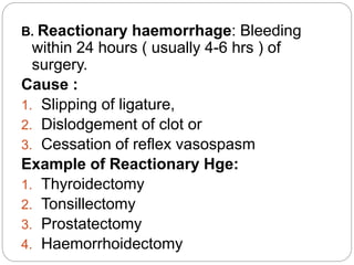
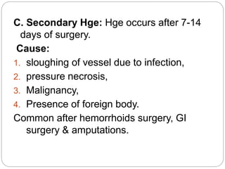
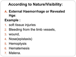
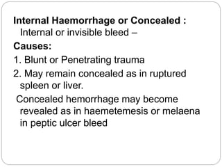
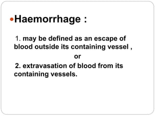
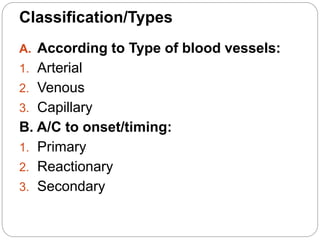
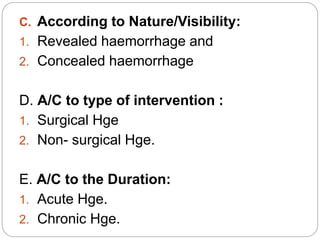
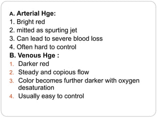
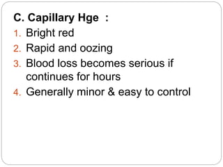
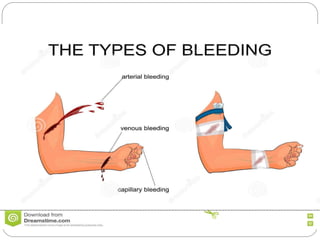
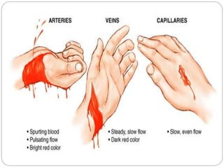
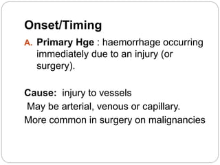
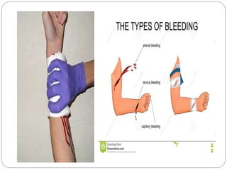
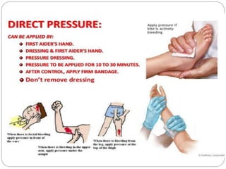
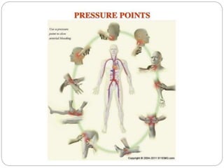
Dr. Md. Majedul Islam
FCPS(Surgery)
Asst Professor of JIMCH
Haemorrhage
Haemorrhage :
1. may be defined as an escape of
blood outside its containing vessel ,
or
2. extravasation of blood from its
containing vessels.
Classification/Types
A. According to Type of blood vessels:
1. Arterial
2. Venous
3. Capillary
B. A/C to onset/timing:
1. Primary
2. Reactionary
3. Secondary
C. According to Nature/Visibility:
1. Revealed haemorrhage and
2. Concealed haemorrhage
D. A/C to type of intervention :
1. Surgical Hge
2. Non- surgical Hge.
E. A/C to the Duration:
1. Acute Hge.
2. Chronic Hge.
A. Arterial Hge:
1. Bright red
2. mitted as spurting jet
3. Can lead to severe blood loss
4. Often hard to control
B. Venous Hge :
1. Darker red
2. Steady and copious flow
3. Color becomes further darker with oxygen
desaturation
4. Usually easy to control
C. Capillary Hge :
1. Bright red
2. Rapid and oozing
3. Blood loss becomes serious if
continues for hours
4. Generally minor & easy to control
Onset/Timing
A. Primary Hge : haemorrhage occurring
immediately due to an injury (or
surgery).
Cause: injury to vessels
May be arterial, venous or capillary.
More common in surgery on malignancies
B. Reactionary haemorrhage: Bleeding
within 24 hours ( usually 4-6 hrs ) of
surgery.
Cause :
1. Slipping of ligature,
2. Dislodgement of clot or
3. Cessation of reflex vasospasm
Example of Reactionary Hge:
1. Thyroidectomy
2. Tonsillectomy
3. Prostatectomy
4. Haemorrhoidectomy
C. Secondary Hge: Hge occurs after 7-14
days of surgery.
Cause:
1. sloughing of vessel due to infection,
2. pressure necrosis,
3. Malignancy,
4. Presence of foreign body.
Common after hemorrhoids surgery, GI
surgery & amputations.
According to Nature/Visibility:
A. External Haemorrhage or Revealed
Hge:
Example :
1. soft tissue injuries
2. Bleeding from the limb vessels,
3. wound,
4. Nose(epistaxis)
5. Hemoptysis
6. Hematemesis
7. Malena.
Internal Haemorrhage or Concealed :
Internal or invisible bleed –
Causes:
1. Blunt or Penetrating trauma
2. May remain concealed as in ruptured
spleen or liver.
Concealed hemorrhage may become
revealed as in haemetemesis or melaena
in peptic ulcer bleed
 Example of internal Hge:
1. Haemocranium(Blood in side the cranium
by trauma) i.e: EDH, SDH, ICH
2. Haemothorax(Blood in the thoracic cavity
by blunt trauma)
3. Hemoperitoneum(blood in side the
peritoneal cavity) , liver, spleen or
mesenteric injury or rupture ectopic
pregnancy.
4. Retroperitoneal Hge: Injury to kidney,
rupture aortic aneurysm.
5. Fracture long bone: Blood collect inside
Revision
Questions of Professional Examination:
Lets check it
Out
Clinical Feature of Hge
 Depends upon 2 factor:
1. Amount of Blood loss.
2. Speed of Blood Loss.
Acute Blood Loss(internal bleeding/Concealed
Hge)
1. Increasing Pallor
2. Increasing Pulse
3. Increasing restlessness
4. Deep respiration
5. Sign of Shock
Sign of Shock
1. Cold clammy skin
2. Tachycardia
3. Hypotension
4. Reduced Urine output
5. Collapse peripheral vein
6. Altered consciousness level
Feature of Chronic Blood Loss :
Anemia
Monitoring of Hge(Acute):
1. Pulse Rate:
10% Blood Loss, No problem – Just Tachycardia
50% Blood Loss - Pulse Absent.
Measure every 15/30 min Interval
When Pt is Stable – Monitor 4 hourly.
2. Blood Pressure. Blood pressure is usually well
maintained and only falls after 30–40 per cent of
circulating volume has been lost.
3. Urine Output.
Normal Urine output 1ml/kg/hr or 1ml/min
Half than normal is Ok
But less than 30 ml/hr – Oliguria
If blood loss > 50% - Anuria develop.
Control Of Hge
Control bleeding using these techniques:
A. Non Surgical Technique
1. Direct pressure (“pressure dressing”) and Packing
2. Elevation
3. Pressure points(Brachial artery,Femoral artery)
4. Tourniquets Application
5. Adrenaline soaked gauze/pad
6. Warm mop/Hot mop.
B. Surgical :
1. Catch the bleeding point by artery forceps.
2. Ligation of bleeding vessels
3. Electro-cauterization.
4. Reapir(liver injry, kidney injury, great vessels).
5. Excision (soid organ where repair not possible i.e –
spleen, kidney)
C. Endoscopic technique:
1. Sclerotherapy
2. Cauterization
3. Ligation
D. Modern technique:
1. Transarterial chemo-
embolisation/Angioembolisation.
Others:
1. Gel foam/Gelatin sponge.
2. Spongiostant.
3. Muscle
4. Bone wax.
 How you will restore blood volume:
1. Blood Transfusion
2. Hemaccel(artificial Plasma solution)
3. I/V fluid(Hartman/Ringer’s Lactate)
Haemorrhage
Haemorrhage
Haemorrhage
Haemorrhage

Haemorrhage

  • 1.
    Dr. Md. MajedulIslam FCPS(Surgery) Asst Professor of JIMCH Haemorrhage
  • 2.
    Haemorrhage : 1. maybe defined as an escape of blood outside its containing vessel , or 2. extravasation of blood from its containing vessels.
  • 3.
    Classification/Types A. According toType of blood vessels: 1. Arterial 2. Venous 3. Capillary B. A/C to onset/timing: 1. Primary 2. Reactionary 3. Secondary
  • 4.
    C. According toNature/Visibility: 1. Revealed haemorrhage and 2. Concealed haemorrhage D. A/C to type of intervention : 1. Surgical Hge 2. Non- surgical Hge. E. A/C to the Duration: 1. Acute Hge. 2. Chronic Hge.
  • 5.
    A. Arterial Hge: 1.Bright red 2. mitted as spurting jet 3. Can lead to severe blood loss 4. Often hard to control B. Venous Hge : 1. Darker red 2. Steady and copious flow 3. Color becomes further darker with oxygen desaturation 4. Usually easy to control
  • 6.
    C. Capillary Hge: 1. Bright red 2. Rapid and oozing 3. Blood loss becomes serious if continues for hours 4. Generally minor & easy to control
  • 9.
    Onset/Timing A. Primary Hge: haemorrhage occurring immediately due to an injury (or surgery). Cause: injury to vessels May be arterial, venous or capillary. More common in surgery on malignancies
  • 10.
    B. Reactionary haemorrhage:Bleeding within 24 hours ( usually 4-6 hrs ) of surgery. Cause : 1. Slipping of ligature, 2. Dislodgement of clot or 3. Cessation of reflex vasospasm Example of Reactionary Hge: 1. Thyroidectomy 2. Tonsillectomy 3. Prostatectomy 4. Haemorrhoidectomy
  • 11.
    C. Secondary Hge:Hge occurs after 7-14 days of surgery. Cause: 1. sloughing of vessel due to infection, 2. pressure necrosis, 3. Malignancy, 4. Presence of foreign body. Common after hemorrhoids surgery, GI surgery & amputations.
  • 12.
    According to Nature/Visibility: A.External Haemorrhage or Revealed Hge: Example : 1. soft tissue injuries 2. Bleeding from the limb vessels, 3. wound, 4. Nose(epistaxis) 5. Hemoptysis 6. Hematemesis 7. Malena.
  • 13.
    Internal Haemorrhage orConcealed : Internal or invisible bleed – Causes: 1. Blunt or Penetrating trauma 2. May remain concealed as in ruptured spleen or liver. Concealed hemorrhage may become revealed as in haemetemesis or melaena in peptic ulcer bleed
  • 14.
     Example ofinternal Hge: 1. Haemocranium(Blood in side the cranium by trauma) i.e: EDH, SDH, ICH 2. Haemothorax(Blood in the thoracic cavity by blunt trauma) 3. Hemoperitoneum(blood in side the peritoneal cavity) , liver, spleen or mesenteric injury or rupture ectopic pregnancy. 4. Retroperitoneal Hge: Injury to kidney, rupture aortic aneurysm. 5. Fracture long bone: Blood collect inside
  • 15.
    Revision Questions of ProfessionalExamination: Lets check it Out
  • 16.
    Clinical Feature ofHge  Depends upon 2 factor: 1. Amount of Blood loss. 2. Speed of Blood Loss. Acute Blood Loss(internal bleeding/Concealed Hge) 1. Increasing Pallor 2. Increasing Pulse 3. Increasing restlessness 4. Deep respiration 5. Sign of Shock
  • 17.
    Sign of Shock 1.Cold clammy skin 2. Tachycardia 3. Hypotension 4. Reduced Urine output 5. Collapse peripheral vein 6. Altered consciousness level Feature of Chronic Blood Loss : Anemia
  • 18.
    Monitoring of Hge(Acute): 1.Pulse Rate: 10% Blood Loss, No problem – Just Tachycardia 50% Blood Loss - Pulse Absent. Measure every 15/30 min Interval When Pt is Stable – Monitor 4 hourly. 2. Blood Pressure. Blood pressure is usually well maintained and only falls after 30–40 per cent of circulating volume has been lost. 3. Urine Output. Normal Urine output 1ml/kg/hr or 1ml/min Half than normal is Ok But less than 30 ml/hr – Oliguria If blood loss > 50% - Anuria develop.
  • 19.
    Control Of Hge Controlbleeding using these techniques: A. Non Surgical Technique 1. Direct pressure (“pressure dressing”) and Packing 2. Elevation 3. Pressure points(Brachial artery,Femoral artery) 4. Tourniquets Application 5. Adrenaline soaked gauze/pad 6. Warm mop/Hot mop. B. Surgical : 1. Catch the bleeding point by artery forceps. 2. Ligation of bleeding vessels 3. Electro-cauterization. 4. Reapir(liver injry, kidney injury, great vessels). 5. Excision (soid organ where repair not possible i.e – spleen, kidney)
  • 20.
    C. Endoscopic technique: 1.Sclerotherapy 2. Cauterization 3. Ligation D. Modern technique: 1. Transarterial chemo- embolisation/Angioembolisation. Others: 1. Gel foam/Gelatin sponge. 2. Spongiostant. 3. Muscle 4. Bone wax.
  • 21.
     How youwill restore blood volume: 1. Blood Transfusion 2. Hemaccel(artificial Plasma solution) 3. I/V fluid(Hartman/Ringer’s Lactate)