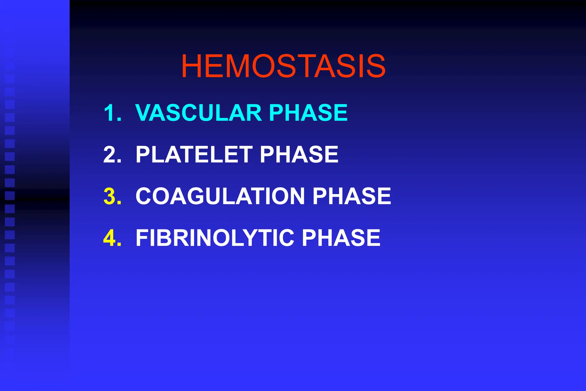
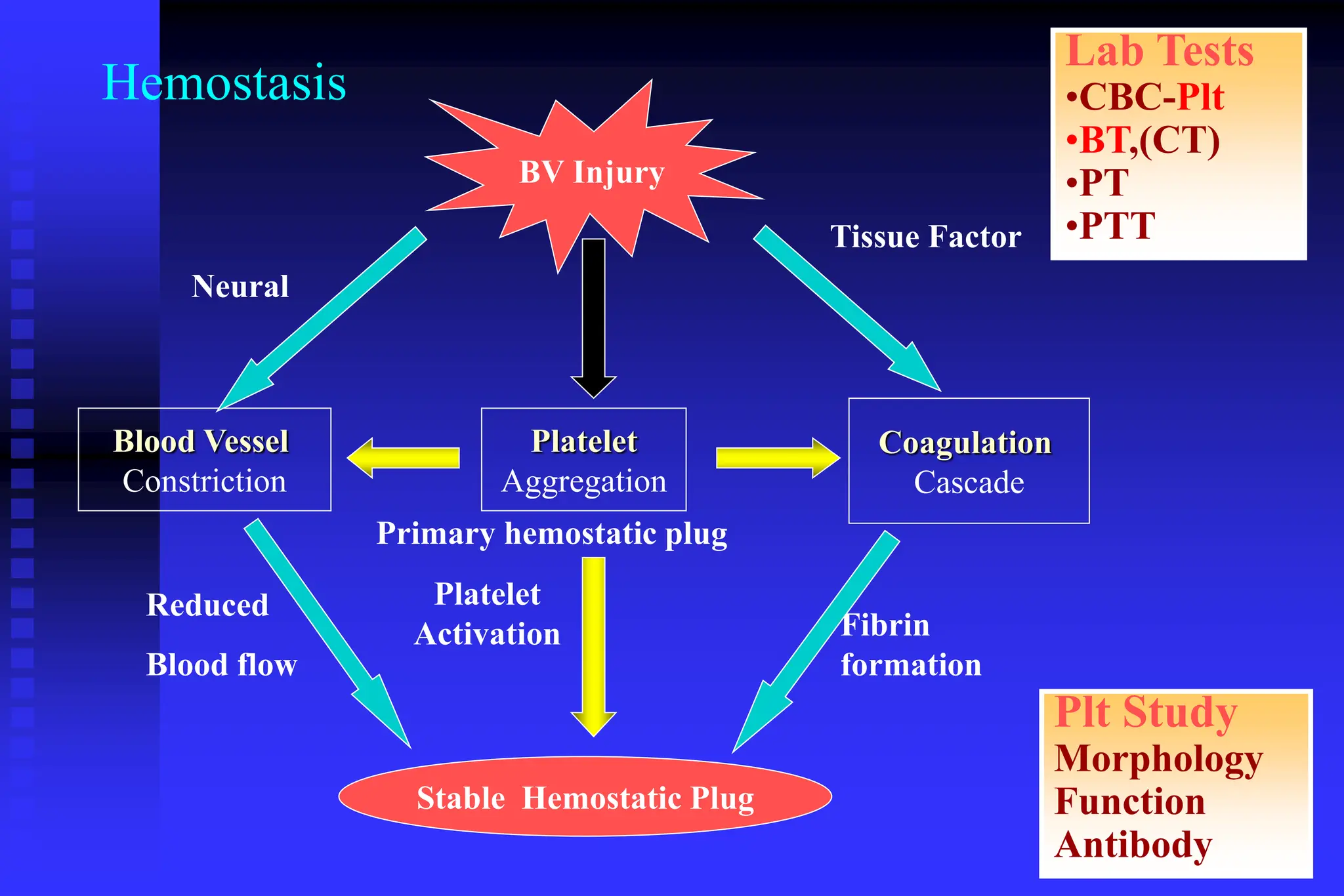
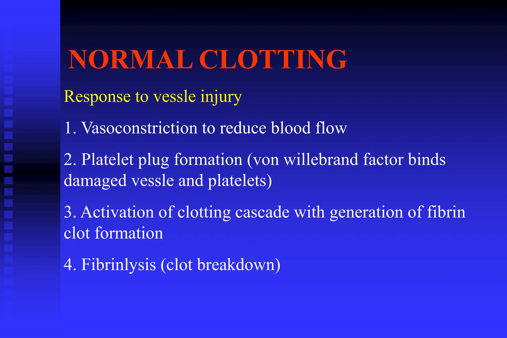
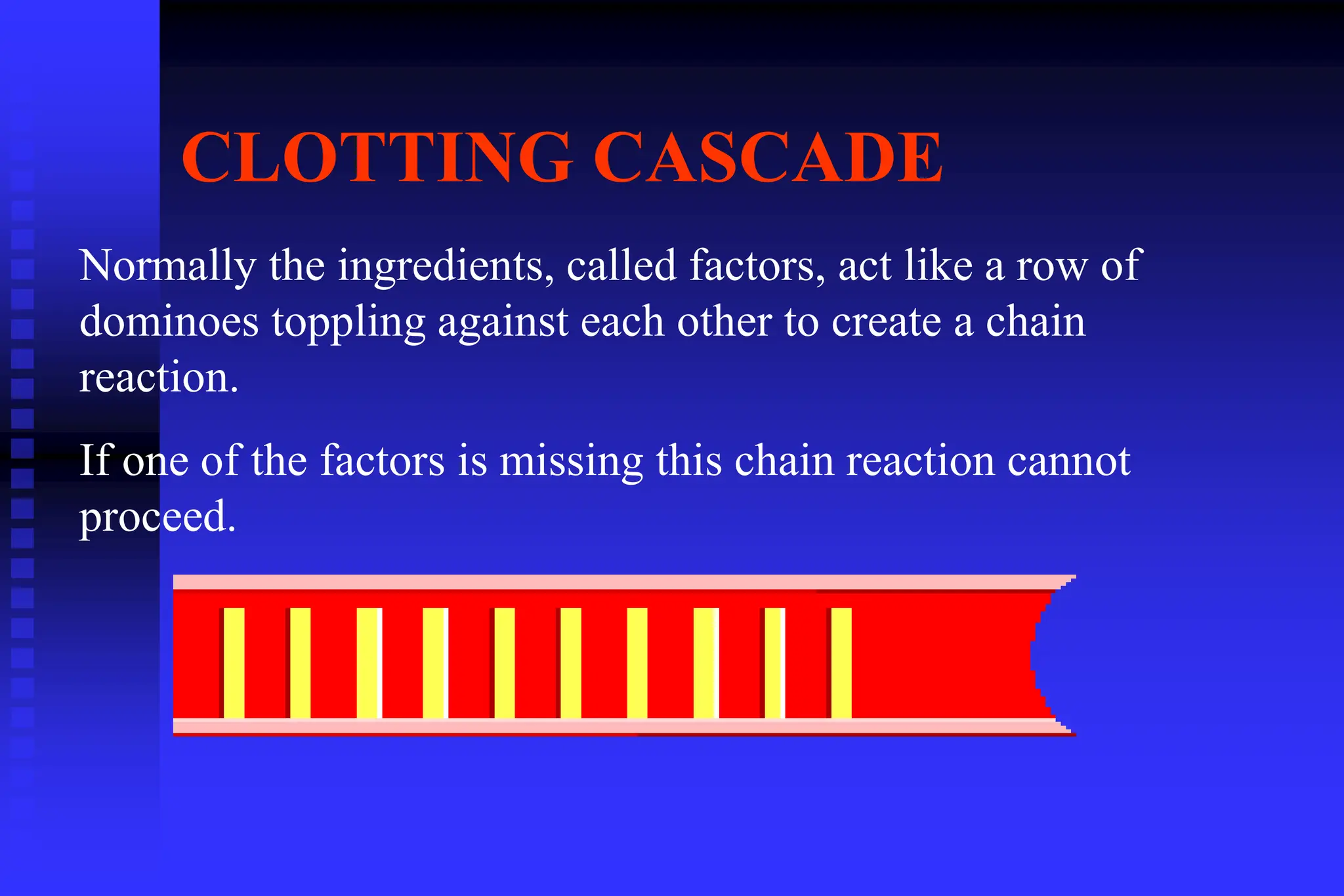
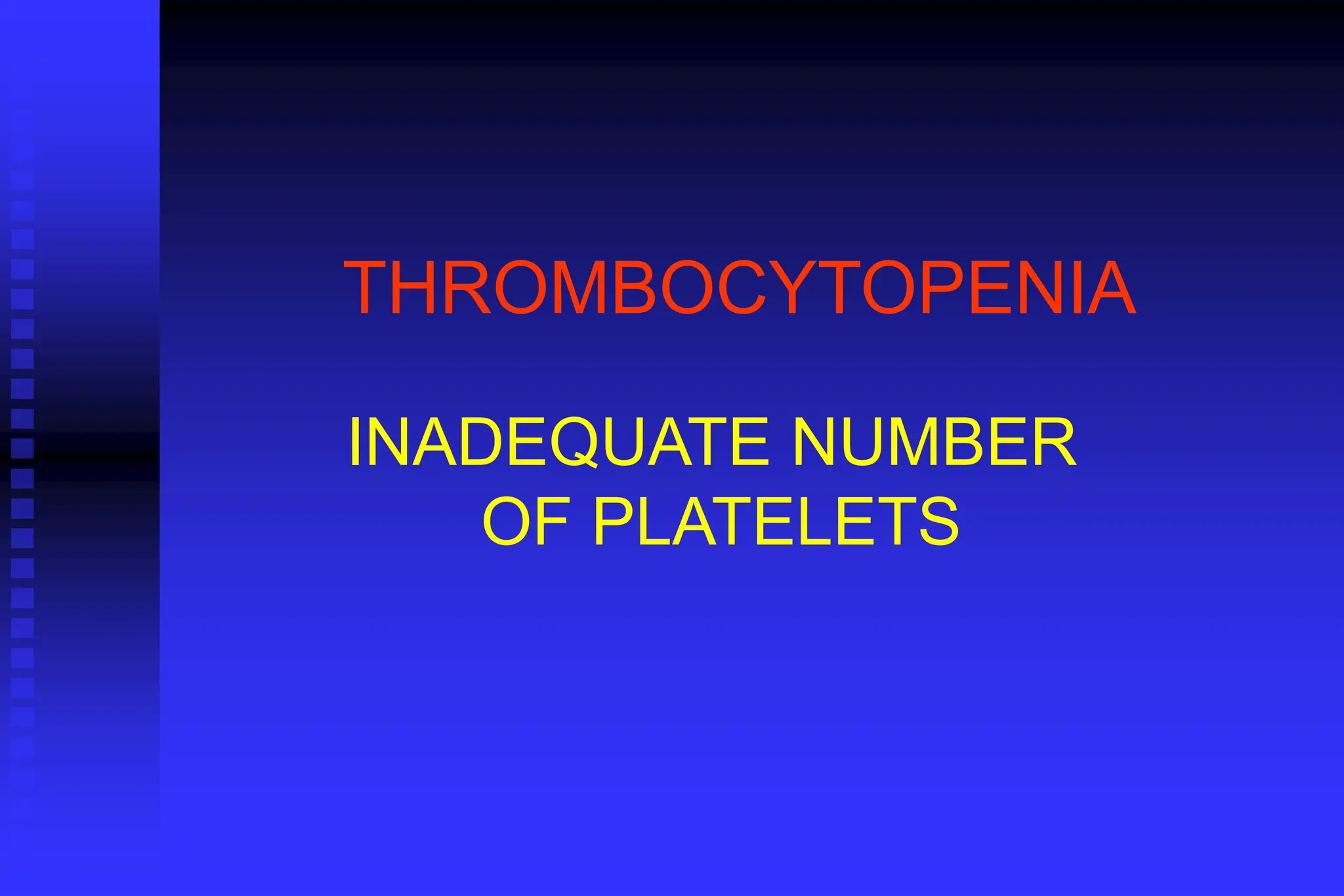
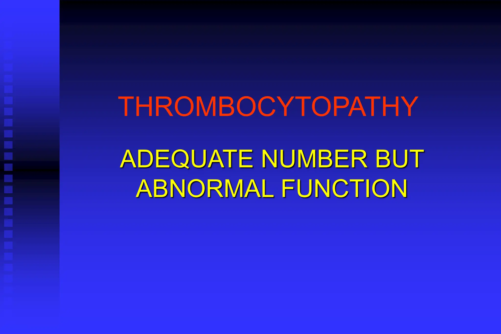
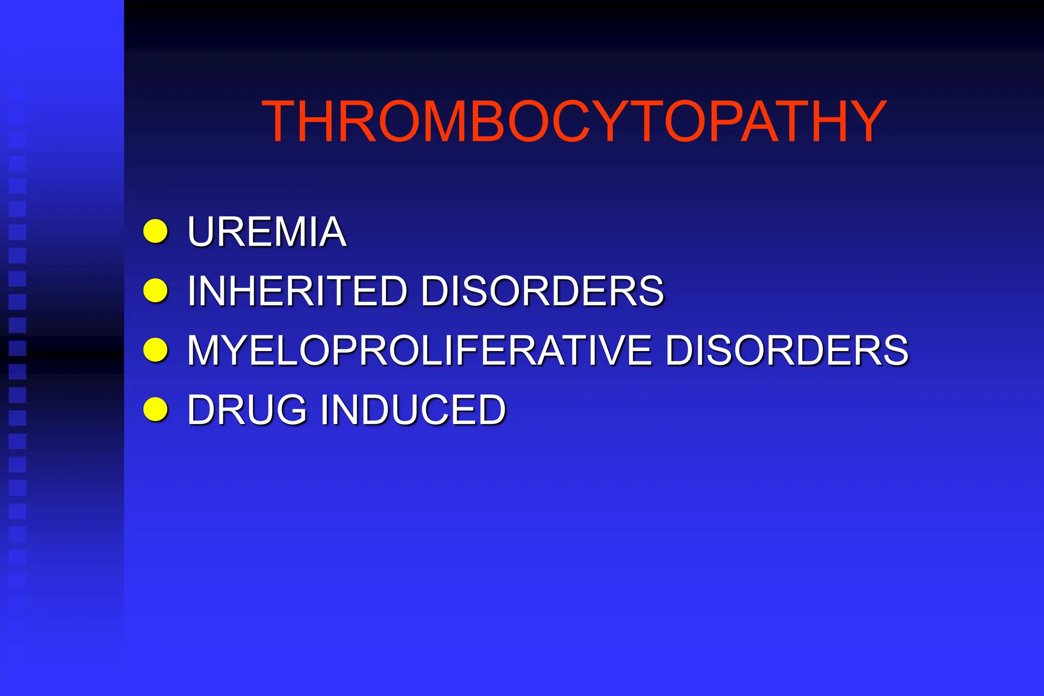
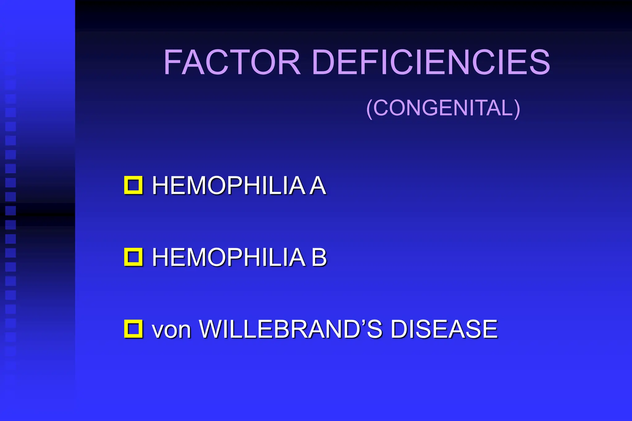
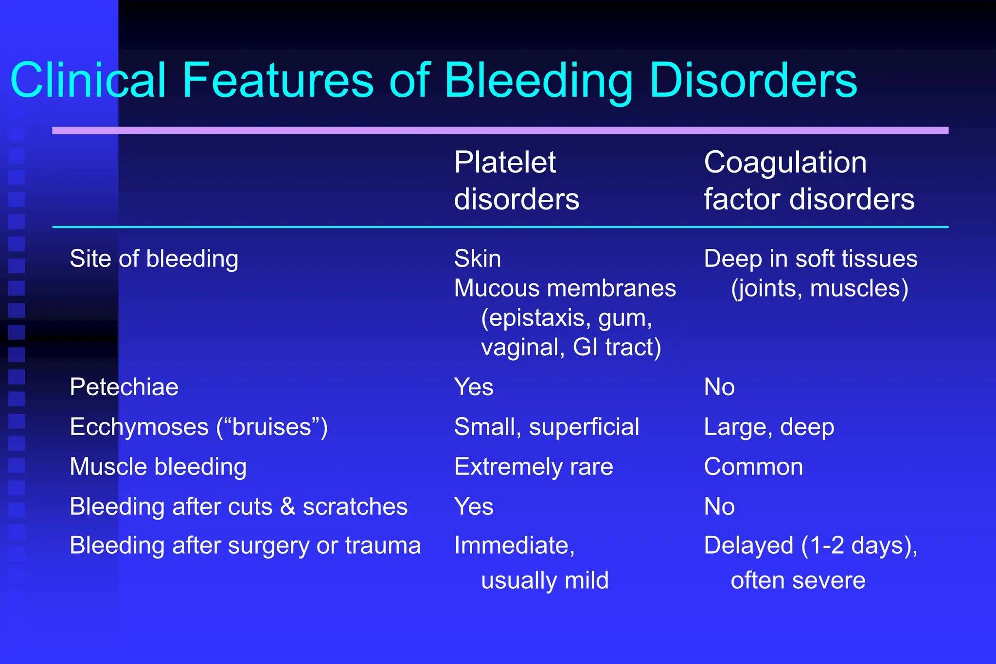
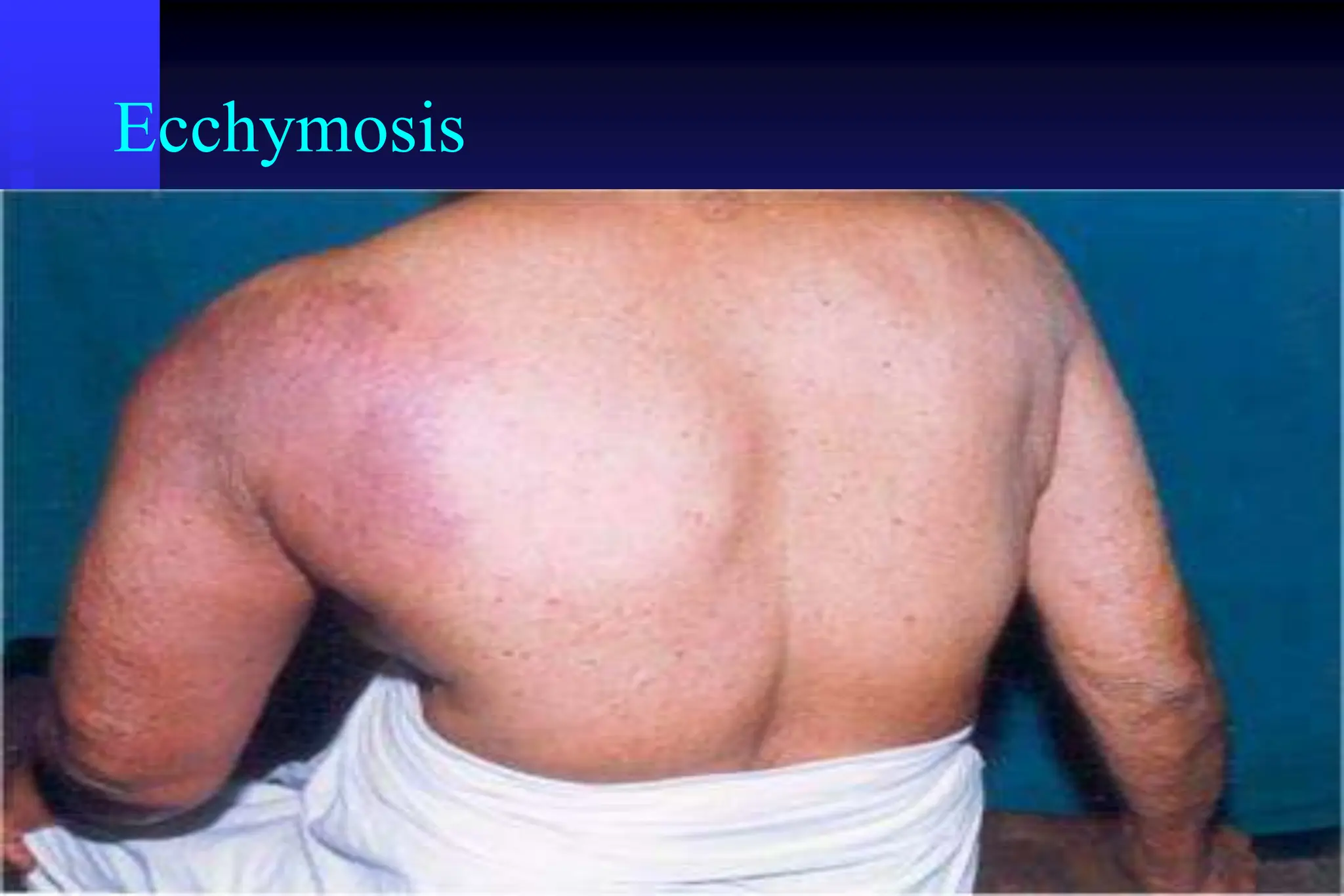
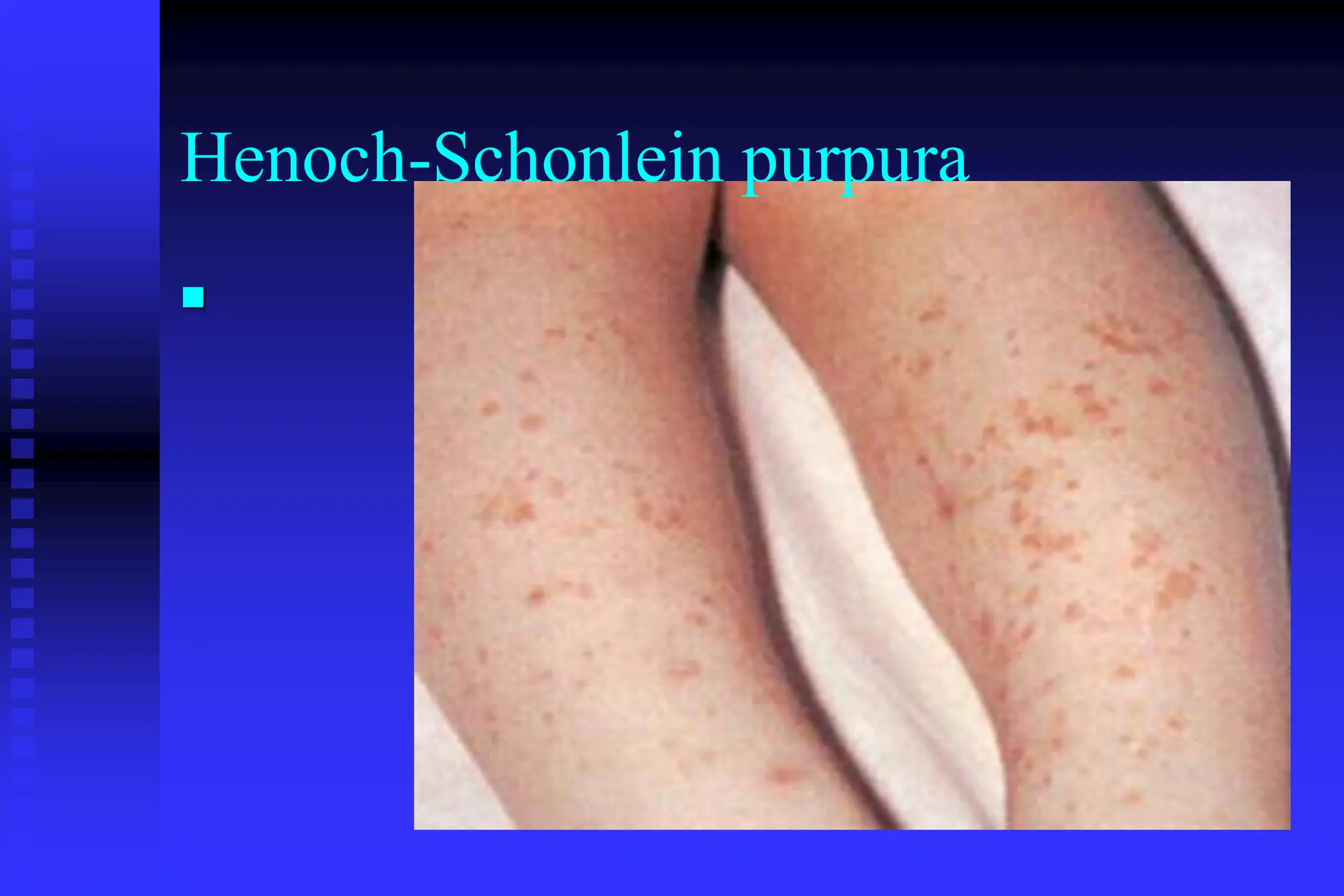
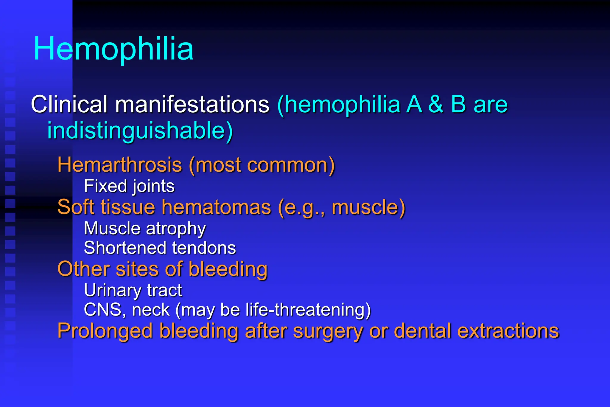
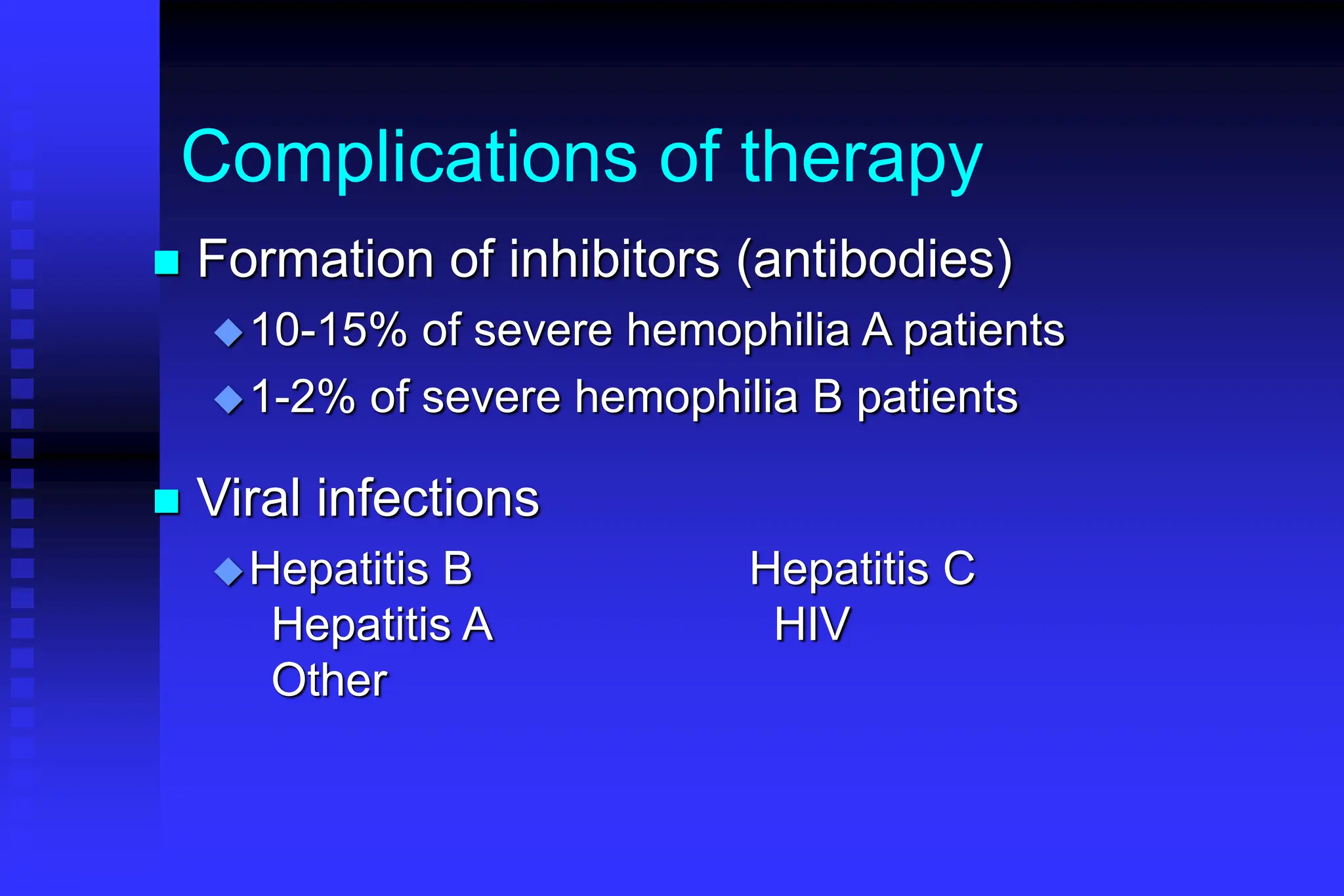
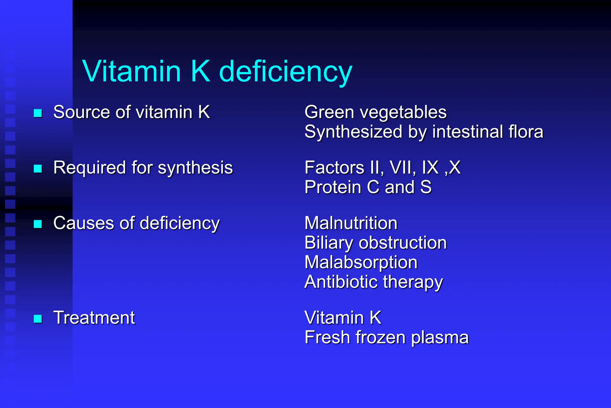
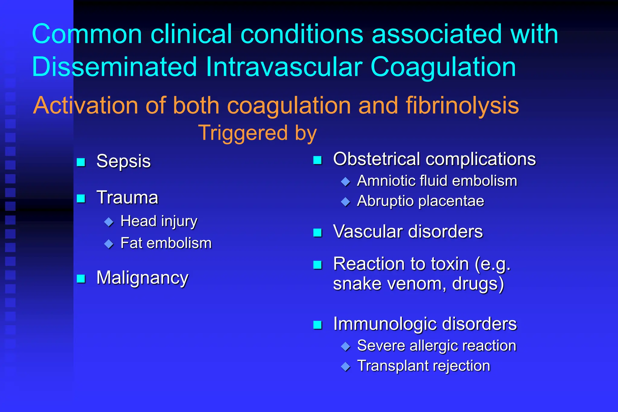
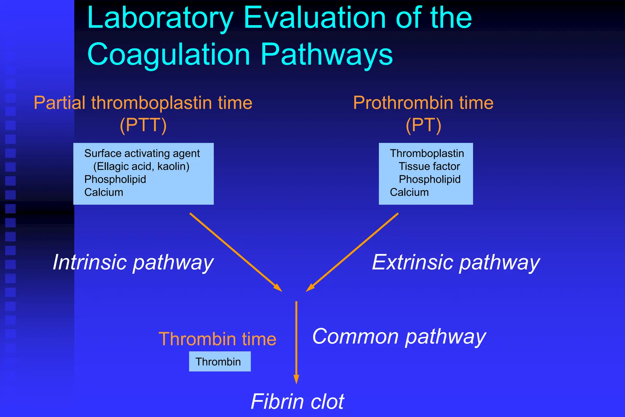
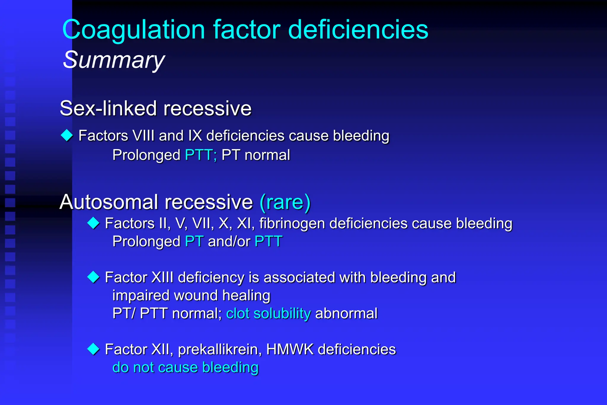
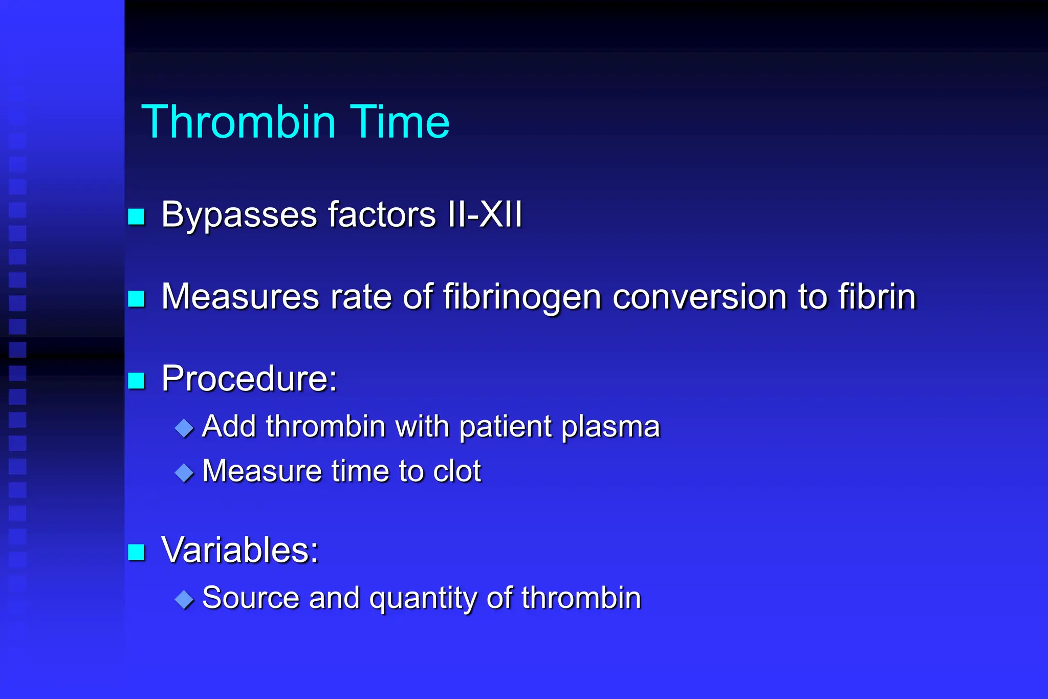
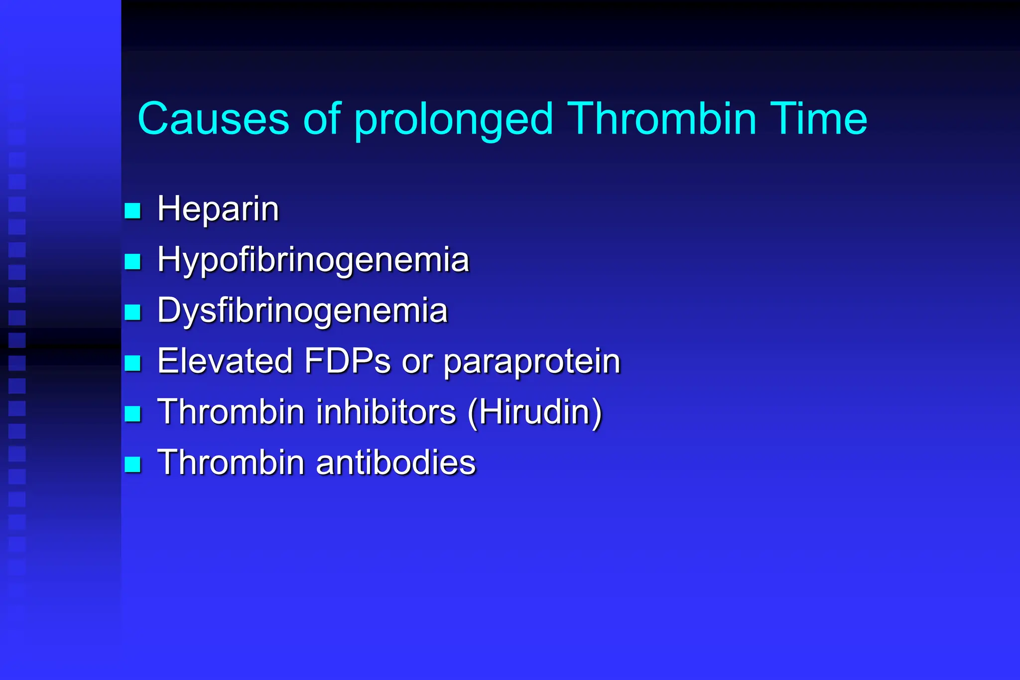
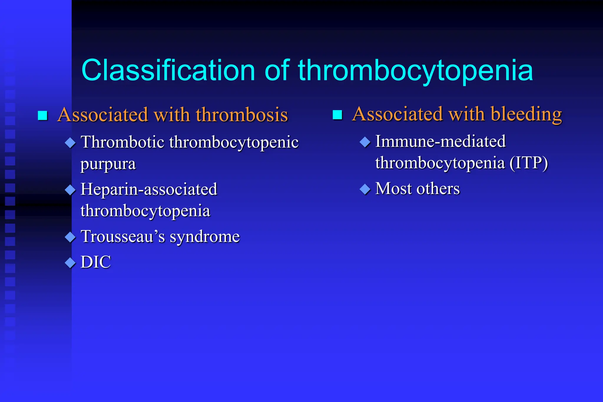
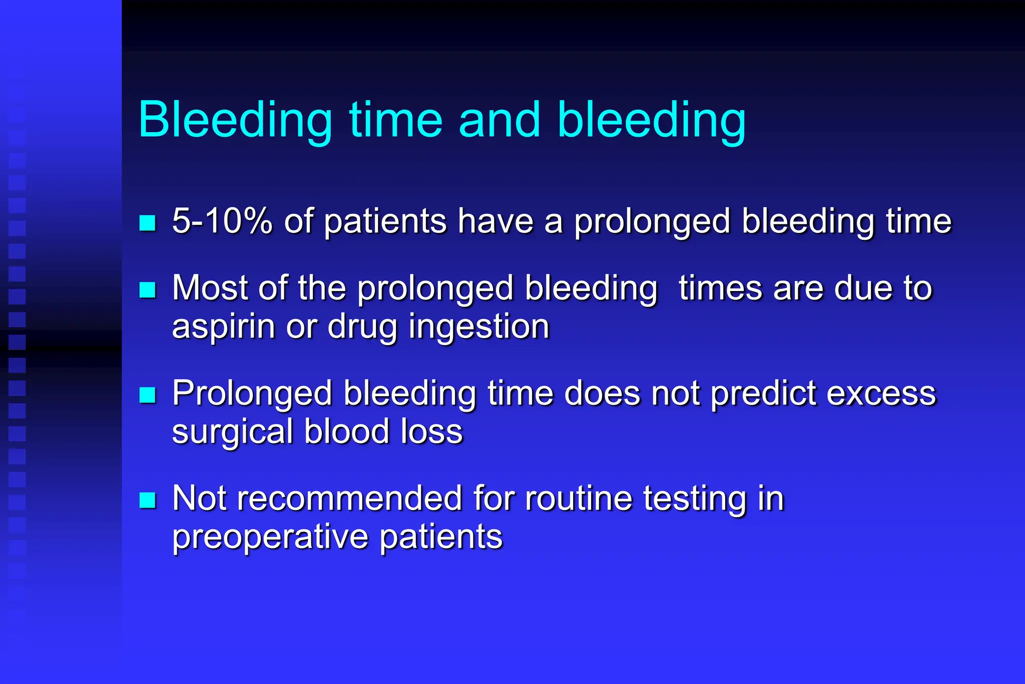
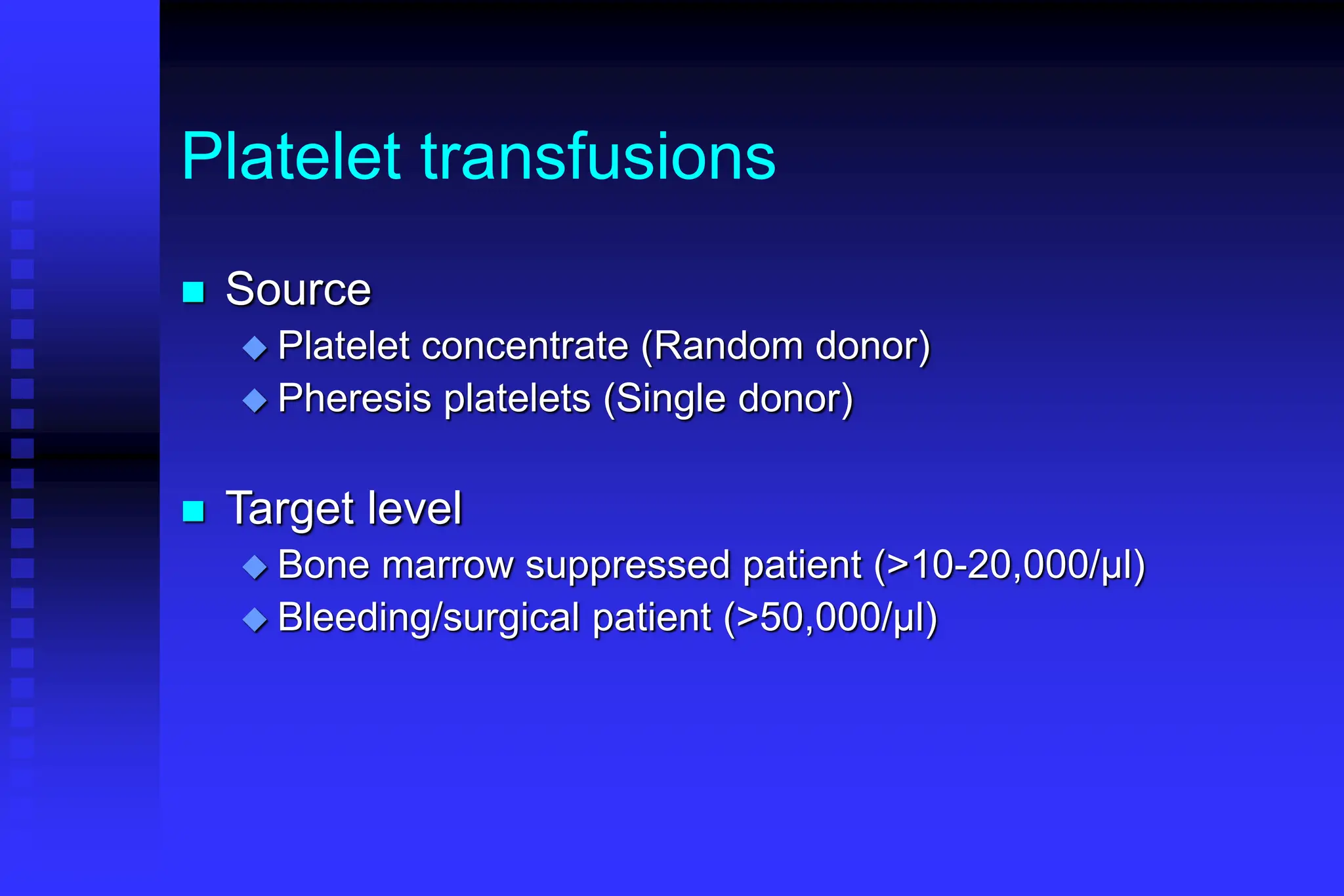
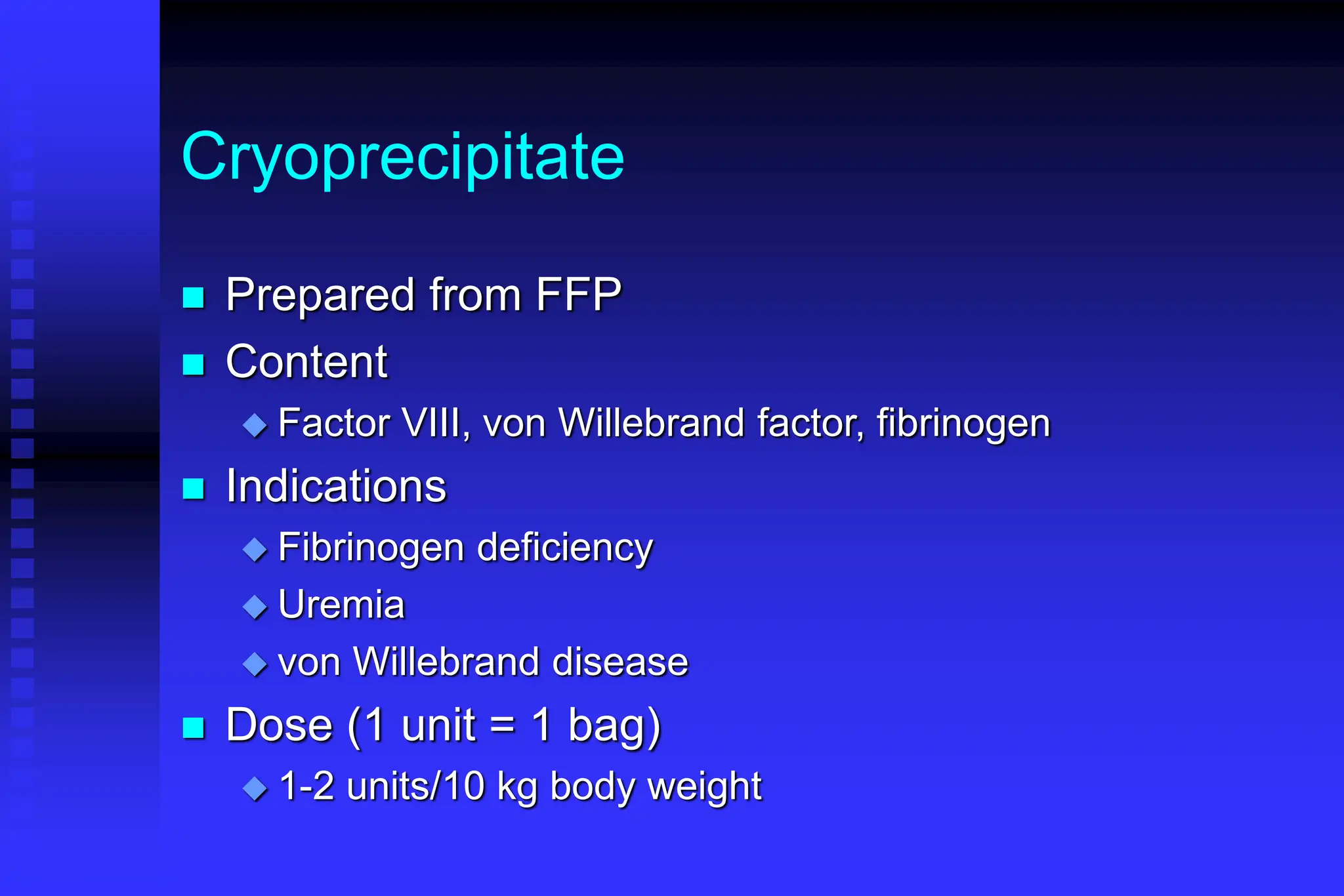
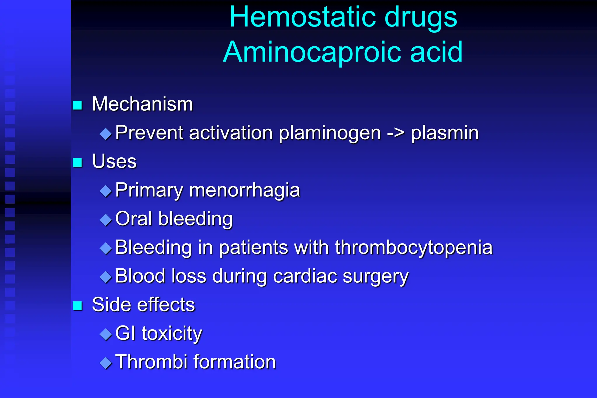
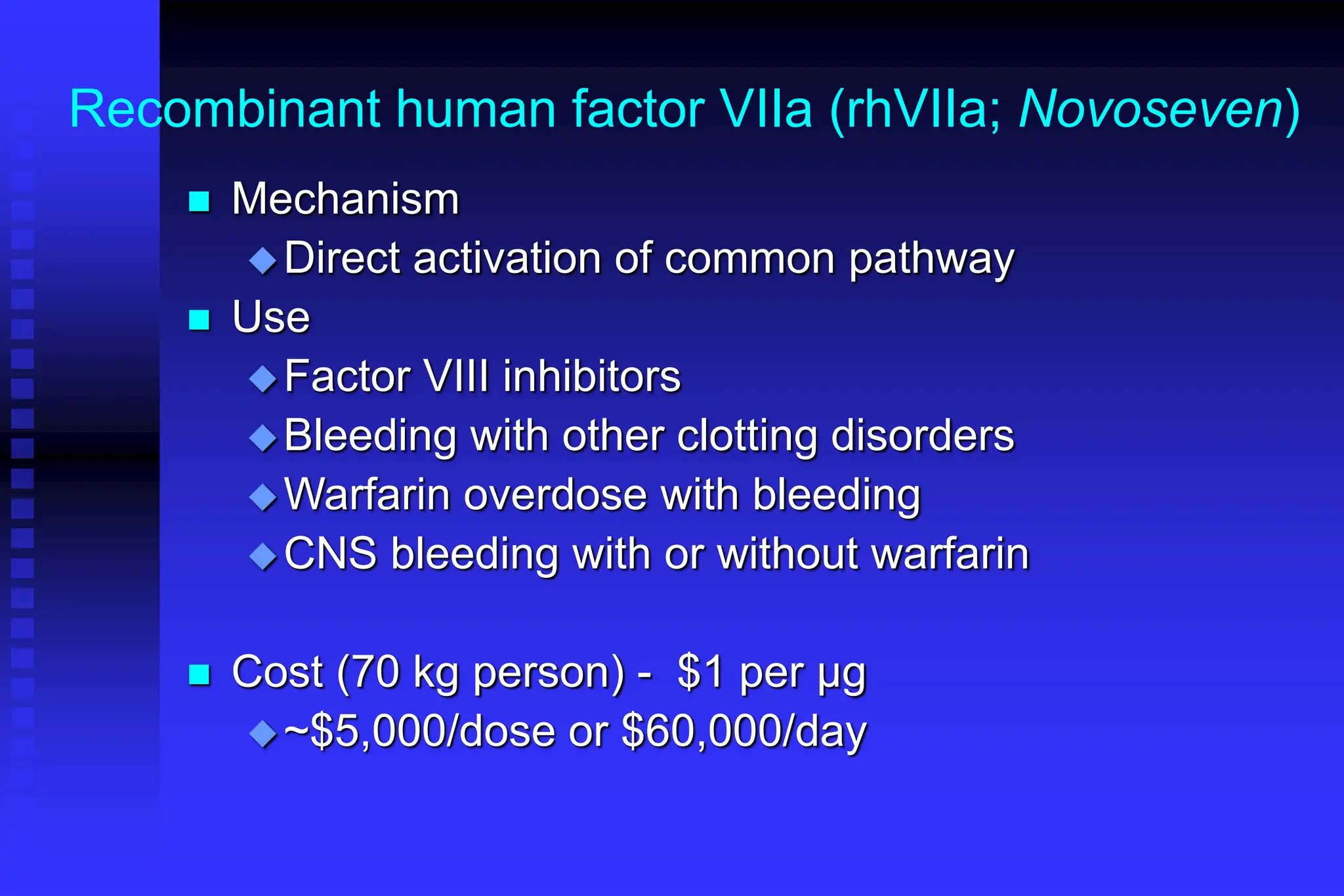
BLEEDING DISORDERS
HEMOSTASIS
1. VASCULAR PHASE
2. PLATELET PHASE
3. COAGULATION PHASE
4. FIBRINOLYTIC PHASE
Hemostasis
BV Injury
Platelet
Aggregation
Platelet
Activation
Blood Vessel
Constriction
Coagulation
Cascade
Stable Hemostatic Plug
Fibrin
formation
Reduced
Blood flow
Tissue Factor
Primary hemostatic plug
Neural
Lab Tests
•CBC-Plt
•BT,(CT)
•PT
•PTT
Plt Study
Morphology
Function
Antibody
NORMAL CLOTTING
Response to vessle injury
1. Vasoconstriction to reduce blood flow
2. Platelet plug formation (von willebrand factor binds
damaged vessle and platelets)
3. Activation of clotting cascade with generation of fibrin
clot formation
4. Fibrinlysis (clot breakdown)
Normally the ingredients, called factors, act like a row of
dominoes toppling against each other to create a chain
reaction.
If one of the factors is missing this chain reaction cannot
proceed.
CLOTTING CASCADE
VASCULAR PHASE
WHEN A BLOOD VESSEL IS
DAMAGED, VASOCONSTRICTION
RESULTS.
PLATELET PHASE
PLATELETS ADHERE TO THE
DAMAGED SURFACE AND FORM A
TEMPORARY PLUG.
COAGULATION PHASE
THROUGH TWO SEPARATE
PATHWAYS THE CONVERSION OF
FIBRINOGEN TO FIBRIN IS
COMPLETE.
THE CLOTTING MECHANISM
INTRINSIC EXTRINSIC
PROTHROMBIN THROMBIN
FIBRINOGEN
FIBRIN
(II) (III)
(I)
V
X
Tissue Thromboplastin
Collagen
VII
XII
XI
IX
VIII
FIBRINOLYTIC PHASE
ANTICLOTTING MECHANISMS ARE
ACTIVATED TO ALLOW CLOT
DISINTEGRATION AND REPAIR OF
THE DAMAGED VESSEL.
HEMOSTASIS
DEPENDENT UPON:
 Vessel Wall Integrity
 Adequate Numbers of Platelets
 Proper Functioning Platelets
 Adequate Levels of Clotting Factors
 Proper Function of Fibrinolytic Pathway
LABORATORY EVALUATION
PLATELET COUNT
BLEEDING TIME (BT)
PROTHROMBIN TIME (PT)
PARTIAL THROMBOPLASTIN TIME (PTT)
THROMBIN TIME (TT)
PLATELET COUNT
 NORMAL 100,000 - 400,000 CELLS/MM3
< 100,000 Thrombocytopenia
50,000 - 100,000 Mild Thrombocytopenia
< 50,000 Sev Thrombocytopenia
BLEEDING TIME
PROVIDES ASSESSMENT OF PLATELET
COUNT AND FUNCTION
NORMAL VALUE
2-8 MINUTES
PROTHROMBIN TIME
Measures Effectiveness of the Extrinsic
Pathway
Mnemonic - PET
NORMAL VALUE
10-15 SECS
PARTIAL THROMBOPLASTIN TIME
 Measures Effectiveness of the Intrinsic
Pathway
Mnemonic - PITT
NORMAL VALUE
25-40 SECS
THROMBIN TIME
 Time for Thrombin To Convert
Fibrinogen Fibrin
 A Measure of Fibrinolytic Pathway
NORMAL VALUE
9-13 SECS
So What Causes Bleeding
Disorders?
VESSEL DEFECTS
PLATELET DISORDERS
FACTOR DEFICIENCIES
OTHER DISORDERS
?
?
VESSEL DEFECTS
 VITAMIN C DEFICIENCY
 BACTERIAL & VIRAL INFECTIONS
 ACQUIRED &
HEREDITARY CONDITIONS
Vascular defect - cont.
 Infectious and hypersensitivity vasculitides
- Rickettsial and meningococcal infections
- Henoch-Schonlein purpura (immune)
PLATELET DISORDERS
 THROMBOCYTOPENIA
 THROMBOCYTOPATHY
THROMBOCYTOPENIA
INADEQUATE NUMBER
OF PLATELETS
THROMBOCYTOPATHY
ADEQUATE NUMBER BUT
ABNORMAL FUNCTION
THROMBOCYTOPENIA
DRUG INDUCED
BONE MARROW FAILURE
HYPERSPLENISM
OTHER CAUSES
OTHER CAUSES
Lymphoma
HIV Virus
Idiopathic Thrombocytopenia Purpura (ITP)
THROMBOCYTOPATHY
 UREMIA
 INHERITED DISORDERS
 MYELOPROLIFERATIVE DISORDERS
 DRUG INDUCED
FACTOR DEFICIENCIES
(CONGENITAL)
 HEMOPHILIA A
 HEMOPHILIA B
 von WILLEBRAND’S DISEASE
FACTOR DEFICIENCIES
HEMOPHILIA A (Classic Hemophilia)
80-85% of all Hemophiliacs
Deficiency of Factor VIII
Lab Results - Prolonged PTT
HEMOPHILIA B (Christmas Disease)
10-15% of all Hemophiliacs
Deficiency of Factor IX
Lab Test - Prolonged PTT
FACTOR DEFICIENCIES
VON WILLEBRAND’S DISEASE
Deficiency of VWF & amount of Factor VIII
Lab Results - Prolonged BT, PTT
OTHER DISORDERS
(ACQUIRED)
 ORAL ANTICOAGULANTS
Warfarin
HEPARIN
 LIVER DISEASE
 MALABSORPTION
 BROAD-SPECTRUM ANTIBIOTICS
INHIBITORS
30% of people with haemophilia develop an antibody to the
clotting factor they are receiving for treatment. These
antibodies are known as inhibitors.
These patients are treated with high does of FVIIa for bleeds or
surgery. This overrides defect in FVIII or FIX deficiency.
Longterm management involves attempting to eradicate
inhibitors by administering high dose FVIII (or FIX) in a
process called immune tolerance
Bleeding Disorders
Clinical Features of Bleeding Disorders
Platelet Coagulation
disorders factor disorders
Site of bleeding Skin Deep in soft tissues
Mucous membranes (joints, muscles)
(epistaxis, gum,
vaginal, GI tract)
Petechiae Yes No
Ecchymoses (“bruises”) Small, superficial Large, deep
Muscle bleeding Extremely rare Common
Bleeding after cuts & scratches Yes No
Bleeding after surgery or trauma Immediate, Delayed (1-2 days),
usually mild often severe
Platelet Coagulation
Petechiae, Purpura Hematoma, Joint bl.
Petechiae
Do not blanch with pressure
(cf. angiomas)
Not palpable
(cf. vasculitis)
(typical of platelet disorders)
Hemarthrosis
Hematoma
Petechiae
Purpura
Ecchymosis
Senile Purpura
Petechiae in patient
with Rocky Mountain
Spotted Fever
Henoch-Schonlein purpura

CT scan showing large hematoma
of right psoas muscle
Coagulation factor disorders
 Inherited bleeding
disorders
 Hemophilia A and B
 vonWillebrands disease
 Other factor deficiencies
 Acquired bleeding
disorders
 Liver disease
 Vitamin K
deficiency/warfarin
overdose
 DIC
Hemophilia A and B
Hemophilia A Hemophilia B
Coagulation factor deficiency Factor VIII Factor IX
Inheritance X-linked X-linked
recessive recessive
Incidence 1/10,000 males 1/50,000 males
Severity Related to factor level
<1% - Severe - spontaneous bleeding
1-5% - Moderate - bleeding with mild injury
5-25% - Mild - bleeding with surgery or trauma
Complications Soft tissue bleeding
Hemophilia
Clinical manifestations (hemophilia A & B are
indistinguishable)
Hemarthrosis (most common)
Fixed joints
Soft tissue hematomas (e.g., muscle)
Muscle atrophy
Shortened tendons
Other sites of bleeding
Urinary tract
CNS, neck (may be life-threatening)
Prolonged bleeding after surgery or dental extractions
Hemarthrosis (acute)
Treatment of hemophilia A
 Intermediate purity plasma products
 May contain von Willebrand factor
 High purity (monoclonal) plasma products
 No functional von Willebrand factor
 Recombinant factor VIII
 Virus free/No apparent risk
 Adjunctive therapy
 Aminocaproic acid
Complications of therapy
 Formation of inhibitors (antibodies)
10-15% of severe hemophilia A patients
1-2% of severe hemophilia B patients
 Viral infections
Hepatitis B Hepatitis C
Hepatitis A HIV
Other
Treatment of hemophilia B
 Agent
High purity factor IX
Recombinant human factor IX
 Dose
Initial dose: 100U/kg
Subsequent: 50U/kg every 24 hours
von Willebrand Disease: Clinical Features
 von Willebrand factor
 Synthesis in endothelium and megakaryocytes
 Forms large multimer
 Carrier of factor VIII
 Anchors platelets to subendothelium
 Bridge between platelets
 Inheritance - autosomal dominant
 Incidence - 1/10,000
 Clinical features - mucocutaneous bleeding
Laboratory evaluation of
von Willebrand disease
 Classification
 Type 1 Partial quantitative deficiency
 Type 2 Qualitative deficiency
 Type 3 Total quantitative deficiency
 Diagnostic tests:
vonWillebrand type
Assay 1 2 3
vWF antigen  Normal 
vWF activity   
Multimer analysis Normal Normal Absent
Treatment of von Willebrand Disease
 Cryoprecipitate
 Source of fibrinogen, factor VIII and VWF
 Only plasma fraction that consistently contains VWF multimers
 DDAVP (deamino-8-arginine vasopressin)
  plasma VWF levels by stimulating secretion from endothelium
 Duration of response is variable
 Not generally used in type 2 disease
 Dosage 0.3 µg/kg q 12 hr IV
 Factor VIII concentrate (Intermediate purity)
Vitamin K deficiency
 Source of vitamin K Green vegetables
Synthesized by intestinal flora
 Required for synthesis Factors II, VII, IX ,X
Protein C and S
 Causes of deficiency Malnutrition
Biliary obstruction
Malabsorption
Antibiotic therapy
 Treatment Vitamin K
Fresh frozen plasma
Common clinical conditions associated with
Disseminated Intravascular Coagulation
 Sepsis
 Trauma
 Head injury
 Fat embolism
 Malignancy
 Obstetrical complications
 Amniotic fluid embolism
 Abruptio placentae
 Vascular disorders
 Reaction to toxin (e.g.
snake venom, drugs)
 Immunologic disorders
 Severe allergic reaction
 Transplant rejection
Activation of both coagulation and fibrinolysis
Triggered by
Disseminated Intravascular Coagulation (DIC)
Mechanism
Systemic activation
of coagulation
Intravascular
deposition of fibrin
Depletion of platelets
and coagulation factors
Bleeding
Thrombosis of small
and midsize vessels
with organ failure
Pathogenesis of DIC
Coagulation Fibrinolysis
Fibrinogen
Fibrin
Monomers
Fibrin
Clot
(intravascular)
Fibrin(ogen)
Degradation
Products
Plasmin
Thrombin Plasmin
Release of
thromboplastic
material into
circulation
Consumption of
coagulation factors;
presence of FDPs
 aPTT
 PT
 TT
 Fibrinogen
Presence of plasmin
 FDP
Intravascular clot
 Platelets
Schistocytes
Disseminated Intravascular Coagulation
Treatment approaches
 Treatment of underlying disorder
 Anticoagulation with heparin
 Platelet transfusion
 Fresh frozen plasma
 Coagulation inhibitor concentrate (ATIII)
Classification of platelet disorders
 Quantitative disorders
 Abnormal distribution
 Dilution effect
 Decreased production
 Increased destruction
 Qualitative disorders
 Inherited disorders (rare)
 Acquired disorders
 Medications
 Chronic renal failure
 Cardiopulmonary bypass
Thrombocytopenia
Immune-mediated
Idioapthic
Drug-induced
Collagen vascular disease
Lymphoproliferative disease
Sarcoidosis
Non-immune mediated
DIC
Microangiopathic hemolytic anemia
Liver Disease and Hemostasis
1. Decreased synthesis of II, VII, IX, X, XI, and
fibrinogen
2. Dietary Vitamin K deficiency (Inadequate
intake or malabsortion)
3. Dysfibrinogenemia
4. Enhanced fibrinolysis (Decreased alpha-2-
antiplasmin)
5. DIC
6. Thrombocytoepnia due to hypersplenism
Management of Hemostatic
Defects in Liver Disease
Treatment for prolonged PT/PTT
 Vitamin K 10 mg SQ x 3 days - usually
ineffective
 Fresh-frozen plasma infusion
 25-30% of plasma volume (1200-1500 ml)
 immediate but temporary effect
Treatment for low fibrinogen
 Cryoprecipitate (1 unit/10kg body weight)
Treatment for DIC (Elevated D-dimer, low factor
VIII, thrombocytopenia
 Replacement therapy
Vitamin K deficiency due to warfarin overdose
Managing high INR values
Clinical situation Guidelines
INR therapeutic-5 Lower or omit next dose;
Resume therapy when INR is therapeutic
INR 5-9; no bleeding Lower or omit next dose;
Resume therapy when INR is therapeutic
Omit dose and give vitamin K (1-2.5 mg po)
Rapid reversal: vitamin K 2-4 mg po (repeat)
INR >9; no bleeding Omit dose; vitamin K 3-5 mg po; repeat as necessary
Resume therapy at lower dose when INR therapeutic
Chest 2001:119;22-38s (supplement)
Vitamin K deficiency due to warfarin overdose
Managing high INR values in bleeding patients
Clinical situation Guidelines
INR > 20; serious bleeding Omit warfarin
Vitamin K 10 mg slow IV infusion
FFP or PCC (depending on urgency)
Repeat vitamin K injections every 12 hrs as needed
Any life-threatening bleeding Omit warfarin
Vitamin K 10 mg slow IV infusion
PCC ( or recombinant human factor VIIa)
Repeat vitamin K injections every 12 hrs as needed
Chest 2001:119;22-38s (supplement)
Approach to Post-operative bleeding
1. Is the bleeding local or due to a hemostatic failure?
1. Local: Single site of bleeding usually rapid with
minimal coagulation test abnormalities
2. Hemostatic failure: Multiple site or unusual pattern
with abnormal coagulation tests
2. Evaluate for causes of peri-operative hemostatic failure
1. Preexisting abnormality
2. Special cases (e.g. Cardiopulmonmary bypass)
3. Diagnosis of hemostatic failure
1. Review pre-operative testing
2. Obtain updated testing
Laboratory Evaluation of Bleeding
Overview
CBC and smear Platelet count Thrombocytopenia
RBC and platelet morphology TTP, DIC, etc.
Coagulation Prothrombin time Extrinsic/common pathways
Partial thromboplastin time Intrinsic/common pathways
Coagulation factor assays Specific factor deficiencies
50:50 mix Inhibitors (e.g., antibodies)
Fibrinogen assay Decreased fibrinogen
Thrombin time Qualitative/quantitative
fibrinogen defects
FDPs or D-dimer Fibrinolysis (DIC)
Platelet function von Willebrand factor vWD
Bleeding time In vivo test (non-specific)
Platelet function analyzer (PFA) Qualitative platelet disorders
and vWD
Platelet function tests Qualitative platelet disorders
Laboratory Evaluation of the
Coagulation Pathways
Partial thromboplastin time
(PTT)
Prothrombin time
(PT)
Intrinsic pathway Extrinsic pathway
Common pathway
Thrombin time
Thrombin
Surface activating agent
(Ellagic acid, kaolin)
Phospholipid
Calcium
Thromboplastin
Tissue factor
Phospholipid
Calcium
Fibrin clot
Coagulation factor deficiencies
Summary
Sex-linked recessive
 Factors VIII and IX deficiencies cause bleeding
Prolonged PTT; PT normal
Autosomal recessive (rare)
 Factors II, V, VII, X, XI, fibrinogen deficiencies cause bleeding
Prolonged PT and/or PTT
 Factor XIII deficiency is associated with bleeding and
impaired wound healing
PT/ PTT normal; clot solubility abnormal
 Factor XII, prekallikrein, HMWK deficiencies
do not cause bleeding
Thrombin Time
 Bypasses factors II-XII
 Measures rate of fibrinogen conversion to fibrin
 Procedure:
 Add thrombin with patient plasma
 Measure time to clot
 Variables:
 Source and quantity of thrombin
Causes of prolonged Thrombin Time
 Heparin
 Hypofibrinogenemia
 Dysfibrinogenemia
 Elevated FDPs or paraprotein
 Thrombin inhibitors (Hirudin)
 Thrombin antibodies
Classification of thrombocytopenia
 Associated with bleeding
 Immune-mediated
thrombocytopenia (ITP)
 Most others
 Associated with thrombosis
 Thrombotic thrombocytopenic
purpura
 Heparin-associated
thrombocytopenia
 Trousseau’s syndrome
 DIC
Bleeding time and bleeding
 5-10% of patients have a prolonged bleeding time
 Most of the prolonged bleeding times are due to
aspirin or drug ingestion
 Prolonged bleeding time does not predict excess
surgical blood loss
 Not recommended for routine testing in
preoperative patients
 Drugs and blood products used for
bleeding
Treatment Approaches to
the Bleeding Patient
 Red blood cells
 Platelet transfusions
 Fresh frozen plasma
 Cryoprecipitate
 Amicar
 DDAVP
 Recombinant Human factor VIIa
RBC transfusion therapy
Indications
 Improve oxygen carrying capacity of blood
Bleeding
Chronic anemia that is symptomatic
Peri-operative management
Red blood cell transfusions
Adverse reactions
Immunologic reactions
Hemolysis RBC incompatibility
Anaphylaxis Usually unknown; rarely against IgA
Febrile reaction Antibody to neutrophils
Urticaria Antibody to donor plasma proteins
Non-cardiogenic Donor antibody to leukocytes
pulmonary edema
Red blood cell transfusions
Adverse reactions
Non-immunologic reactions
Congestive heart failure Volume overload
Fever and shock Bacterial contamination
Hypocalcemia Massive transfusion
Platelet transfusions
 Source
 Platelet concentrate (Random donor)
 Pheresis platelets (Single donor)
 Target level
 Bone marrow suppressed patient (>10-20,000/µl)
 Bleeding/surgical patient (>50,000/µl)
Platelet transfusions - complications
 Transfusion reactions
 Higher incidence than in RBC transfusions
 Related to length of storage/leukocytes/RBC mismatch
 Bacterial contamination
 Platelet transfusion refractoriness
 Alloimmune destruction of platelets (HLA antigens)
 Non-immune refractoriness
 Microangiopathic hemolytic anemia
 Coagulopathy
 Splenic sequestration
 Fever and infection
 Medications (Amphotericin, vancomycin, ATG, Interferons)
Fresh frozen plasma
 Content - plasma (decreased factor V and VIII)
 Indications
 Multiple coagulation deficiencies (liver disease, trauma)
 DIC
 Warfarin reversal
 Coagulation deficiency (factor XI or VII)
 Note
 Viral screened product
 ABO compatible
Cryoprecipitate
 Prepared from FFP
 Content
 Factor VIII, von Willebrand factor, fibrinogen
 Indications
 Fibrinogen deficiency
 Uremia
 von Willebrand disease
 Dose (1 unit = 1 bag)
 1-2 units/10 kg body weight
Hemostatic drugs
Aminocaproic acid
 Mechanism
Prevent activation plaminogen -> plasmin
 Uses
Primary menorrhagia
Oral bleeding
Bleeding in patients with thrombocytopenia
Blood loss during cardiac surgery
 Side effects
GI toxicity
Thrombi formation
Hemostatic drugs
Desmopressin
 Mechanism
Increased release of VWF from endothelium
 Uses
Most patients with von Willebrand disease
Mild hemophilia A
 Side effects
Facial flushing and headache
Water retention and hyponatremia
Recombinant human factor VIIa (rhVIIa; Novoseven)
 Mechanism
Direct activation of common pathway
 Use
Factor VIII inhibitors
Bleeding with other clotting disorders
Warfarin overdose with bleeding
CNS bleeding with or without warfarin
 Cost (70 kg person) - $1 per µg
~$5,000/dose or $60,000/day

different bleeding_disorders presentation

  • 1.
  • 2.
    HEMOSTASIS 1. VASCULAR PHASE 2.PLATELET PHASE 3. COAGULATION PHASE 4. FIBRINOLYTIC PHASE
  • 3.
    Hemostasis BV Injury Platelet Aggregation Platelet Activation Blood Vessel Constriction Coagulation Cascade StableHemostatic Plug Fibrin formation Reduced Blood flow Tissue Factor Primary hemostatic plug Neural Lab Tests •CBC-Plt •BT,(CT) •PT •PTT Plt Study Morphology Function Antibody
  • 4.
    NORMAL CLOTTING Response tovessle injury 1. Vasoconstriction to reduce blood flow 2. Platelet plug formation (von willebrand factor binds damaged vessle and platelets) 3. Activation of clotting cascade with generation of fibrin clot formation 4. Fibrinlysis (clot breakdown)
  • 5.
    Normally the ingredients,called factors, act like a row of dominoes toppling against each other to create a chain reaction. If one of the factors is missing this chain reaction cannot proceed. CLOTTING CASCADE
  • 6.
    VASCULAR PHASE WHEN ABLOOD VESSEL IS DAMAGED, VASOCONSTRICTION RESULTS.
  • 7.
    PLATELET PHASE PLATELETS ADHERETO THE DAMAGED SURFACE AND FORM A TEMPORARY PLUG.
  • 8.
    COAGULATION PHASE THROUGH TWOSEPARATE PATHWAYS THE CONVERSION OF FIBRINOGEN TO FIBRIN IS COMPLETE.
  • 9.
    THE CLOTTING MECHANISM INTRINSICEXTRINSIC PROTHROMBIN THROMBIN FIBRINOGEN FIBRIN (II) (III) (I) V X Tissue Thromboplastin Collagen VII XII XI IX VIII
  • 10.
    FIBRINOLYTIC PHASE ANTICLOTTING MECHANISMSARE ACTIVATED TO ALLOW CLOT DISINTEGRATION AND REPAIR OF THE DAMAGED VESSEL.
  • 11.
    HEMOSTASIS DEPENDENT UPON:  VesselWall Integrity  Adequate Numbers of Platelets  Proper Functioning Platelets  Adequate Levels of Clotting Factors  Proper Function of Fibrinolytic Pathway
  • 12.
    LABORATORY EVALUATION PLATELET COUNT BLEEDINGTIME (BT) PROTHROMBIN TIME (PT) PARTIAL THROMBOPLASTIN TIME (PTT) THROMBIN TIME (TT)
  • 13.
    PLATELET COUNT  NORMAL100,000 - 400,000 CELLS/MM3 < 100,000 Thrombocytopenia 50,000 - 100,000 Mild Thrombocytopenia < 50,000 Sev Thrombocytopenia
  • 14.
    BLEEDING TIME PROVIDES ASSESSMENTOF PLATELET COUNT AND FUNCTION NORMAL VALUE 2-8 MINUTES
  • 15.
    PROTHROMBIN TIME Measures Effectivenessof the Extrinsic Pathway Mnemonic - PET NORMAL VALUE 10-15 SECS
  • 16.
    PARTIAL THROMBOPLASTIN TIME Measures Effectiveness of the Intrinsic Pathway Mnemonic - PITT NORMAL VALUE 25-40 SECS
  • 17.
    THROMBIN TIME  Timefor Thrombin To Convert Fibrinogen Fibrin  A Measure of Fibrinolytic Pathway NORMAL VALUE 9-13 SECS
  • 18.
    So What CausesBleeding Disorders? VESSEL DEFECTS PLATELET DISORDERS FACTOR DEFICIENCIES OTHER DISORDERS ? ?
  • 19.
    VESSEL DEFECTS  VITAMINC DEFICIENCY  BACTERIAL & VIRAL INFECTIONS  ACQUIRED & HEREDITARY CONDITIONS
  • 20.
    Vascular defect -cont.  Infectious and hypersensitivity vasculitides - Rickettsial and meningococcal infections - Henoch-Schonlein purpura (immune)
  • 21.
  • 22.
  • 23.
  • 24.
    THROMBOCYTOPENIA DRUG INDUCED BONE MARROWFAILURE HYPERSPLENISM OTHER CAUSES
  • 25.
  • 26.
    THROMBOCYTOPATHY  UREMIA  INHERITEDDISORDERS  MYELOPROLIFERATIVE DISORDERS  DRUG INDUCED
  • 27.
    FACTOR DEFICIENCIES (CONGENITAL)  HEMOPHILIAA  HEMOPHILIA B  von WILLEBRAND’S DISEASE
  • 28.
    FACTOR DEFICIENCIES HEMOPHILIA A(Classic Hemophilia) 80-85% of all Hemophiliacs Deficiency of Factor VIII Lab Results - Prolonged PTT HEMOPHILIA B (Christmas Disease) 10-15% of all Hemophiliacs Deficiency of Factor IX Lab Test - Prolonged PTT
  • 29.
    FACTOR DEFICIENCIES VON WILLEBRAND’SDISEASE Deficiency of VWF & amount of Factor VIII Lab Results - Prolonged BT, PTT
  • 30.
    OTHER DISORDERS (ACQUIRED)  ORALANTICOAGULANTS Warfarin HEPARIN  LIVER DISEASE  MALABSORPTION  BROAD-SPECTRUM ANTIBIOTICS
  • 31.
    INHIBITORS 30% of peoplewith haemophilia develop an antibody to the clotting factor they are receiving for treatment. These antibodies are known as inhibitors. These patients are treated with high does of FVIIa for bleeds or surgery. This overrides defect in FVIII or FIX deficiency. Longterm management involves attempting to eradicate inhibitors by administering high dose FVIII (or FIX) in a process called immune tolerance
  • 32.
  • 33.
    Clinical Features ofBleeding Disorders Platelet Coagulation disorders factor disorders Site of bleeding Skin Deep in soft tissues Mucous membranes (joints, muscles) (epistaxis, gum, vaginal, GI tract) Petechiae Yes No Ecchymoses (“bruises”) Small, superficial Large, deep Muscle bleeding Extremely rare Common Bleeding after cuts & scratches Yes No Bleeding after surgery or trauma Immediate, Delayed (1-2 days), usually mild often severe
  • 34.
  • 35.
    Petechiae Do not blanchwith pressure (cf. angiomas) Not palpable (cf. vasculitis) (typical of platelet disorders)
  • 36.
  • 37.
  • 38.
  • 39.
  • 40.
  • 41.
  • 42.
    Petechiae in patient withRocky Mountain Spotted Fever
  • 43.
  • 44.
    CT scan showinglarge hematoma of right psoas muscle
  • 45.
    Coagulation factor disorders Inherited bleeding disorders  Hemophilia A and B  vonWillebrands disease  Other factor deficiencies  Acquired bleeding disorders  Liver disease  Vitamin K deficiency/warfarin overdose  DIC
  • 46.
    Hemophilia A andB Hemophilia A Hemophilia B Coagulation factor deficiency Factor VIII Factor IX Inheritance X-linked X-linked recessive recessive Incidence 1/10,000 males 1/50,000 males Severity Related to factor level <1% - Severe - spontaneous bleeding 1-5% - Moderate - bleeding with mild injury 5-25% - Mild - bleeding with surgery or trauma Complications Soft tissue bleeding
  • 47.
    Hemophilia Clinical manifestations (hemophiliaA & B are indistinguishable) Hemarthrosis (most common) Fixed joints Soft tissue hematomas (e.g., muscle) Muscle atrophy Shortened tendons Other sites of bleeding Urinary tract CNS, neck (may be life-threatening) Prolonged bleeding after surgery or dental extractions
  • 48.
  • 49.
    Treatment of hemophiliaA  Intermediate purity plasma products  May contain von Willebrand factor  High purity (monoclonal) plasma products  No functional von Willebrand factor  Recombinant factor VIII  Virus free/No apparent risk  Adjunctive therapy  Aminocaproic acid
  • 50.
    Complications of therapy Formation of inhibitors (antibodies) 10-15% of severe hemophilia A patients 1-2% of severe hemophilia B patients  Viral infections Hepatitis B Hepatitis C Hepatitis A HIV Other
  • 51.
    Treatment of hemophiliaB  Agent High purity factor IX Recombinant human factor IX  Dose Initial dose: 100U/kg Subsequent: 50U/kg every 24 hours
  • 52.
    von Willebrand Disease:Clinical Features  von Willebrand factor  Synthesis in endothelium and megakaryocytes  Forms large multimer  Carrier of factor VIII  Anchors platelets to subendothelium  Bridge between platelets  Inheritance - autosomal dominant  Incidence - 1/10,000  Clinical features - mucocutaneous bleeding
  • 53.
    Laboratory evaluation of vonWillebrand disease  Classification  Type 1 Partial quantitative deficiency  Type 2 Qualitative deficiency  Type 3 Total quantitative deficiency  Diagnostic tests: vonWillebrand type Assay 1 2 3 vWF antigen  Normal  vWF activity    Multimer analysis Normal Normal Absent
  • 54.
    Treatment of vonWillebrand Disease  Cryoprecipitate  Source of fibrinogen, factor VIII and VWF  Only plasma fraction that consistently contains VWF multimers  DDAVP (deamino-8-arginine vasopressin)   plasma VWF levels by stimulating secretion from endothelium  Duration of response is variable  Not generally used in type 2 disease  Dosage 0.3 µg/kg q 12 hr IV  Factor VIII concentrate (Intermediate purity)
  • 55.
    Vitamin K deficiency Source of vitamin K Green vegetables Synthesized by intestinal flora  Required for synthesis Factors II, VII, IX ,X Protein C and S  Causes of deficiency Malnutrition Biliary obstruction Malabsorption Antibiotic therapy  Treatment Vitamin K Fresh frozen plasma
  • 56.
    Common clinical conditionsassociated with Disseminated Intravascular Coagulation  Sepsis  Trauma  Head injury  Fat embolism  Malignancy  Obstetrical complications  Amniotic fluid embolism  Abruptio placentae  Vascular disorders  Reaction to toxin (e.g. snake venom, drugs)  Immunologic disorders  Severe allergic reaction  Transplant rejection Activation of both coagulation and fibrinolysis Triggered by
  • 57.
    Disseminated Intravascular Coagulation(DIC) Mechanism Systemic activation of coagulation Intravascular deposition of fibrin Depletion of platelets and coagulation factors Bleeding Thrombosis of small and midsize vessels with organ failure
  • 58.
    Pathogenesis of DIC CoagulationFibrinolysis Fibrinogen Fibrin Monomers Fibrin Clot (intravascular) Fibrin(ogen) Degradation Products Plasmin Thrombin Plasmin Release of thromboplastic material into circulation Consumption of coagulation factors; presence of FDPs  aPTT  PT  TT  Fibrinogen Presence of plasmin  FDP Intravascular clot  Platelets Schistocytes
  • 59.
    Disseminated Intravascular Coagulation Treatmentapproaches  Treatment of underlying disorder  Anticoagulation with heparin  Platelet transfusion  Fresh frozen plasma  Coagulation inhibitor concentrate (ATIII)
  • 60.
    Classification of plateletdisorders  Quantitative disorders  Abnormal distribution  Dilution effect  Decreased production  Increased destruction  Qualitative disorders  Inherited disorders (rare)  Acquired disorders  Medications  Chronic renal failure  Cardiopulmonary bypass
  • 61.
    Thrombocytopenia Immune-mediated Idioapthic Drug-induced Collagen vascular disease Lymphoproliferativedisease Sarcoidosis Non-immune mediated DIC Microangiopathic hemolytic anemia
  • 62.
    Liver Disease andHemostasis 1. Decreased synthesis of II, VII, IX, X, XI, and fibrinogen 2. Dietary Vitamin K deficiency (Inadequate intake or malabsortion) 3. Dysfibrinogenemia 4. Enhanced fibrinolysis (Decreased alpha-2- antiplasmin) 5. DIC 6. Thrombocytoepnia due to hypersplenism
  • 63.
    Management of Hemostatic Defectsin Liver Disease Treatment for prolonged PT/PTT  Vitamin K 10 mg SQ x 3 days - usually ineffective  Fresh-frozen plasma infusion  25-30% of plasma volume (1200-1500 ml)  immediate but temporary effect Treatment for low fibrinogen  Cryoprecipitate (1 unit/10kg body weight) Treatment for DIC (Elevated D-dimer, low factor VIII, thrombocytopenia  Replacement therapy
  • 64.
    Vitamin K deficiencydue to warfarin overdose Managing high INR values Clinical situation Guidelines INR therapeutic-5 Lower or omit next dose; Resume therapy when INR is therapeutic INR 5-9; no bleeding Lower or omit next dose; Resume therapy when INR is therapeutic Omit dose and give vitamin K (1-2.5 mg po) Rapid reversal: vitamin K 2-4 mg po (repeat) INR >9; no bleeding Omit dose; vitamin K 3-5 mg po; repeat as necessary Resume therapy at lower dose when INR therapeutic Chest 2001:119;22-38s (supplement)
  • 65.
    Vitamin K deficiencydue to warfarin overdose Managing high INR values in bleeding patients Clinical situation Guidelines INR > 20; serious bleeding Omit warfarin Vitamin K 10 mg slow IV infusion FFP or PCC (depending on urgency) Repeat vitamin K injections every 12 hrs as needed Any life-threatening bleeding Omit warfarin Vitamin K 10 mg slow IV infusion PCC ( or recombinant human factor VIIa) Repeat vitamin K injections every 12 hrs as needed Chest 2001:119;22-38s (supplement)
  • 66.
    Approach to Post-operativebleeding 1. Is the bleeding local or due to a hemostatic failure? 1. Local: Single site of bleeding usually rapid with minimal coagulation test abnormalities 2. Hemostatic failure: Multiple site or unusual pattern with abnormal coagulation tests 2. Evaluate for causes of peri-operative hemostatic failure 1. Preexisting abnormality 2. Special cases (e.g. Cardiopulmonmary bypass) 3. Diagnosis of hemostatic failure 1. Review pre-operative testing 2. Obtain updated testing
  • 67.
    Laboratory Evaluation ofBleeding Overview CBC and smear Platelet count Thrombocytopenia RBC and platelet morphology TTP, DIC, etc. Coagulation Prothrombin time Extrinsic/common pathways Partial thromboplastin time Intrinsic/common pathways Coagulation factor assays Specific factor deficiencies 50:50 mix Inhibitors (e.g., antibodies) Fibrinogen assay Decreased fibrinogen Thrombin time Qualitative/quantitative fibrinogen defects FDPs or D-dimer Fibrinolysis (DIC) Platelet function von Willebrand factor vWD Bleeding time In vivo test (non-specific) Platelet function analyzer (PFA) Qualitative platelet disorders and vWD Platelet function tests Qualitative platelet disorders
  • 68.
    Laboratory Evaluation ofthe Coagulation Pathways Partial thromboplastin time (PTT) Prothrombin time (PT) Intrinsic pathway Extrinsic pathway Common pathway Thrombin time Thrombin Surface activating agent (Ellagic acid, kaolin) Phospholipid Calcium Thromboplastin Tissue factor Phospholipid Calcium Fibrin clot
  • 69.
    Coagulation factor deficiencies Summary Sex-linkedrecessive  Factors VIII and IX deficiencies cause bleeding Prolonged PTT; PT normal Autosomal recessive (rare)  Factors II, V, VII, X, XI, fibrinogen deficiencies cause bleeding Prolonged PT and/or PTT  Factor XIII deficiency is associated with bleeding and impaired wound healing PT/ PTT normal; clot solubility abnormal  Factor XII, prekallikrein, HMWK deficiencies do not cause bleeding
  • 70.
    Thrombin Time  Bypassesfactors II-XII  Measures rate of fibrinogen conversion to fibrin  Procedure:  Add thrombin with patient plasma  Measure time to clot  Variables:  Source and quantity of thrombin
  • 71.
    Causes of prolongedThrombin Time  Heparin  Hypofibrinogenemia  Dysfibrinogenemia  Elevated FDPs or paraprotein  Thrombin inhibitors (Hirudin)  Thrombin antibodies
  • 72.
    Classification of thrombocytopenia Associated with bleeding  Immune-mediated thrombocytopenia (ITP)  Most others  Associated with thrombosis  Thrombotic thrombocytopenic purpura  Heparin-associated thrombocytopenia  Trousseau’s syndrome  DIC
  • 73.
    Bleeding time andbleeding  5-10% of patients have a prolonged bleeding time  Most of the prolonged bleeding times are due to aspirin or drug ingestion  Prolonged bleeding time does not predict excess surgical blood loss  Not recommended for routine testing in preoperative patients
  • 74.
     Drugs andblood products used for bleeding
  • 75.
    Treatment Approaches to theBleeding Patient  Red blood cells  Platelet transfusions  Fresh frozen plasma  Cryoprecipitate  Amicar  DDAVP  Recombinant Human factor VIIa
  • 76.
    RBC transfusion therapy Indications Improve oxygen carrying capacity of blood Bleeding Chronic anemia that is symptomatic Peri-operative management
  • 77.
    Red blood celltransfusions Adverse reactions Immunologic reactions Hemolysis RBC incompatibility Anaphylaxis Usually unknown; rarely against IgA Febrile reaction Antibody to neutrophils Urticaria Antibody to donor plasma proteins Non-cardiogenic Donor antibody to leukocytes pulmonary edema
  • 78.
    Red blood celltransfusions Adverse reactions Non-immunologic reactions Congestive heart failure Volume overload Fever and shock Bacterial contamination Hypocalcemia Massive transfusion
  • 79.
    Platelet transfusions  Source Platelet concentrate (Random donor)  Pheresis platelets (Single donor)  Target level  Bone marrow suppressed patient (>10-20,000/µl)  Bleeding/surgical patient (>50,000/µl)
  • 80.
    Platelet transfusions -complications  Transfusion reactions  Higher incidence than in RBC transfusions  Related to length of storage/leukocytes/RBC mismatch  Bacterial contamination  Platelet transfusion refractoriness  Alloimmune destruction of platelets (HLA antigens)  Non-immune refractoriness  Microangiopathic hemolytic anemia  Coagulopathy  Splenic sequestration  Fever and infection  Medications (Amphotericin, vancomycin, ATG, Interferons)
  • 81.
    Fresh frozen plasma Content - plasma (decreased factor V and VIII)  Indications  Multiple coagulation deficiencies (liver disease, trauma)  DIC  Warfarin reversal  Coagulation deficiency (factor XI or VII)  Note  Viral screened product  ABO compatible
  • 82.
    Cryoprecipitate  Prepared fromFFP  Content  Factor VIII, von Willebrand factor, fibrinogen  Indications  Fibrinogen deficiency  Uremia  von Willebrand disease  Dose (1 unit = 1 bag)  1-2 units/10 kg body weight
  • 83.
    Hemostatic drugs Aminocaproic acid Mechanism Prevent activation plaminogen -> plasmin  Uses Primary menorrhagia Oral bleeding Bleeding in patients with thrombocytopenia Blood loss during cardiac surgery  Side effects GI toxicity Thrombi formation
  • 84.
    Hemostatic drugs Desmopressin  Mechanism Increasedrelease of VWF from endothelium  Uses Most patients with von Willebrand disease Mild hemophilia A  Side effects Facial flushing and headache Water retention and hyponatremia
  • 85.
    Recombinant human factorVIIa (rhVIIa; Novoseven)  Mechanism Direct activation of common pathway  Use Factor VIII inhibitors Bleeding with other clotting disorders Warfarin overdose with bleeding CNS bleeding with or without warfarin  Cost (70 kg person) - $1 per µg ~$5,000/dose or $60,000/day