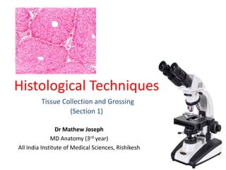
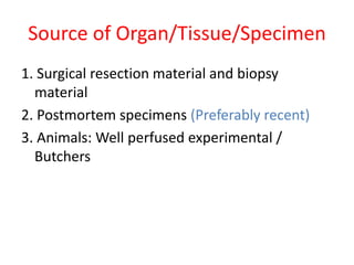
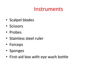
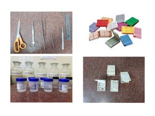
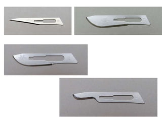
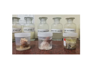
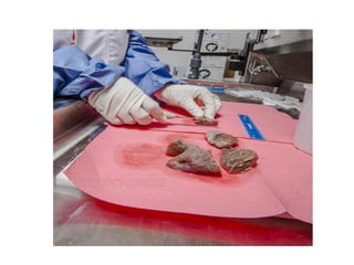
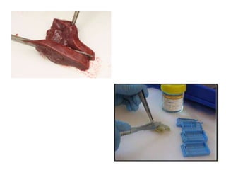
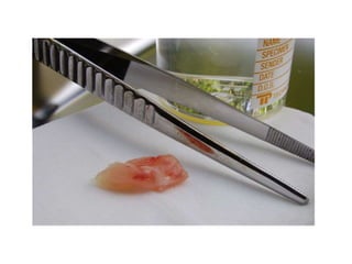
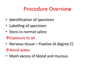
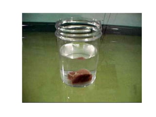
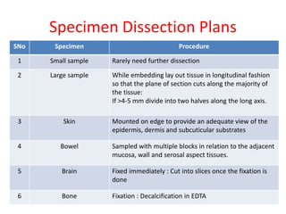
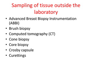
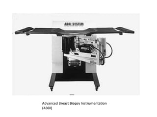
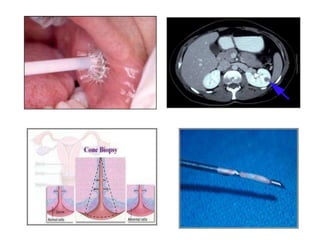
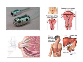
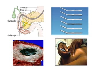
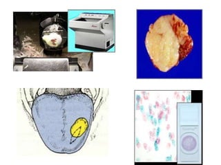
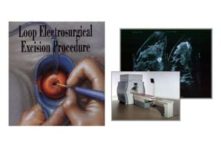
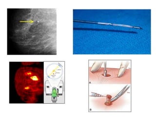
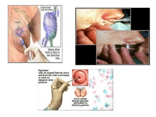
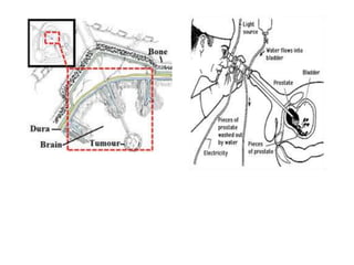
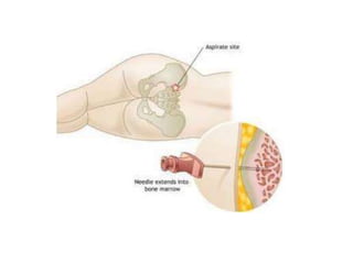
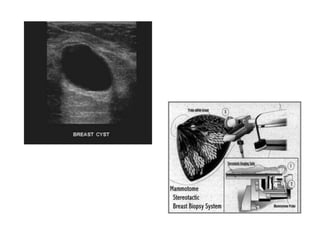
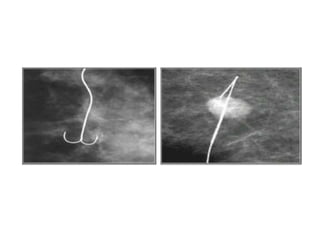
Histological Techniques
Tissue Collection and Grossing
(Section 1)
Dr Mathew Joseph
MD Anatomy (3rd year)
All India Institute of Medical Sciences, Rishikesh
Contents
Tissue collection and Grossing:
• Sources of sampling
• Instruments
• Procedure
• Specimen dissection plan
• Sampling of tissue outside the laboratory
Section 1
Tissue Collection and Grossing
Source of Organ/Tissue/Specimen
1. Surgical resection material and biopsy
material
2. Postmortem specimens (Preferably recent)
3. Animals: Well perfused experimental /
Butchers
Workstation
• Well illuminated room
• Ventilation and exhaust fan
• Appropriate sink
• Waste disposal unit
• Firm, even and clean surface
Instruments
• Scalpel blades
• Scissors
• Probes
• Stainless steel ruler
• Forceps
• Sponges
• First-aid box with eye wash bottle
Procedure Overview
• Identification of specimen
• Labelling of specimen
• Store in normal saline
Exposure to air
• Nervous tissue – Fixative (4 degree C)
Avoid water
• Wash excess of blood and mucous
Specimen Dissection Plans
SNo Specimen Procedure
1 Small sample Rarely need further dissection
2 Large sample While embedding lay out tissue in longitudinal fashion
so that the plane of section cuts along the majority of
the tissue:
If >4-5 mm divide into two halves along the long axis.
3 Skin Mounted on edge to provide an adequate view of the
epidermis, dermis and subcuticular substrates
4 Bowel Sampled with multiple blocks in relation to the adjacent
mucosa, wall and serosal aspect tissues.
5 Brain Fixed immediately : Cut into slices once the fixation is
done
6 Bone Fixation : Decalcification in EDTA
Sampling of tissue outside the
laboratory
• Advanced Breast Biopsy Instrumentation
(ABBI)
• Brush biopsy
• Computed tomography (CT)
• Cone biopsy
• Core biopsy
• Crosby capsule
• Curettings
Sampling of tissue outside the
laboratory
• Ductal lavage
• Endoscopic biopsy
• Endoscopic retrograde
cholangiopancreatography (ERCP)
• Evacuation
• Excision biopsy
• Fine needle aspiration
• Fluroscopy
Sampling of tissue outside the
laboratory
• Frozen section
• Imprint
• Incision biopsy
• Liquid based cytology (LBC)
• Loop electrosurgical excison procedure (LEEP)
• Magnetic resonance imaging (MRI)
• Mammography
• Needle biopsy
Sampling of tissue outside the
laboratory
• Positron emission tomography with
fluorodeoxy- glucose (PET with FDG)
• Punch biopsy
• Sentinel node biopsy
• Shave biopsy
• Smears
• Stereotactic biopsy
• Transurethral resection
Sampling of tissue outside the
laboratory
• Trephine (bone marrow) biopsy
• Ultrasound
• Vacuum-assisted biopsies
• Wire localisation biopsy
Advanced Breast Biopsy Instrumentation
(ABBI)
Thank You

Histological Techniques: Section 1:Tissue collection and Grossing

  • 1.
    Histological Techniques Tissue Collectionand Grossing (Section 1) Dr Mathew Joseph MD Anatomy (3rd year) All India Institute of Medical Sciences, Rishikesh
  • 2.
    Contents Tissue collection andGrossing: • Sources of sampling • Instruments • Procedure • Specimen dissection plan • Sampling of tissue outside the laboratory
  • 4.
  • 5.
    Source of Organ/Tissue/Specimen 1.Surgical resection material and biopsy material 2. Postmortem specimens (Preferably recent) 3. Animals: Well perfused experimental / Butchers
  • 6.
    Workstation • Well illuminatedroom • Ventilation and exhaust fan • Appropriate sink • Waste disposal unit • Firm, even and clean surface
  • 8.
    Instruments • Scalpel blades •Scissors • Probes • Stainless steel ruler • Forceps • Sponges • First-aid box with eye wash bottle
  • 15.
    Procedure Overview • Identificationof specimen • Labelling of specimen • Store in normal saline Exposure to air • Nervous tissue – Fixative (4 degree C) Avoid water • Wash excess of blood and mucous
  • 17.
    Specimen Dissection Plans SNoSpecimen Procedure 1 Small sample Rarely need further dissection 2 Large sample While embedding lay out tissue in longitudinal fashion so that the plane of section cuts along the majority of the tissue: If >4-5 mm divide into two halves along the long axis. 3 Skin Mounted on edge to provide an adequate view of the epidermis, dermis and subcuticular substrates 4 Bowel Sampled with multiple blocks in relation to the adjacent mucosa, wall and serosal aspect tissues. 5 Brain Fixed immediately : Cut into slices once the fixation is done 6 Bone Fixation : Decalcification in EDTA
  • 18.
    Sampling of tissueoutside the laboratory • Advanced Breast Biopsy Instrumentation (ABBI) • Brush biopsy • Computed tomography (CT) • Cone biopsy • Core biopsy • Crosby capsule • Curettings
  • 19.
    Sampling of tissueoutside the laboratory • Ductal lavage • Endoscopic biopsy • Endoscopic retrograde cholangiopancreatography (ERCP) • Evacuation • Excision biopsy • Fine needle aspiration • Fluroscopy
  • 20.
    Sampling of tissueoutside the laboratory • Frozen section • Imprint • Incision biopsy • Liquid based cytology (LBC) • Loop electrosurgical excison procedure (LEEP) • Magnetic resonance imaging (MRI) • Mammography • Needle biopsy
  • 21.
    Sampling of tissueoutside the laboratory • Positron emission tomography with fluorodeoxy- glucose (PET with FDG) • Punch biopsy • Sentinel node biopsy • Shave biopsy • Smears • Stereotactic biopsy • Transurethral resection
  • 22.
    Sampling of tissueoutside the laboratory • Trephine (bone marrow) biopsy • Ultrasound • Vacuum-assisted biopsies • Wire localisation biopsy
  • 23.
    Advanced Breast BiopsyInstrumentation (ABBI)
  • 35.