Downloaded 48 times


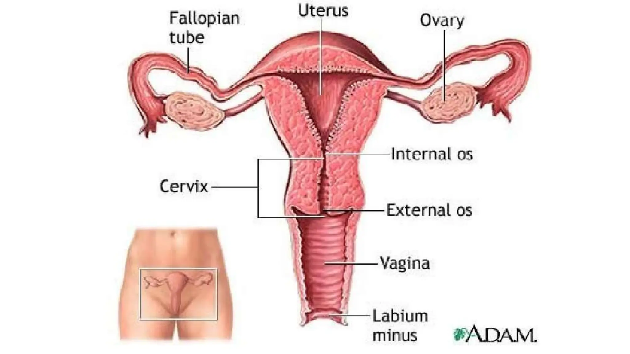
![Female Reproductive System: Major Organs
• Vagina
• Cervix
• Uterus
• Uterine tubes [fallopian tubes]
• Ovaries [gonads]](https://image.slidesharecdn.com/anatomyoffemalereproductivesystem-240227043218-44f8cb97/75/Anatomy-of-Female-Reproductive-System-pptx-6-2048.jpg)


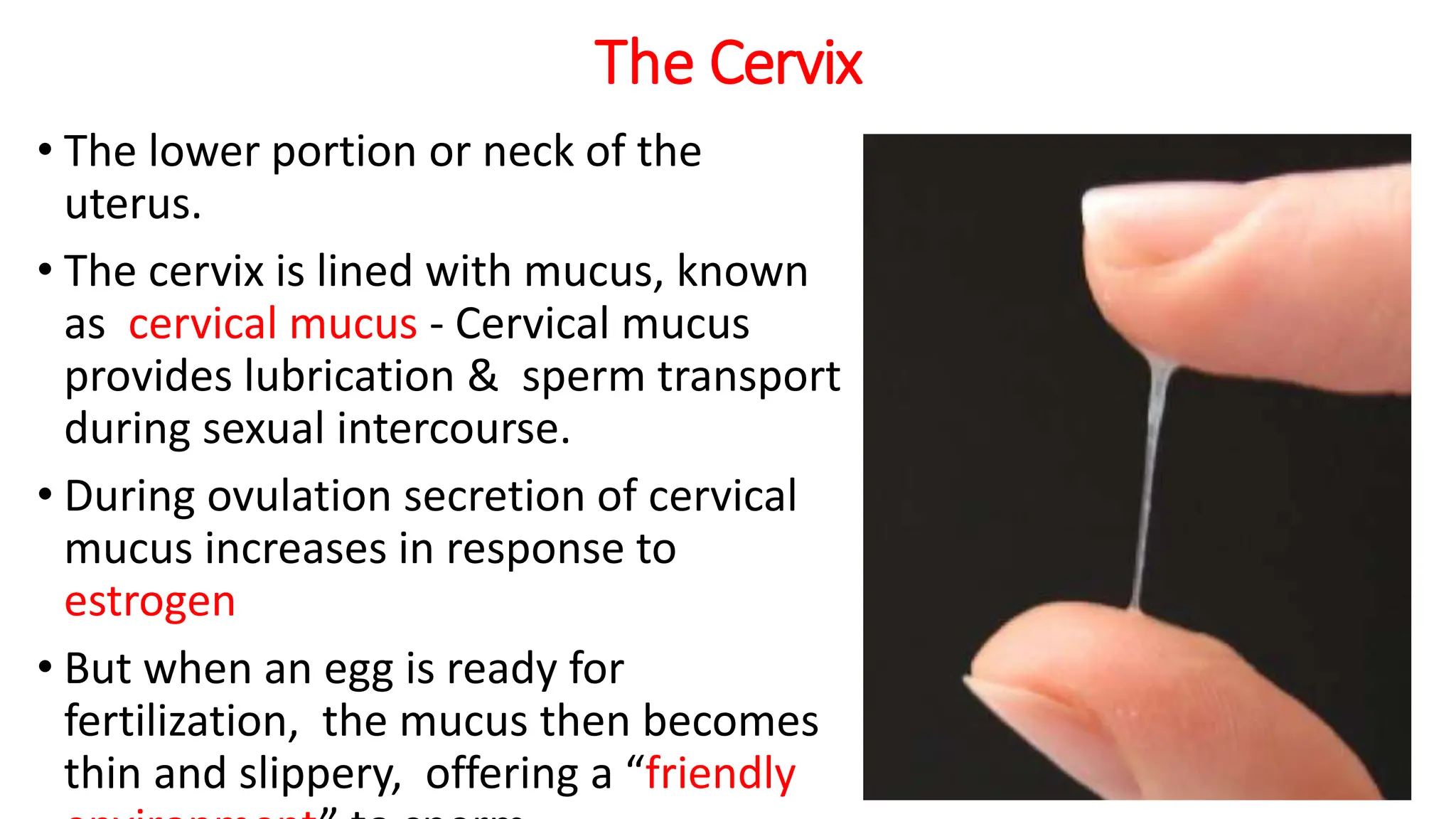


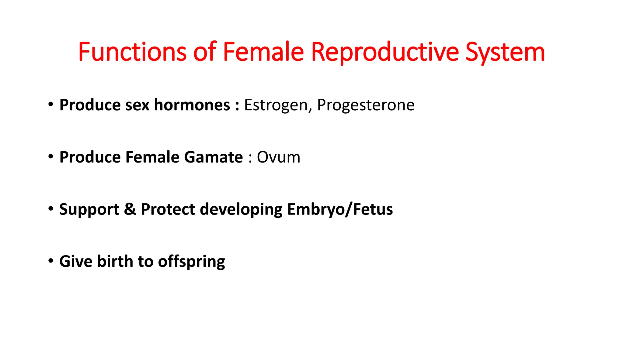
The document provides a comprehensive overview of the female reproductive system, detailing its major organs including the vagina, cervix, uterus, fallopian tubes, and ovaries, along with their functions. It describes the anatomy and physiological roles of these components, such as hormone production, gamete formation, and supporting pregnancy. Additionally, it covers aspects like blood supply, lymphatic drainage, and nerve supply associated with the reproductive organs.


