Learning Objectives
By theend of this session students are
expected to be able to:
Define common terms used in
diagnostic Histopathological
specimen (Autopsy, Biopsy, Cytology,
Pathology, Pathogenesis,
histopathology, Cytopathology)
List types of biopsies (Incisional,
excisional, punch biopsies, curetting,
chips, wide bore needle, and fine
needle aspiration)
3.
Learning objectives
Explain eachtype of biopsy
(Incisional, excisional, curetting,
chips, wide bore needle, and fine
needle aspiration)
CONT…..
Autopsy/post mortem ornecropsy ;
Examination of dead body to
determine the cause of death.
Autopsy specimen ;
Tissue specimen taken from a dead
body.
Pathology;
The study of structural and
functional causes of diseases.
Pathogenesis;
Is the mechanisms by which
6.
Cont..
Soft tissue;
Refers toany non-epithelial tissue
other than bone, cartilage, CNS,
haematopoietic, and lymphoid tissue
7.
Types of biopsies
•To diagnose medical condition a sample
of tissue has to be taken from the
patient and send to the
histopathology/Cytopathology for
processing and examination.
• The procedure /technique of collecting
the specimen give biopsy their names.
Excisional biopsy
Incisional biopsy
Endoscopic biopsy
1. Excisional biopsy:
–Involvesthe removal of whole organ
or affected areas at operation so
as to allow accurate diagnosis.
–For example breast lump, spleen and
lymph nodes are obtained as
excisional biopsies.
–Excisional biopsy helps to
determine whether the surgical
treatment was effective by
examining the distance of
10.
Cont..
– the lesionor tumour from the
margins of the excisized tissue or
organ.
11.
2. Incisional biopsy:
–Involves the removal of only a portion of
organ or affected area so as to allow
accurate diagnosis.
– This type of biopsy is commonly used
for tumours of soft tissue (muscle, fat,
connective) to distinguish benign
conditions from malignant soft tissue
tumours.
– A benign means an abnormal but
non cancerous
12.
3. Endoscopic biopsy;
–The endoscope is done by inserting
the device through natural body
orifice or a small surgical incision.this
involves the use of fibre optic
endoscope (long ,thin tube) in which
device is inserted into the lumen of
an organ for visualization of an
abnormal area on the lining of an
organ in order to obtain small
amount of tissue for study.
13.
definition
Endoscopy a proceduredone to examine
structure inside your body by using long thin
tube(endoscope) inside your body until it reach
the organ or area they need to check.
14.
CONT…
• Endoscope ismainly done to GIT
(alimentary tract endoscopy), urinary
bladder (cystoscopy), abdominal cavity
( laparoscopy ), joint cavity
(anthroscopy), bronchial system
(laryngoscopy and bronchoscopy) and
mid portion of the chest
(mediastinoscopy)
15.
definition
• Pap testa procedure in which a small brush
is used to gently remove cells from the
surface of cervix to under microscope for
cervical cancer.
16.
4. Colposcopic biopsy:
•Involves pinching of the biopsy from
the abnormal area on the cervix with
the help of “focusing telescope” that is
inserted into the female genitalia.
Normally is done to a patient after
abnormal Pap- smear is obtained.
• One the advantage of colposcopy is
that a physician will be able to see in
detail abnormal areas in cervix of the
uterus so that good representation of
the abnormal area can be removed.
17.
5. Stereotactic biopsy
–Isa new technique used
for evaluating breast
lesions.
–Thepatient lies on her
abdomen so that breast
hangs down into a
space that can be x-rayed
by a computerized
imaging device.
18.
Cont..
–The computer displaysthe
mammographic image on a
screen.
– Radiologist identifies the
abnormality and marks it
electronically on the screen.
19.
CONT…..
–Computer then positionsa movable
arm directly over the abnormal area.
A biopsy device is attached to the
arm, and the spring loaded gun
quickly inserts a hollow biopsy
needle into the breast.
–The needle is removed and the
tissue it contains is sent to the
laboratory.
20.
6. Punch biopsy
–This technique is used to sample
skin rashes and small masses.
–After a local anaesthetic is injected
cookie cutter (3 or 4 mm
diameter), is used to cut out a
cylindrical piece of skin.
–The hole is typically closed with
the suture(stitch) and heals with
minimal scarring.
21.
7. Bone marrowbiopsy
• Bone marrow biopsy is taken from
the posterior(back) superior iliac
spine.
• The patient lies on his or her stomach
and local anaesthesia is applied.
• needle is then inserted deeper to
deaden surface membrane covering the
bone.
• A larger rigid syringe with a very
sharp point is then introduced into the
marrow space.
22.
Cont..
• A syringeis attached to the needle
and suction is applied. The marrow
cells are then drawn into the syringe.
• The contents of the syringe (which
looks like blood with tiny chunks of
fat floating around in it) is put onto a
glass slide and smeared out.
• After staining, the cells are
examined under a microscope by
pathologist or haematologist
23.
8. Core needleaspirations (Wide bore
needle biopsy)
• This performed to bone marrow.
• Normally after aspirating material as
bone marrow biopsy a slight larger
needle is used to extract core of bone.
• The calcium is removed from the bone
to make it soft, then processed and
tissue sections are made. Core biopsy
is also called trephine biopsy.
24.
definition
• Fine needleaspirate is used to remove sample
from or abnormal mass to examine it under
microscope.
25.
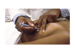
9. Fine needleaspirations
– Is a sample of suspicious mass
removed from the body with the
help of needle (of 22 – 25 gauge)
and cytological gun for diagnostic
purposes.
– Routine injection with that gauge size
of needle is inserted into a tumour
and few tens to thousands of cells
are drawn up into syringe then are
smeared on the slide, stained and
examined under a microscope by the
pathologist or cytopathologist.
26.
Cont..
–The target canbe superficial
“lump (uvimbe)or bump” or
radiologically imaged.
superficial “lump or bump” can be
found in thyroid, lymph nodes,
salivary gland, breasts etc.
– Deep seated and radiologically
imaged can be done to the lung,
liver, pancreas, kidney and
retro-peritoneum
27.
1o. Curettings
–Are uterinematerials obtained by
scrapping endometrial
wall(lining of uterus). This can be
done to look for endometrial
cancer, ovarian cancer, ectopic
pregnancy or miscarriage.
–Normally a surgical instrument
shaped like a scoop or spoon is
used to remove tissue or growth
from the uterine wall.
28.
11. Chips
• Aresmall soft tissues fragments obtain
by curetting from prostate.
30.
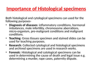
Importance of Histologicalspecimens
Both histological and cytological specimens can used for the
following purposes-
• Diagnosis of diseases: inflammatory conditions, hormonal
imbalances, male infertility, chromosome aberrations,
micro-organism, pre-malignant conditions and malignant
conditions.
• Teaching: Gross tissues specimen and stained slides can be
used for teaching purposes.
• Research: Collected cytological and histological specimens
and archived specimens are used in research works.
• Forensic: Histological and cytological specimens can be
used in determining the cause of death and legal issue e.g.
determining a murder, rape cases, paternity dispute.
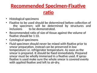
Recommended Specimen-Fixative
ratio
• Histologicalspecimens
• Fixative to be used should be determined before collection of
the specimen; will be determined by structures and
inclusions to be demonstrated.
• Recommended ratio of size of tissue against the volume of
fixative should be 1:10.
• Cytological specimens
• Fluid specimen should never be mixed with fixative prior to
smear preparation, instead can be preserved in low
temperature i.e. refrigerator temperature. As soon as the
smear is prepared. It should be fixed immediately. Prepared
smear must be wholly immersed in a fixative used. If spray-
fixative is used make sure the whole smear is covered evenly
with applied fixative and left to air-dry.
34.
Key points
• Histologicalspecimens includes; Inclusion biopsy,
Exclusion biopsy and Curretings.
• Cytological specimens includes: Needle aspirates,
Smears, Washings/lavage, Effusions, Urine and Sputum
• Recommend containers for histological and cytological
specimens are histological bottles and universal bottle
respectively; however smears are prepared on glass
slides.
• To histological specimens, recommended ratio of tissue
size against volume of fixative is 1:10. To cytological
specimen make sure it is evenly covered or wholly
immersed in the fixative used.
35.
Key Points
• Histopathologyis a study of
abnormal or diseased tissue.
• Cytology is a study of
structure, composition and
function of cells.
• Eleven common types of biopsies
includes Excisional biopsy Incisional,
Endoscopic, Colposcopy, Stereotactic
Punch, Bone marrow, Core needle
aspirations (Wide bore needle biopsy),
Fine needle aspirations, Curetting and
Chips.


![What is histopathology[1]](https://cdn.slidesharecdn.com/ss_thumbnails/whatishistopathology1-190401112812-thumbnail.jpg?width=640&height=640&fit=bounds)


![BIOPSY IN SURGERY 1 [Auto-savedd] 3.pptx](https://cdn.slidesharecdn.com/ss_thumbnails/biopsyinsurgery1auto-saved3-251018145913-707215d9-thumbnail.jpg?width=640&height=640&fit=bounds)


