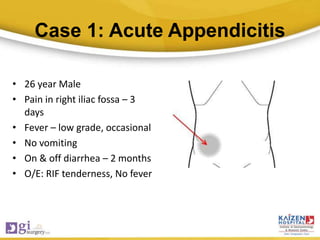
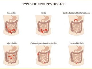
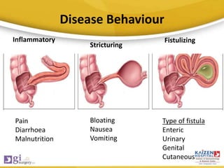
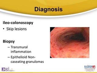
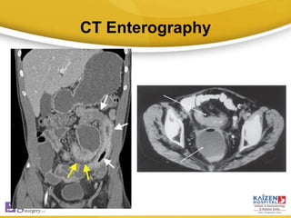
Crohn’s Disease
Are we missing the diagnosis
?
Dr. Harsh Shah
MS, FMAS, DNB, MCh(GI)
GI & HPB Surgeon
Kaizen Hospital,
Institute of GE, Ahmedabad, India
Inflammatory bowel disease
• Crohn’s disease
• Ulcerative colitis
• Indeterminate colitis
Case 1: Acute Appendicitis
• 26 year Male
• Pain in right iliac fossa – 3
days
• Fever – low grade, occasional
• No vomiting
• On & off diarrhea – 2 months
• O/E: RIF tenderness, No fever
Investigation
• Hb – 9.2 g%
• TLC – 11000
• P78, L20
• ESR – 60 mm/1st hour
• Urine, RFT - Normal
USG Abdomen
• Inflammed appendix. Probe
tenderness present.
• Thickened wall of terminal
ileum & cecum
• CT Scan not done
• Appendicectomy
performed.
Post-operative course
• Uneventful
• Patient discharged on day 2
• HPE – Acute appendicitis
Follow-up
• Presented to OPD with wound infection on POD-5
• Stitch opened  Fecal discharge
• No fever. Taking diet. Passing motions.
Fecal fistula draining 150ml per day
What went wrong ?
• History of diarrhea for 2 months
• Hb - Low
• Thickened cecum & terminal ileum on USG
• CT Scan: Thickened terminal ileum/cecum.
• Colonoscopy : Biopsy from cecum & terminal ileum
Crohn’s Disease
CT Scan
Learning Point
• Careful history taking in acute appendicitis
• When in doubt – CT scan
Case 2: Fistula in Ano
• 38 year/Male
• Perianal pus discharge - 3
months
• Taken antibiotics (Ciplox +
Metro) course twice
O/E:
• Low perianal fistula
• Internal opening at 6 o’clock
• Operated for Fistulotomy
Post-operative course
• Uneventful
• Wound healed in 1 month
Follow-up
• Two fistula opening in perianal
region away from previous site
• One was high fistula other was
low fistula on MR Fistulogram
Further treatment
• Underwent seton placement for high fistula &
fistulotomy for low fistula
3 months later
• Patient developed one more high fistula
What should be done ?
• Biopsy of the fistula tract
Crohn’s Disease
Learning Point
• Recurrent perianal fistula – Biopsy of the
fistula tract
Case 3: Intestinal Tuberculosis
• 68 year female
• Pain abdomen, wt loss x 6 months
• USG Abdomen: Thickening in ileo-cecal region
• Started on ATT empirically, No biopsy
• Abdominal distension, Vomiting x 2 months
Clinical Examination
• Signs of small bowel
obstruction
• X-ray
Surgery
• Laparotomy
• Dilated small bowel
• Terminal ileum &
cecum resected
• Anastomosis done
Post-operative course
• Uneventful
• Discharged on POD-6
• Biopsy:
– Non-caseating granuloma
– AFB stain negative
– s/o crohn’s disease
• Started on Azathioprine
• Doing well
Learning Point
• Never start ATT without biopsy confirmation
CROHN’S DISEASE
Also Known as regional ileitis
Overview
• Dr Bernard Crohn, 1932
• Chronic non-specific inflammation of GIT involving all the layers
• Course is relapsing & remitting
• Can involve any part from mouth to anus
• Propensity for ileo-cecal region
• F>M, Bimodal age distribution
Aetiology & Pathogenesis
Genetic
susceptibility
Environmental
factors
Host
Immune
Response
Crohn’s Disease
Clinical features
• Pain abdomen
• Diarrhoea
• Anemia
• Low grade fever
• Weight loss
• Fatigue/malaise
Disease Behaviour
Pain
Diarrhoea
Malnutrition
Bloating
Nausea
Vomiting
Type of fistula
Enteric
Urinary
Genital
Cutaneous
Inflammatory
Stricturing
Fistulizing
Diagnosis
Ileo-colonoscopy
• Skip lesions
Biopsy
– Transmural
inflammation
– Epithelioid Non-
caseating granulomas
CT Enterography
Ancillary Investigations
• ESR/CRP
• ASCA (Anti-saccharomyces cerevisae)
• Fecal calprotectin
Treatment
• Disease can be controlled, not cured
• Relapses are common
• Preserve the bowel
Medical Treatment
• Induction of remission
– Steroids
– Anti-TNF(Infliximab)
– Antibiotics
• Maintenance of remission
– Azathioprine
– Infliximab
• Nutrition
Surgical Treatment
Stricturoplasty
Side to side anastomosis
Take Home messages
• Ileocecal region most common site
• Site & behaviour variable
• Relapsing course
• Commonly confused with intestinal TB
• Colonoscopic biopsy confirmatory
• Immunomodulators & judicious use of surgery

Crohn's Disease